Opioid receptors are members of the seven transmembrane G-protein-coupled receptor (GPCR) superfamily (Ref. Reference Kieffer1). There are three types of opioid receptors: mu opioid receptor (MOR), kappa opioid receptor (KOR) and delta opioid receptor (DOR). At the cellular level, opioid receptors are coupled to G i/G o proteins and their activation leads to inhibition of adenylyl cyclase activity and voltage-gated Ca2+ channels, increases in mitogen-activated protein kinase (MAPK) phosphorylation and in the activity of inwardly rectifying K+ channels and phospholipase C beta (Ref. Reference Waldhoer, Bartlett and Whistler2). The signalling cascades initiated by the activation of opioid receptors induce the transcription of genes that regulate cellular differentiation, proliferation and survival (Ref. Reference Chen, Law and Loh3). At the systems level, opioid receptor activation leads to a number of physiological outcomes including analgesia, feelings of euphoria and anxiety, respiratory depression, constipation, immunosuppression and changes in feeding and locomotor activity (Ref. Reference Waldhoer, Bartlett and Whistler2). Despite the large number of physiological outcomes associated with opioid signalling, these receptors are largely valued for their role in inducing analgesia.
In general, studies show that MOR- or DOR-selective agonists can induce both analgesia and reward (Ref. Reference Waldhoer, Bartlett and Whistler2). The use of transgenic mice has provided greater insight into the potential roles of individual opioid receptor subtypes. These studies indicate that the analgesic effects of clinical opioids such as morphine and the rewarding properties associated with nonopiate drugs of abuse such as marijuana (Ref. Reference Filbey4) are primarily mediated by the activation of MOR (Ref. Reference Matthes5). However, DOR has been shown to have a role in the regulation of emotional responses associated with opioid use (Refs Reference Waldhoer, Bartlett and Whistler2, Reference Kieffer and Evans6). Despite this knowledge, the major clinical limitation of opioids – tolerance and development of addiction following chronic use – has not yet been overcome (Refs Reference Kieffer1, Reference Waldhoer, Bartlett and Whistler2, Reference Matthes5, Reference Smith and Milligan7). To better understand how tolerance and addiction can be dissociated from the desirable outcome of analgesia, the field has begun to take a closer look at the interactions between MOR and DOR, and more recently on the role of the MOR–DOR heteromer.
Over the last decade, several studies have shown that GPCRs can form homomers (associations between same receptor subtype) or heteromers (associations between different receptor subtypes or between different GPCRs) (Ref. Reference Smith and Milligan7). Such receptor interactions have been shown to modulate ligand binding, to affect interactions with intracellular scaffolding proteins, to alter signalling cascades induced following receptor activation and to alter receptor trafficking (reviewed in Refs Reference Gomes8, Reference Satake and Sakai9, Reference Gonzalez-Maeso10). In the case of opioid receptors, several studies have reported receptor heteromers that show receptor binding, signalling and trafficking properties that are distinct from those of receptor monomers or homomers (Refs Reference Smith and Milligan7, Reference Gomes11, Reference Rozenfeld and Devi12, Reference Rozenfeld and Devi13, Reference Williams and Devi14, Reference Chakrabarti, Liu and Gintzler15, Reference Gupta16). In addition, studies have shown that regulation of heteromer formation at the cellular level has the potential to modulate opioid signalling both in vitro and in vivo (Refs Reference Rozenfeld and Devi12, Reference Rozenfeld and Devi13). Finally, a recent study suggested that opioid receptor signalling could be modulated not only by the levels and types of heteromers present in a cell, but also by the activation state of both the opioid receptor and its partner GPCR (Ref. Reference Gomes11). This suggests the intriguing possibility of tissue- or disease-specific expression of heteromeric complexes that could be targeted to develop drugs that specifically target the heteromer in a disease state. In this review, we discuss MOR–DOR heteromers and the potential for targeting these complexes to achieve antinociception or analgesia without the adverse effects of tolerance, dependence and addiction associated with chronic opioid use.
Opioid receptor complexes
The existence of mu–delta ‘opioid receptor complexes’ was postulated as early as 1981 (Ref. Reference Rothman and Westfall17), and the possibility that allosteric binding could contribute to the heterogeneity of opioid receptor interactions was considered even earlier (Ref. Reference Lee and Smith18). Such hypotheses were drawn from the results of studies probing the activity of opioid peptides in radiolabelled binding assays and in guinea pig ileum and mouse vas deferens contraction assays (Ref. Reference Lord19). These studies revealed a surprising diversity in opioid receptor activity that, at the time, was attributed to ligand binding at several sites of a single receptor or binding to altered forms of the receptor (Ref. Reference Lord19). These opioid ligands also demonstrated heterogeneity in binding characteristics, under a range of ligand concentrations and in the presence of endogenous opioids (Ref. Reference Lord19).
Support for MOR–DOR complex formation was provided by competitive binding assays that measured the ability of a test drug to displace the binding of a known radiolabelled ligand in cellular membrane preparations (Ref. Reference Rothman and Westfall20). In this type of assay, inhibition of radiolabelled ligand binding is usually attributed to competitive displacement of the ligand by the test drug. However, studies examining competitive displacement of radiolabelled Leu-enkephalin by unlabelled Leu-enkephalin (a DOR receptor agonist) in the absence or presence of different concentrations of morphine (recently shown to exert its effects through MOR using transgenic animals) found that the latter shifted the Leu-enkephalin binding kinetics from being competitive to noncompetitive (Refs Reference Rothman and Westfall17, Reference Rothman and Westfall20). This suggested that morphine, bound to a receptor not labelled by radiolabelled Leu-enkephalin, allosterically induced an apparent loss of Leu-enkephalin-labelled receptors (Refs Reference Rothman and Westfall17, Reference Rothman and Westfall20).
Studies using the tail-flick assay to measure the analgesic properties of different opioid ligands in vivo showed that their potency was consistently greater than what would have been predicted from data obtained from in vitro binding assays (Ref. Reference Barrett and Vaught21). For example, subanalgesic doses of the DOR-selective agonist, Leu-enkephalin, could potentiate morphine-mediated analgesia (Ref. Reference Vaught, Rothman and Westfall22). This suggested the possibility of interactions between opioid receptor subtypes and provided a potential mechanism by which the site-specific activity of paired receptor complexes could dictate the outcome of opioid drug treatment (Ref. Reference Vaught, Rothman and Westfall22). This model assumed that the availability of opioid receptors and their endogenous ligands is regionally localised and regulated. The observed effect of any given opioid treatment would therefore depend on the interaction, or interference, of region-specific factors in the microenvironment of the receptors. Thus, it becomes apparent that in vitro binding assay measurements cannot necessarily predict the in vivo analgesic utility of opioid ligands (Ref. Reference Vaught, Rothman and Westfall22). Furthermore, both in vitro and in vivo assays could be affected by differences in the ratio of MOR to DOR as well as the presence or absence of endogenous ligands (Ref. Reference Gomes11).
Studies examining receptor expression and interaction at the cellular level have implicated MOR in the regulation of DOR expression at the plasma membrane (Refs Reference Gendron23, Reference Morinville24, Reference Cahill25). For example, chronic exposure to morphine leads to an increase in the surface expression of DOR in cultured cortical neurons and in neurons in the dorsal horn of the spinal cord in vivo (Refs Reference Morinville24, Reference Cahill25); this does not occur in MOR-deficient animals (Ref. Reference Morinville24). Similarly, increases in the surface expression of DOR were also described in cultured dorsal root ganglion (DRG) neurons following chronic exposure to morphine in vitro (Ref. Reference Gendron23). These studies indicate a role for MOR in mediating DOR trafficking and cell surface availability, thereby providing further support for interactions between MOR and DOR. Such results suggest that DOR regulates morphine dependence – possibly by modulating MOR expression – and reciprocally, that MOR regulates DOR expression and availability in response to chronic morphine. Although MOR–DOR signalling and the definitive contributions of each receptor to heteromer-mediated signalling are still under scrutiny, these studies make clear the functional relevance of MOR–DOR interactions in pain and tolerance development. Substantiating these findings through investigation of the physical interaction between MOR and DOR has become essential to delineate the contribution of each receptor towards MOR–DOR interactions. Studies examining the coexpression of these receptors, as well as their direct physical interaction, are discussed below.
MOR and DOR transgenic mice
Studies using transgenic mice with genetic deletions of either MOR or DOR provided tremendous insight into the functional roles of these receptors in antinociception and other physiological and pharmacological outcomes such as tolerance (Refs Reference Matthes5, Reference Zhu26, Reference Kieffer and Gaveriaux-Ruff27, Reference Billa, Xia and Moron28). These transgenic mice served as appropriate controls to determine the contribution of each opioid receptor subtype to a given physiological response. In addition, these transgenic mice have enabled studies to assess the contribution of receptor–receptor interactions to opioid-receptor-mediated signalling and behavioural responses (Ref. Reference Kieffer and Gaveriaux-Ruff27).
Early investigations by Matthes and colleagues found that MOR-deficient mice did not show morphine-mediated analgesia, physical dependence or conditioned place preference (Ref. Reference Matthes5). These results indicated the unique requirement for MOR in morphine-mediated antinociceptive responses and suggested that MOR is the target of morphine in vivo and indeed the receptor responsible for morphine-induced antinociception and behaviour. However, this finding was unexpected, given previous studies showing that, at high doses, morphine can also bind to DOR and KOR and that all three opioid receptor subtypes are involved in the blockade of pain at the level of the spinal cord (Ref. Reference Dickenson29). In addition, administration of DOR-selective agonists to MOR-deficient mice led to reduced antinociception in the tail-flick test (Refs Reference Matthes5, Reference Matthes30). This suggested that either the predicted involvement of DOR in spinal analgesia was incorrect, or that the availability of MOR was required for DOR-mediated spinal analgesia. One possibility for reduced DOR-mediated antinociception in MOR-deficient mice could be due to impairment of DOR-mediated activity. However, examination of signalling responses using G-protein binding and adenylyl cyclase assays found DOR-mediated signalling to be intact in these animals (Ref. Reference Matthes30). In addition, DOR agonists continued to mediate a peripheral effect, such as inhibition of muscle contraction (using the vas deferens twitch assay) in MOR-deficient mice (Ref. Reference Matthes30). Taken together, these results indicated a normal DOR function in MOR-deficient mice and suggested that optimal DOR-mediated antinociception requires the presence of MOR.
In the case of DOR-deficient animals, studies with Dor1 mutant mice generated by deletion of exon 2 demonstrated a strong reduction in antinociceptive responses to intrathecally administered DOR-selective agonists compared with wild-type animals (Ref. Reference Zhu26). However, these agonists showed antinociceptive effects following intracerebroventricular administration, suggesting that they exerted their supraspinal analgesic effects at a receptor other than DOR (Ref. Reference Zhu26). Interestingly, these DOR-deficient mice showed normal morphine-mediated antinociceptive responses though the development of tolerance to morphine was abolished (Ref. Reference Zhu26). Thus, these studies suggest interactions between MOR and DOR and that the latter has a role in the development of tolerance to morphine.
Further support for functional interactions between MOR and DOR comes from a study examining the coupling of MOR to voltage-gated Ca2+ channels in DRG neurons from wild type and from animals lacking DOR (Ref. Reference Walwyn31). This work found that the MOR-selective agonists, morphine and DAMGO, were less effective at inhibiting the activity of voltage-gated Ca2+ channels in neurons lacking DOR compared with wild-type neurons (Ref. Reference Walwyn31). These effects were neither because of decrease in the density and function of voltage-gated Ca2+ channels nor because of changes in MOR mRNA levels; this suggests functional interactions between MOR and DOR at the level of inhibition of voltage-gated Ca2+ channel activity.
MOR–DOR interacting complexes – anatomical and molecular evidence
Demonstration of MOR–DOR heteromer formation requires that MOR and DOR be present not only in the same cell, but also in the same subcellular compartment. Studies investigating the distribution of MOR and DOR in the dorsal horn of the rat spinal cord using dual immunocytochemical analysis and electron microscopy revealed the presence of both MOR and DOR in the same somatodendritic compartments, both in discrete areas of the plasma membrane and in organelles (Ref. Reference Chengm, Liu-Chen and Pickel32). Expression of MOR and DOR in the same cells was also revealed using in situ hybridisation (Ref. Reference Wang and Wessendorf33). These studies detected coexpression of mRNA encoding MOR and DOR in spinally projecting neurons of the rostral ventromedial medulla (RVM) (Ref. Reference Wang and Wessendorf33). These data provided one of the first demonstrations that MOR and DOR colocalised in neurons of central nervous system regions associated with nociception.
The existence of MOR–DOR interacting complexes has been demonstrated more directly through the use of heterologous expression systems (Refs Reference Gomes34, Reference Gomes, Filipovska and Devi35, Reference George36, Reference Gomes37). Early work from our laboratory revealed that interacting complexes could be isolated both from heterologous cells expressing recombinant receptors as well as from endogenous tissue expressing native opioid receptors (Refs Reference Gomes34, Reference Gomes37). Furthermore, close proximity to form interacting complexes between MOR and DOR was demonstrated by coimmunoprecipitation experiments (Refs Reference Gomes34, Reference Gomes37). In these studies, cells were transfected with either FLAG-tagged MOR, Myc-tagged DOR or both epitope-tagged receptors. The lysates from these cells were then immunoprecipitated with antibodies directed against the Myc epitope. The resulting immunoprecipitates were probed with anti-FLAG antibodies by Western blot, revealing a distinct band at ~150 kDa only in cells coexpressing both receptors (Ref. Reference Gomes37). Bioluminescence resonance energy transfer assays using MOR–luciferase- and DOR–YFP (yellow fluorescent protein)-tagged receptors in heterologous live cells supported the existence of receptors that are in close enough proximity (<5–10 nm apart), to form interacting complexes (Ref. Reference Gomes34).
However, it is important to note that there is a debate on whether MOR–DOR heteromerisation occurs, given a recently published study challenging the presence of MOR and DOR in the same cells (Ref. Reference Scherrer38). This study used a knock-in mouse model with DOR tagged with enhanced green fluorescent protein (eGFP) and used antibodies directed against GFP or MOR to examine colocalisation between the eGFP-tagged DOR and endogenous MOR. The authors reported colocalisation in <5% of DRG neurons in vivo (Ref. Reference Scherrer38); these data contradicted results from other studies reporting substantial MOR–DOR colocalisation (Refs Reference Wang and Wessendorf33, Reference Wang39). This might be attributable to a number of important factors. Of note is the relatively higher level of expression of DOR in these knock-in mice (Ref. Reference Scherrer40) and the possibility that there is an increase in surface expression of DOR in these mice as a direct consequence of the incorporated C-terminal GFP tag (Ref. Reference Wang41). In addition, the antibody directed against GFP used in this study for the visualisation of eGFP-tagged DOR could exhibit higher avidity for GFP than the MOR-directed antibody exhibits for MOR (Ref. Reference Scherrer40). Finally, a recent study reported that increased surface expression of DOR attenuated the maturation of MOR (Ref. Reference Decaillot42). These factors could contribute to an underestimation of MOR–DOR colocalisation in vivo.
The dynamics underlying the formation of MOR–DOR heteromers, as well as the molecular mechanisms underlying their trafficking and surface expression, remains an issue of significant interest to the field. It has been hypothesised that GPCR heteromers are formed intracellularly before trafficking and insertion at the plasma membrane (Refs Reference Wang39, Reference Decaillot42). Early reports using cells that constitutively express MOR and where DOR expression was induced by ponasterone A treatment suggested that the formation of MOR–DOR complexes occurred at the cell surface and required interactions with Gi/o proteins (Ref. Reference Law43). This study, however, examined trafficking of biotinylated cell surface receptors and so could not examine whether the induced DOR was associated with pre-existing MOR (which is constitutively expressed) or with newly synthesised MOR nor did it examine colocalisation of MOR and DOR in various cellular compartments. Work from our own laboratory has showed that putative MOR–DOR heteromers are retained in the Golgi complex and require the presence of a specific receptor transport protein such as RTP4 (Ref. Reference Decaillot42). This transport protein prevents ubiquitylation and proteasomal degradation of MOR–DOR complexes, leading to enhanced heteromer trafficking to the cell surface (Ref. Reference Decaillot42). We further used antibodies that selectively recognise MOR–DOR heteromers to show that long-term morphine treatment leads to an increase in heteromer levels in cells endogenously expressing both receptors (Ref. Reference Gupta16). Taken together with a report showing that morphine could help rescue cell surface expression of mutant MOR (Ref. Reference Chaipatikul44), our findings indicate that morphine can function as a pharmaco-chaperone that promotes MOR–DOR heteromer trafficking from the Golgi to the cell surface. This possibility is interesting in the light of studies showing that prolonged morphine treatment increases cell surface availability of DOR (Refs Reference Gendron23, Reference Morinville24, Reference Cahill25). Additional studies will be needed to further elucidate molecular mechanisms underlying maturation and trafficking of the MOR–DOR heteromers.
Although not much is known about the mechanism of MOR–DOR heteromer formation, there is strong molecular and immunohistochemical evidence showing that DOR is coexpressed with MOR in the same cellular environment. Cells expressing both MOR and DOR have been identified in the GABAergic neurons of the hippocampus (Ref. Reference Stumm45). Additionally, small DRG neurons were found to coexpress MOR and DOR at the single-cell level (Ref. Reference Wang39) and functional studies indicated a role for MOR–DOR interaction in synaptic transmission and opioid analgesia (Ref. Reference Wang39). Although the debate over the existence of MOR–DOR heteromers continues, the recent generation of tools such as monoclonal antibodies that selectively recognise MOR–DOR heteromers (Ref. Reference Gupta16) or of agents that disrupt the heteromer in vivo (Ref. Reference He46) represents an important step towards the resolution of these findings. These important experimental results will be discussed in greater detail below.
Biochemical characterisation of the MOR–DOR heteromer
Numerous investigations have contributed to our current understanding of the biochemical characteristics of MOR–DOR complexes through the use of recombinant systems (reviewed in Ref. Reference Smith and Milligan7). These studies indicate that MOR–DOR heteromers show a pharmacological profile that is distinct from that of either MOR or DOR alone (Fig. 1). Such work has provided direct evidence for the formation of MOR–DOR heteromers, heteromer-induced alteration of ligand-binding properties, alteration of cyclic adenosine monophosphate (cAMP) regulation and signalling, and for changes in the induction of MAPK phosphorylation (Refs Reference Gomes34, Reference Gomes, Filipovska and Devi35, Reference George36, Reference Gomes37). Furthermore, MOR–DOR heteromers have been found to couple to distinct signalling pathways (Refs Reference George36, Reference Rozenfeld and Devi47). One study reported that the MOR–DOR heteromer couples to pertussis-toxin-insensitive inhibitory G protein (Gαz) (Ref. Reference George36), whereas another study found that the heteromers are constitutively coupled to β-arrestin-2 (Ref. Reference Rozenfeld48). Although these findings appear divergent, they are not necessarily in opposition, given the potential for the MOR–DOR heteromer to couple to and signal through different pathways in different brain regions and under a variety of different conditions. Indeed, such distinct coupling might be one of several ways in which MOR–DOR heteromerisation can contribute to alterations in the induction of tolerance in response to morphine and other opiate compounds.
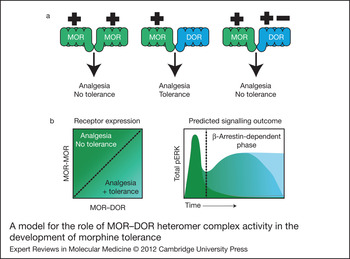
A model for the role of MOR–DOR heteromer complex activity in the development of morphine tolerance. (a) Signalling of MOR in the absence of DOR (left pair), as found in Dor1 mutant mice, is not sufficient to the development of tolerance. The presence of DOR (centre pair) leads to the development of tolerance, but the occupancy of DOR (right pair) by agonist (+) or antagonist (−) in the context of MOR–DOR is sufficient to abrogate this effect. (b) Increased expression of MOR–DOR heteromer complexes produces a sustained increase in pERK that is associated with β-arrestin-dependent signalling and tolerance induction (dark green). Reduction of MOR–DOR pairs (blue/green) reduces the duration of pERK signalling and subsequent tolerance formation. When there is an abundance of MOR homomers (light blue), there is no secondary, sustained pERK generation over time and tolerance is not developed. Abbreviations: DOR, delta opioid receptor; MOR, mu opioid receptor; pERK, phosphorylated extracellular signal-regulated kinases.

The MOR–DOR heteromerisation-mediated switch from a G-protein to a β-arrestin-2-mediated signalling pathway (Ref. Reference Rozenfeld and Devi47) is intriguing because β-arrestin-2 is found to colocalise at the plasma membrane in cells coexpressing MOR–DOR, but not in cells where either MOR or DOR were expressed alone (Ref. Reference Rozenfeld and Devi47). Furthermore, β-arrestin-2 was found to coimmunoprecipitate with MOR–DOR only in cells coexpressing MOR and DOR (Refs Reference Rozenfeld and Devi47, Reference Rozenfeld48). Accompanying the shift from G-protein- to β-arrestin-2-dependent signalling were marked alterations in the temporal dynamics of extracellular signal-regulated kinases (ERK) phosphorylation in cells coexpressing MOR and DOR, relative to the kinetics observed for cells expressing only MOR. In particular, stimulation with the MOR-selective agonist, DAMGO, produced a slow, but sustained, increase in phosphorylated ERK (pERK) levels in cells coexpressing MOR and DOR; this is in contrast to a rapid, yet transient, increase in pERK levels in cells that expressed only MOR (Ref. Reference Rozenfeld and Devi47). Of critical importance was the observation that the expression level of the MOR–DOR heteromer correlated directly to the duration of the sustained increase in pERK: as the ratio of MOR–DOR heteromers to individual receptor homomers increased, so did the duration of ERK phosphorylation. Additionally, it was shown that this sustained phase of pERK induction was a direct consequence of MOR–DOR heteromer signalling in a β-arrestin-2-dependent manner, because siRNA against β-arrestin-2 shifted the temporal dynamics and kinetics of DAMGO-mediated pERK formation to that observed in cells expressing only MOR (Ref. Reference Rozenfeld and Devi47). These findings are relevant, considering that β-arrestin-2 has been implicated in modulation of analgesia and in the development of tolerance. In particular, studies have shown that β-arrestin-2-knockout mice show enhanced morphine-induced analgesia (Refs Reference Bohn49, Reference Bohn50) and do not develop tolerance to morphine (Refs Reference Bohn49, Reference Bohn50). Taken together, these studies suggest a role for the MOR–DOR heteromer in analgesia and tolerance induction through modulation of β-arrestin-2-dependent signalling pathways. Thus, the unique binding and signalling properties of the MOR–DOR heteromer (Refs Reference Gomes34, Reference Gomes, Filipovska and Devi35, Reference George36, Reference Gomes37) suggest that it represents a novel functional unit distinct from either MOR or DOR alone and reveals the dynamic nature of signalling through these receptors, which might offer insights into the development of opiate-induced tolerance.
MOR–DOR heteromers and chronic opiate administration
Evidence suggests that there is an increased abundance of MOR–DOR heteromers in pathophysiological conditions such as chronic pain or subsequent to chronic exposure to opiate drugs such as morphine, relative to the normal or naive state (Ref. Reference Gupta16). In the case of morphine, several studies have previously demonstrated that chronic exposure increases the total abundance of opioid receptors (Refs Reference Gupta16, Reference Gendron23, Reference Cahill25). An increase in the colocalisation of MOR and DOR at the plasma membrane and within intracellular compartments was observed in cultured DRG neurons, following chronic exposure to morphine (Ref. Reference Gupta16). In addition, using an antibody that selectively recognises MOR–DOR heteromers, we showed that exposure of mice to escalating dose of morphine administration known to reliably induce both tolerance and physical dependence (Ref. Reference Trang51) resulted in increased heteromer expression in both the medial nucleus of the trapezoid body (MNTB) and the RVM (Ref. Reference Gupta16), brain regions implicated in the processing of painful stimuli. The mRNA encoding MOR and DOR, as shown in the Allen Brain Atlas (http://www.brain-map.org/), exist in these brain regions, consistent with the presence of both proteins in these regions. Furthermore, an increase in the abundance of MOR–DOR heteromers was detected by ELISA in membranes prepared from cortex, MNTB, RVM, nucleus accumbens and the ventral tegmental area of animals subjected to chronic morphine treatment, compared with saline-injected controls (Ref. Reference Gupta16). Because the increases in MOR–DOR heteromer levels following chronic morphine administration are not observed in saline-injected controls or in animals acutely administered with morphine, this would suggest that upregulation of MOR–DOR heteromers represents a compensatory homeostatic response to chronic morphine-mediated signalling (Ref. Reference Gupta16).
New tools for the study of opioid heteromers in vivo
Many in vivo studies have focused on the induction of tolerance to morphine at the cellular level, because the action of morphine is unique from that of other opioid receptor agonists. Classical models predict that following receptor activation, opioid receptors are usually desensitised, endocytosed and then either recycled to the cell surface or targeted for degradation (Ref. Reference Lefkowitz52). It is generally thought that cellular tolerance develops when the number of functional receptors at the cell surface is reduced, thereby limiting the cell's ability to signal in response to a drug. However, morphine has been shown to elicit significantly less desensitisation and endocytosis of MOR (Ref. Reference He53) even in tolerant animals (Ref. Reference Williams, Christie and Manzoni54). Instead, tolerance to morphine has been attributed to superactivation of the cAMP signalling pathway (reviewed in Refs Reference Nestler55, Reference Finn and Whistler56); this is supported by studies showing that MOR mutations that increase morphine-mediated receptor endocytosis reduce cAMP superactivation and development of tolerance (Ref. Reference Finn and Whistler56). Studies have also reported that MOR–DOR heteromerisation induces alterations in protomer trafficking (Refs Reference Decaillot42, Reference He53), which might lead to changes in receptor availability, and as a result, contribute to the alteration of signalling pathways and regulation of the development of tolerance.
Findings to date strongly suggest that DOR receptor availability is required for tolerance induction. In vivo, the development of tolerance has been found to correlate with enhanced surface expression of DOR in neurons expressing MOR (Refs Reference Morinville24, Reference Zhu26, Reference Guan57, Reference Shippenberg, Chefer and Thompson58). Indeed, an increase in MOR–DOR complex formation and subsequent increase in heteromer-specific signalling might accompany tolerance induction (Ref. Reference Gomes8). The development of MOR–DOR heteromer-specific agonists and antibodies that specifically recognise this heteromer has greatly contributed to the understanding of the role of MOR–DOR heteromers in tolerance.
Bivalent ligands of the MOR agonist–DOR antagonist series can be used to directly test the effect of MOR–DOR heteromer-selective activation on the development of tolerance (Refs Reference Daniels59, Reference Lenard60, Reference Lenard and Roerig61). These bivalent ligands, containing an MOR agonist and a DOR antagonist pharmacophore bound by a linker bridge, were shown to suppress tolerance while preserving antinociceptive response in the tail-flick test (Refs Reference Daniels59, Reference Lenard60, Reference Lenard and Roerig61). These findings support the concept of the MOR–DOR as a functional unit. Interestingly, varying the spacer length of these bivalent compounds was found to alter the profiles of tolerance and conditioned-place preference as well as the severity of side effects such as gastrointestinal transit (Refs Reference Daniels59, Reference Lenard60, Reference Lenard and Roerig61), indicating that different mechanisms of heteromer complex activity might contribute to the development of morphine-induced behaviours.
Recently, our group generated a novel heteromer-specific antibody to directly assess the formation and function of MOR–DOR heteromers (Ref. Reference Gupta16). This antibody recognises an epitope found in membranes of wild type but not MOR- or DOR-knockout animals. Interestingly, this heteromer-specific antibody also selectively inhibits heteromer signalling, as indicated by its ability to block signalling potentiation of MOR agonist DAMGO by a low dose of the DOR-antagonist TIPPψ (Ref. Reference Gupta16). We used this MOR–DOR heteromer-specific antibody to assess the level of MOR–DOR in the brain following tolerance induction. The antibody detected enhanced levels of MOR–DOR in neurons after chronic morphine treatment (associated with tolerance induction), but not acute morphine treatment (not associated with tolerance induction). MOR–DOR heteromer abundance was increased in the RVM (Ref. Reference Gupta16), a brain region that has been implicated in the development of neuropathic pain, a condition characterised by supersensitivity to painful stimuli (hyperalgesia) and painful sensation by normally nonpainful stimuli (tactile allodynia) (Refs Reference Ren and Dubner62, Reference Porreca, Ossipov and Gebhart63). Interestingly, a recent study using in situ hybridisation, single-cell PCR and immunostaining demonstrated expression of both MOR and DOR in the small neurons of the DRG, which convey nociceptive signals to the spinal cord (Ref. Reference Wang39). These findings indicate an abundance of MOR–DOR heteromers in neural regions associated with descending inhibitory pain pathways, which is of interest in the light of the unclear role of DOR in mediating pain sensitivity in the MOR-deficient mouse (Refs Reference Matthes30, Reference Sora64).
Another recently developed tool for probing the functioning of the MOR–DOR heteromer is a peptide able to selectively target the heteromer (Ref. Reference He46). In this study, the authors demonstrated that the heteromeric interaction between MOR and DOR could be disrupted in cultured DRG neurons by the expression of an interfering fusion protein, consisting of the first transmembrane region of MOR and the TAT peptide (Ref. Reference He46). This novel peptide tool was used to show that the cotrafficking of MOR and DOR to postendocytotic degradation pathways that typically occur subsequent to DOR activation could be disrupted by expression of the fusion protein in DRG cells, in turn leading to the desensitisation of MOR (Ref. Reference He46). More importantly, the small interfering peptide had the ability to disrupt MOR–DOR heteromer-mediated activity in the spinal cord and in small DRG neurons in vivo (Ref. Reference He46). As a result of this disruption, animals treated with the small interfering peptide showed an enhanced analgesic response to morphine administration, as well as a concomitant reduction in tolerance to the drug (Ref. Reference He46). Although these results are fascinating, it is clear that further study of MOR–DOR heteromers in preclinical models will be necessary to understand more fully the role of these receptor complexes in analgesia and tolerance induction.
MOR and DOR interactions with other analgesic receptors
Several heteromer-like interactions between MOR, DOR and other GPCRs have been observed and experimentally verified (Refs Reference Gomes8, Reference Rozenfeld and Devi12, Reference Rozenfeld and Devi13). Of specific interests are those heteromers associated with antinociception, because these receptor complexes might share signalling pathways in common with those of the MOR–DOR heteromer. Included among these complexes are heteromers consisting of the cannabinoid receptor type 1(CB1R) and either MOR or DOR (Ref. Reference Bushlin, Rozenfeld and Devi65). Because cannabinoids are known to mediate analgesic effects, it is of clinical relevance to determine the capacity for both cannabinoid and opioid receptor activity to modulate pain relief without side effects.
Several studies suggested the possibility of interactions between CB1R and MOR (extensively reviewed in Ref. Reference Robledo66). In this section, we will describe a few of the studies supporting the possibility of CB1R–MOR interactions. Tetrahydrocannabinol (THC)-mediated activation of dopamine outflow from the nucleus accumbens can be blocked by the opioid antagonist, naloxone (Refs Reference Chen67, Reference Tanda, Pontieri and Di Chiara68). In studies examining the effects of morphine and THC on antinociception it was observed that nonanalgesic doses of THC enhanced the potency of morphine-mediated antinociception (Refs Reference Cichewicz and McCarthy69, Reference Cichewicz70) and that nonanalgesic doses of morphine could enhance THC-mediated antinociception (Ref. Reference Reche, Fuentes and Ruiz-Gayo71). In addition, administration of the CB1R antagonist, SR 141716A, precipitates withdrawal in morphine-dependent animals (Ref. Reference Navarro72). Interestingly, administration of either THC or the endogenous cannabinoid, anandamide, decreased the somatic signs of naloxone-precipitated withdrawal symptoms in mice chronically treated with morphine (Refs Reference Valverde73, Reference Lichtman74, Reference Vela, Ruiz-Gayo and Fuentes75). Several studies demonstrated that the somatic signs of morphine withdrawal are markedly decreased in CB1R-knockout mice (Refs Reference Lichtman74, Reference Ledent76), but conflicting results were reported in studies using MOR-knockout animals. One study reported no change in the severity of cannabinoid withdrawal effects (Ref. Reference Ghozland77), whereas another reported a decrease (Ref. Reference Lichtman74). Interestingly, the somatic signs of cannabinoid withdrawal were reduced in mice lacking both MOR and DOR (Ref. Reference Castane78), which would suggest that these two receptors have a role in the development of tolerance to and dependence on cannabinoids. Conflicting results were also reported with studies examining CB1R levels following chronic morphine administration. One study reported that chronic morphine administration decreased CB1R density and function as measured using a G-protein activity assay (Ref. Reference Vigano79), whereas other studies reported either an increase (Refs Reference Rubino80, Reference Gonzalez81, Reference Gonzalez82) or no change in CB1R levels (Refs Reference Thorat and Bhargava83, Reference Romero84). Nevertheless, the expression of CB1R was increased at the spinal level following chronic intrathecal administration of morphine (Ref. Reference Lim, Wang and Mao85). Studies examining the localisation of mRNA encoding CB1R and MOR provide additional support for the possibility that these two receptors could interact as heteromeric complexes in several areas of the central nervous system, including the limbic system, mesencephalon, brain stem and spinal cord (Refs Reference Salio86, Reference Rodriguez, Mackie and Pickel87). Furthermore, studies have reported colocalisation of CB1R and MOR in striatal GABAergic neurons (Refs Reference Rodriguez, Mackie and Pickel87, Reference Hohmann and Herkenham88, Reference Pickel89), in areas of the dorsal horn of the spinal cord (Refs Reference Salio86, Reference Hohmann and Herkenham88, Reference Welch and Stevens90), as well as areas of the brain controlling nociceptive responses such as the periaqueductal grey, raphe nuclei and central-medial thalamic nuclei (Refs Reference Herkenham91, Reference Mansour92, Reference Lichtman, Cook and Martin93). Taken together, these studies are consistent with the hypothesis that CB1R and MOR interact and in some cases they could directly associate to form heteromers.
We used bioluminescence energy transfer assays to demonstrate that opioid and cannabinoid receptors are in close enough proximity (<10 nm apart) to form heteromeric complexes. We found that the CB1R agonist attenuated MOR-mediated signalling and this effect was reciprocal, in that MOR agonists could attenuate CB1R-mediated signalling (Ref. Reference Rios, Gomes and Devi94). Further support for the formation of functional CB1R–MOR heteromers comes from a study using coimmunoprecipitation, fluorescence resonance energy transfer assay and electrophysiology (Ref. Reference Hojo95).
In the case of CB1R–DOR heteromerisation, there are only a small number of studies showing interactions between these two receptors. In vitro studies using heterologous cells have shown that there is a crosstalk between CB1R and DOR at the level of signal desensitisation (Refs Reference Korzh96, Reference Rubovitch, Gafni and Sarne97, Reference Shapira, Gafni and Sarne98, Reference Law99, Reference Dill and Howlett100). In vivo studies showed that a DOR antagonist could block the anxiolytic activity of THC. Research on DOR-knockout mice shows increased levels and functional activity of CB1R in the substantia nigra (Ref. Reference Berrendero101) and CB1R-knockout mice also show increased functional activity of DOR in the caudate putamen (Ref. Reference Uriguen102). We have recently reported that CB1R and DOR directly interact to form heteromers, and this leads to the targeting of CB1R to the cell surface. We found that CB1R–DOR recruits distinct signalling complexes leading to the activation of a novel antiapoptotic signalling pathway that has a role in neuronal survival (Ref. Reference Rozenfeld103). These results suggest that CB1R–DOR heteromerisation is involved in increasing the diversity of endocannabinoid signalling and indicate that heteromerisation has an important role in enhancing the repertoire of GPCR signalling.
Interactions between cannabinoid and opioid receptors have clinical relevance. The Institute of Medicine has reported that cannabis would be useful to treat pain, should there be synergistic interactions with opioid analgesics (Ref. Reference Narang104). In fact, the cannabinoid receptor agonist, THC (Marinol/Dronabinol), is currently in phase III clinical trial as an add-on therapy for patients using opioids in the treatment of chronic pain (Clinical Trial Identifier: NCT00153192). This trial represents a novel use of Marinol, which is generally not indicated for chronic somatic nociceptive pain syndrome. Results from a phase I double-blinded, randomised, placebo-controlled study indicate that patients who received Marinol experienced decreased pain intensity compared with those who received placebo (Ref. Reference Narang104). These results suggest that the use of cannabinoids might benefit patients who continue to experience pain despite treatment with opioid drugs. However, these studies were not designed to test for synergistic effects between the cannabinoid and opioid pathways, or to gauge the influence of cannabinoid receptor agonists on the development of tolerance to opioids. Thus, careful preclinical studies are needed to elucidate the existence and function of CB1R-opioid receptor complexes.
Summary and perspectives
MOR–DOR heteromers represent a novel signalling complex whose distinct pharmacological profile reveals the potential to induce analgesia without tolerance. Modulation of this receptor complex might significantly alter the way clinicians treat pain. Thus, discovery of nontoxic, bioavailable drugs that can mediate this effect in the clinic would be a major advance in the field of pain management. Furthermore, better understanding of the local effect of MOR–DOR heteromers will probably contribute to our knowledge of mechanisms underlying both tolerance development and regulation of peripheral pain.
Indeed, studies have shown that in addition to MOR–DOR, several types of opioid receptor heteromers as well as opioid-cannabinoid receptor heteromers exist. Although we have not discussed KOR–DOR heteromer formation and signalling (Ref. Reference Jordan and Devi105), or that of MOR–nociceptin (Ref. Reference Pan, Bolan and Pasternak106) and MOR–KOR (Refs Reference Jordan and Devi105, Reference Wang107), many groups are working to provide evidence of the physiological and pathophysiological roles of these receptor complexes. Current understanding of these heteromers is yet to establish their active presence in a tissue- or disease-specific manner, although the development of new tools such as heteromer-selective agonists, antagonists and antibodies will greatly aid in the discovery of microenvironment-specific activity and function of these and other receptor complexes.
Acknowledgements and funding
L.A.D. is supported by NIH grants DA008863 and DA019521. C.M.C. is supported by NIH CTSA grant UL1RR029887 awarded to the Mount Sinai School of Medicine.