Introduction
Bipolar disorder type I (BD-I) is a highly heritable psychiatric condition characterized by distinct episodes of depression and mania, associated with impaired emotion processing and mood dysregulation (Sanchez-Moreno et al. Reference Sanchez-Moreno, Martinez-Aran, Tabares-Seisdedos, Torrent, Vieta and Ayuso-Mateos2009; Green et al. Reference Green, Lino, Hwang, Sparks, James and Mitchell2011). Critically, a growing literature shows that these deficits occur even when patients are in a euthymic phase (MacQueen et al. Reference MacQueen, Young and Joffe2001; Fagiolini et al. Reference Fagiolini, Kupfer, Masalehdan, Scott, Houck and Frank2005), suggesting they represent a trait-like feature of the disorder. Most models of emotional processing and regulation in bipolar disorder (BD) posit a dysfunction of the prefrontal-amygdala circuit, in which a hypoactive prefrontal cortex (PFC), a hyperactive amygdala, and/or a disconnection between these two structures, cause enhanced emotional responses and impaired regulation (Strakowski et al. Reference Strakowski, DelBello and Adler2005; Phillips et al. Reference Phillips, Ladouceur and Drevets2008a ; Wolkenstein et al. Reference Wolkenstein, Zwick, Hautzinger and Joormann2014).
Emotion regulation (ER) may be operationally defined as ‘the processes by which individuals influence which emotions they have, when they have them, and how they experience and express these emotions’ (Gross, Reference Gross1998b ). In fact, ER is a broad concept that encompasses a variety of processes and strategies (Gross, Reference Gross1998b , Reference Gross, Lewis, Haviland-Jones and Feldman Barrett2008; Ochsner & Gross, Reference Ochsner and Gross2005; Gross & Thompson, Reference Gross, Thompson and Gross2007). To date, relatively few studies have directly investigated the neural correlates of ER in euthymic BD (Pan et al. Reference Pan, Keener, Hassel and Phillips2009; Townsend & Altshuler, Reference Townsend and Altshuler2012), and most of these focused on inhibition of emotional information (a form of automatic regulation), for instance using emotional versions of the Go/No-go (Elliott et al. Reference Elliott, Ogilvie, Rubinsztein, Calderon, Dolan and Sahakian2004; Wessa et al. Reference Wessa, Houenou, Paillere-Martinot, Berthoz, Artiges, Leboyer and Martinot2007; Hummer et al. Reference Hummer, Hulvershorn, Karne, Gunn, Wang and Anand2012) and Stroop (Malhi et al. Reference Malhi, Lagopoulos, Sachdev, Ivanovski and Shnier2005; Lagopoulos & Malhi, Reference Lagopoulos and Malhi2007) tasks. Most studies yielded significant differences in activation between groups, particularly in the PFC. The direction and location of the effects, however, varies between studies, such as increased (Hummer et al. Reference Hummer, Hulvershorn, Karne, Gunn, Wang and Anand2012) and decreased (Malhi et al. Reference Malhi, Lagopoulos, Sachdev, Ivanovski and Shnier2005) activity in left VLPFC in patients, as well as reduced (Lagopoulos & Malhi, Reference Lagopoulos and Malhi2007) and enhanced (Wessa et al. Reference Wessa, Houenou, Paillere-Martinot, Berthoz, Artiges, Leboyer and Martinot2007) responses in medial PFC [and orbitofrontal cortex (OFC), in the latter case].
One type of ER that has received much attention is reappraisal, which involves cognitively reinterpreting emotional information in order to change an emotional response. The most common paradigm for studying reappraisal, particularly in neuroimaging, involves the presentation of highly arousing negative images under two conditions: in Look trials, subjects are instructed to passively view the image and let their emotions flow spontaneously; in Decrease trials, participants are required to downregulate their emotional responses by creating a new interpretation of the meaning, cause, consequence or personal relevance of the situation depicted in the image (Jackson et al. Reference Jackson, Malmstadt, Larson and Davidson2000; Ochsner et al. Reference Ochsner, Bunge, Gross and Gabrieli2002; Phan et al. Reference Phan, Fitzgerald, Nathan, Moore, Uhde and Tancer2005; Eippert et al. Reference Eippert, Veit, Weiskopf, Erb, Birbaumer and Anders2007; Kim & Hamann, Reference Kim and Hamann2012). When compared to unregulated viewing, downregulation of negative images engages a large portion of the lateral PFC (LPFC) (bilateral, although often more left lateralized) and medial regions of the PFC (MPFC), including the dorsal anterior cingulate cortex (dACC) (Diekhof et al. Reference Diekhof, Geier, Falkai and Gruber2011; Buhle et al. Reference Buhle, Silvers, Wager, Lopez, Onyemekwu, Kober, Weber and Ochsner2014; Frank et al. Reference Frank, Dewitt, Hudgens-Haney, Schaeffer, Ball, Schwarz, Hussein, Smart and Sabatinelli2014). It is generally thought that (some of) these regions inhibit the amygdala, either directly or through other cortical relays (Ray & Zald, Reference Ray and Zald2012). Consistent with this model, many (although not all) studies reported reduced amygdala activation, particularly in the left hemisphere, during ER (Buhle et al. Reference Buhle, Silvers, Wager, Lopez, Onyemekwu, Kober, Weber and Ochsner2014; Frank et al. Reference Frank, Dewitt, Hudgens-Haney, Schaeffer, Ball, Schwarz, Hussein, Smart and Sabatinelli2014).
Two recent studies applied this paradigm to investigate voluntary ER in BD. In one study of 13 BD-I patients (Morris et al. Reference Morris, Sparks, Mitchell, Weickert and Green2012) patients were reported to successfully downregulate their negative emotions, although to a lesser extent than healthy controls (HC). They also engaged PFC regions, including ventrolateral prefrontal cortex (VLPFC), dorsolateral prefrontal cortex (DLPFC) and OFC, similar to HC. However, activation in right VLPFC was stronger in BD patients than in HC. Connectivity analyses with the amygdala as seed revealed significant task-dependent negative correlation with left PFC in HC but not in BD patients. The authors interpreted these results as suggesting that a lack of cortico-amygdala coupling, despite hyperactive VLPFC, underlies the difficulty of these patients to regulate their emotions. The second study (Townsend et al. Reference Townsend, Torrisi, Lieberman, Sugar, Bookheimer and Altshuler2013), which employed a block design, revealed reduced activations during regulation in the BD group, compared to HC, in many regions within the PFC, including VLPFC, DLPFC and ACC. Connectivity analyses revealed weaker negative coupling between left amygdala (seed) and bilateral VLPFC. These findings were interpreted as confirming a reduced cortical control over amygdala during ER, which would underlie the decreased ability in BD to regulate emotions. That is, both studies reported reduced amygdala-PFC connectivity during downregulation of negative affect in BD. Intriguingly, they also reported abnormal patterns of activation in VLPFC, albeit in opposite directions.
A potentially important limitation of both studies is that they only used neutral images as a behavioral control condition that was not included in the main analyses. Moreover, these images were always associated with the Look instruction, potentially confounding the effects of regulation and valence; that is, the Decrease instruction was 100% predictive of a negative image, whereas the Look instruction could be followed by either a negative or neutral image.
Therefore, we decided to study ER and its neural correlates in a group of euthymic BD-I patients using a well-validated functional magnetic resonance imaging (fMRI) paradigm that avoided previous limitations. Critically, we employed a full 2 × 2 factorial event-related design, which allowed us to identify brain activations specifically associated with negative emotion downregulation, by orthogonalizing the valence (Negative/Neutral) and instruction (Look/Decrease) factors.
Method
Participants
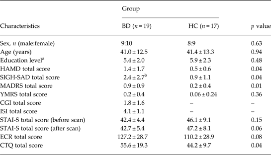
Nineteen patients suffering from BD-I and 17 HC participated in the study. Demographic and clinical information is provided in Table 1. Patients were recruited from the Bipolar Disorders Program at the Douglas Mental Health University Institute and HC from the Montreal community through local advertisements. The Faculty of Medicine Research Ethics Board of McGill University approved the study protocol. All procedures in this study comply with the Helsinki Declaration of 1975, as revised in 2008. Subjects provided written informed consent and were compensated with $150 for their participation.
Demographic and clinical and characteristics of the participants

BD, Bipolar disorder; HC, healthy controls; HAMD, Hamilton Depression Rating Scale; SIGH-SAD, Structured Interview Guide for the Hamilton Depression Rating Scale, Seasonal Affective Disorders; MADRS, Montgomery–Asberg Depression Rating Scale; YMRS, Young Mania Rating Scale; CGI, Clinical Global Impression; ISI, Intensity of Somatotherapy Index; STAI-S, State-Trait Anxiety Inventory, state anxiety; ECR, Experiences in Close Relationships; CTQ, Childhood Trauma Questionnaire.
Values given are mean ± s.d. unless otherwise stated.
a Educational level for each subject was rated on an eight-point scale (1, grade 6 or less; 2, grades 7–12; 3, graduated high school or high school equivalent; 4, part college; 5, graduated 2-year college/technical school; 6, graduated 4-years college; 7, part graduate/professional school; 8, completed graduate/professional school)
b Data from one subject missing.
BD-I diagnosis was initially determined through a clinical interview and confirmed using the Structured Clinical Interview for Psychiatric Disorders of Axis I and II of the Diagnostic and Statistical Manual of the American Psychiatric Association (SCID-I DSM-IV). In addition, inclusion criteria required a well-documented previous manic phase (i.e. requiring hospitalization). The Hamilton Depression Rating Scale (HAMD-29; Hamilton, Reference Hamilton1960), Montgomery–Asberg Depression Rating Scale (MADRS; Montgomery & Asberg, Reference Montgomery and Asberg1979), and the Young Mania Rating Scale (YMRS; Young et al. Reference Young, Biggs, Ziegler and Meyer1978) were administered to ensure patients were in a euthymic state at the time of the study, according to the recommended criteria of scores of HAMD (<7), MADRS (<8), and YMRS (<4) (Ferrier & Thompson, Reference Ferrier and Thompson2002; Mann-Wrobel et al. Reference Mann-Wrobel, Carreno and Dickinson2011).
Exclusion criteria were the presence of any major Axis I psychiatric disorder (control group) or the presence at the time of testing of an active co-morbid disorder associated to the diagnosis of bipolar I disorder (bipolar group). The presence of any personality disorder was also an exclusion criterion in both groups. Other exclusion criteria included a history of alcohol or drug abuse or dependence during the last year, use of benzodiazepines, as well as standard exclusion criteria for MRI scanning (e.g. claustrophobia, metallic implants, pregnancy). In patients, medication use was quantified using the Intensity of Somatotherapy Index (ISI; Bauer et al. Reference Bauer, McBride, Shea, Gavin, Holden and Kendall1997), which combines the different types and daily doses of medication into a composite measure. This index of medication load was then included in the behavioral and fMRI analyses as a nuisance covariate in the models, as this has been suggested to be ‘the most feasible option’ (Phillips et al. Reference Phillips, Travis, Fagiolini and Kupfer2008b ) to account for possible non-specific effects of medication on brain activity (Hafeman et al. Reference Hafeman, Chang, Garrett, Sanders and Phillips2012).
Protocol
The study protocol was divided into three sessions, taking place on different days, within a 1-week period.
During the first session, participants were screened with a medical and a psychiatric interview (as described above). All subjects passed a blood test that included a routine analysis, thyroid function, pregnancy test for women, a toxicology screen and medication levels for bipolar participants. An EKG was also performed, followed by a medical check-up done by a family physician.
Participants were trained during the second visit in the ER task and, after ensuring that they understood the instructions and performed the task as expected (based on ratings and self-report), they completed a 15-min practice test in a mock scanner, using similar (but different) pictures as those employed in the actual fMRI session.
The fMRI experiment was performed during a third session. Subjects arrived 1 h before the starting time in order to do another short practice of the task. After completing the scanning session (described below), participants were shown, on a computer, all the pictures that were required to be reappraised and were asked to rate how much effort they did in order to reappraise each of them, using a scale ranging from 1 (minimum effort) to 10 (maximum effort).
ER paradigm
The ER task was based on the ‘situational focused strategy’. It consists in using a cognitive procedure to regulate negative emotions in which participants have to think that the event they are looking at will have a positive outcome (e.g. the dramatic scene of a sinking ship is reinterpreted as positive event where all passengers are rescued). A total of 48 negative and 48 neutral images selected from the International Affective Pictorial System (IAPS) were used in the experiment. Mean normative valence ratings and standard deviations (9-point Likert scale: unpleasant to pleasant) were 2.3 ± 0.6 and 5.7 ± 1.2 for the negative and neutral images, respectively, whereas the mean normative ratings for arousal (calm to excited) were 6.1 ± 0.7 and 3.9 ± 1.2 for the negative and neutral stimuli, respectively.
The paradigm was similar to that used in previous studies (Ochsner et al. Reference Ochsner, Bunge, Gross and Gabrieli2002, Reference Ochsner, Ray, Cooper, Robertson, Chopra, Gabrieli and Gross2004), and consisted of a full 2 × 2 factorial design, with valence (Negative/Neutral) and instruction (Look/Decrease) as within-subject orthogonal factors. Each trial started with a 10-s fixation cross, followed by a cue (4 s) instructing subjects to either look at the following picture letting their emotions flow spontaneously or decrease the negative affect induced by it through reappraisal. A picture depicting either a negative or neutral scene was then presented for 10 s, followed by a 7-point rating scale in which participants rated how much negative affect they felt during the presentation of the previous image. The trial ended with a slide, lasting between 4 and 10 s, instructing subjects to relax until the next trial. Sixteen images per category were presented in a pseudo-random order (with no more than two consecutive trials of the same category to avoid anticipation and/or habituation effects), maintaining similar first-order transition matrices within and between trial types. The experiment was divided in two equivalent sessions (i.e. eight trials of each category).
fMRI acquisition and analysis
Scanning was performed at the Montreal Neurological Institute (MNI) on a 3 T Siemens Trio system with a 12-channel head coil. T2*-weighted echo-planar images with blood oxygen-level dependent (BOLD) contrast were acquired in an ascending oblique-axial interleaved fashion (TE/TR: 30/2020 ms; flip angle: 90°; FOV: 256 × 256 mm2; matrix size: 64 × 64; slice thickness: 4 mm) covering the whole head (34 slices). At the end of the functional runs, a high-resolution T1-weighted scan was obtained for anatomical localization (176 sagittal slices, voxel size: 1 mm3, TE/TR = 2.98/2300 ms).
Image preprocessing was performed using SPM8 (Wellcome Trust Centre for Neuroimaging, UK) following standard procedures. Briefly, images were corrected for differences in slice time acquisition, realigned to the first image using a rigid-body transformation, normalized to the MNI template and spatially smoothed with an isotropic Gaussian kernel of 10 mm full-width half maximum. A high-pass filter with a cut-off of 128 s was applied to remove low-frequency temporal drifts.
Data analysis was also performed in SPM8 in the context of an event-related general linear model. For each subject, first-level design matrices were built with four event types representing the four experimental categories (Neutral Look, Neutral Decrease, Negative Look, Negative Decrease). In order to take into account possible temporal differences in evoked responses during the 10-s picture presentation (Ochsner et al. Reference Ochsner, Bunge, Gross and Gabrieli2002, Reference Ochsner, Ray, Cooper, Robertson, Chopra, Gabrieli and Gross2004), each event was modeled as two adjacent 5-s boxcars, corresponding to the first and second halves of the stimulus presentation, convolved with the canonical hemodynamic response function. In addition, the six parameters obtained in the realignment procedure were also included in the model as nuisance covariates. Subject-specific parameter estimates for each of the conditions were taken to a mixed-effects second-level ANOVA with time (Early/Late), instruction (Look/Decrease) and valence (Negative/Neutral) as within-subject factors and group (BD/HC) as the between-subjects factor. Regions significantly activated in more than one contrast or group were obtained using conjunctions (Minimum Statistic compared to the Conjunction Null) (Nichols et al. Reference Nichols, Brett, Andersson, Wager and Poline2005). Effective degrees of freedom were computed taking into account the non-sphericity of the variance-covariance matrix. Statistical significance of activations was determined using a cluster-based correction for multiple comparisons across the whole brain of p < 0.05 (fanily-wise error) (Friston et al. Reference Friston, Worsley, Frackowiak, Mazziotta and Evans1994) obtained through Monte Carlo simulations (voxel-level threshold: p = 0.005; n = 10 000) using a procedure adapted from Slotnick et al. (Reference Slotnick, Moo, Segal and Hart2003) (https://www2.bc.edu/~slotnick/scripts.htm), and taking into account the spatial smoothness estimated from the actual data. This resulted in a cluster size threshold of 220 voxels. No cluster extent threshold was applied for the amygdala, due to its small size.
Results
Behavior
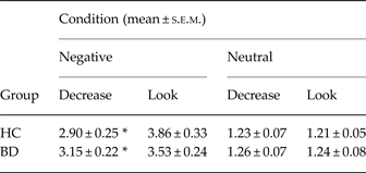
Subjective affect ratings confirmed that both groups successfully regulated their emotional reactions to negative images when instructed to do so, as confirmed by a significant difference in the ratings for Decrease and Look negative images (HC: p = 0.005; BD: p = 0.02; Table 2). There was a trend for a triple interaction, group × valence × instruction, (p = 0.07), suggesting that the BD group were slightly less efficient than HC in regulating their negative emotions. No differences within or between groups were observed for the neutral images (p's > 0.6). Moreover, both groups reported similar perceived effort in regulating negative emotions (HC: 4.3; BD: 4.9; p = 0.25).
Ratings of the intensity of negative emotional experience (1–7 scale) during passive viewing (Look) or active decrease of emotional intensity through reappraisal (Decrease)

BD, Bipolar disorder; HC, healthy controls.
* Significant difference between conditions (p < 0.05).
fMRI
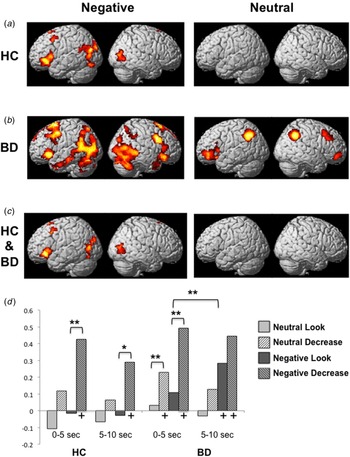
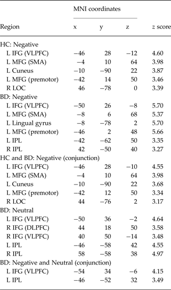
As shown in Fig. 1a, b (left), and confirmed by a conjunction analysis (Fig. 1c , left), the Negative Decrease minus Negative Look contrast revealed, in both groups, the expected activations in the regions previously implicated in ER for this task, including regions in the PFC – such as VLPFC, supplementary motor area and premotor cortex – and occipital cortex (the corresponding coordinates are reported in Table 3). A group comparison for this contrast did not yield any significant activations, even when lowering the threshold of significance (p = 0.01 uncorrected).
Statistical parametric maps (SPMs) of the contrast Negative Decrease minus Negative Look for (a) healthy controls (HC), (b) BD-I patients (BD), and (c) for the conjunction of the two groups. (d) Mean parameter estimates for the ventrolateral prefrontal cortex cluster obtained in the conjunction analysis as a function of condition, group and stimulus onset time. Brackets indicate significant differences between conditions (*p < 0.05, **p < 0.005), whereas ‘+’ indicates conditions with a response significantly different from baseline (p < 0.005).

Coordinates and corresponding brain regions for the contrasts Decrease minus Look as a function of group and stimulus valence

MNI, Montreal Neurological Institute; HC, healthy controls; IFG; inferior frontal gyrus; VLPFC, ventrolateral prefrontal cortex; MFG, middle frontal gyrus; SMA, supplementary motor area; LOC, lateral-occipital complex; BD, bipolar disorder; IPL, inferior parietal lobe; DLPFC, dorsolateral prefrontal cortex; L, left; R, right.
Interestingly, whereas no significant activations were observed in the control group for the contrast Neutral Decrease minus Neutral Look, the BD group revealed significant clusters of activations [Fig. 1b (right) and Table 3] that overlapped with those obtained in the corresponding Negative contrast. Indeed, a conjunction analysis in this group for the negative and neutral Decrease minus Look contrast yielded significant clusters of activation in the left VLPFC (MNI coordinates: x = −50, y = 34, z = −6; z score = 4.15) and inferior parietal lobe (x = −46, y=−52, z = 32; z score = 3.49). The VLPFC response pattern, of particular interest given the previous studies mentioned in the Introduction (Morris et al. Reference Morris, Sparks, Mitchell, Weickert and Green2012; Townsend et al. Reference Townsend, Torrisi, Lieberman, Sugar, Bookheimer and Altshuler2013), was also confirmed by post-hoc statistics on the parameter estimates corresponding to the cluster obtained in the HC and BD conjunction analysis (Fig. 1d ), as well as by a significant three-way interaction (instruction × valence × group) obtained in a whole-brain analysis (x = −50, y = 42, z = −2; z score = 3.32). Importantly, this difference in brain responses between groups was obtained despite similar ratings for the neutral images (see Table 2).
Finally, with regards to the VLPFC, it is worth noting the increased response to Negative Look images in the second half of the stimulus presentation, only for the BD group (Fig. 1d ), confirmed by a statistically significant effect of time (p = 0.003).
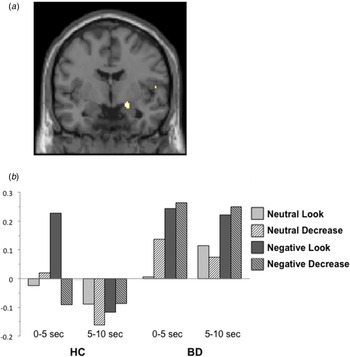
There was also reduced amygdala activation in HC for the Negative Decrease condition, relative to the Negative Look, during the early phase of the stimulus presentation (x = 20, y = −4, z = 14; z score = 2.71) (see Fig. 2a ), as previously reported (Ochsner et al. Reference Ochsner, Ray, Cooper, Robertson, Chopra, Gabrieli and Gross2004). By contrast, the BD group exhibited sustained amygdala response to negative stimuli irrespective of instructions (Fig. 2b ). This difference in amygdala response patterns between patients and HC was confirmed by significant group × instruction (p = 0.01) and group × instruction × time (p = 0.04) interactions. No other significant activations were obtained for this contrast.
(a) Significant amygdala activation for the contrast Negative Look minus Negative Decrease for the healthy control group (HC) (x = 20, y = −4, z = 14); (b) average parameter estimates for this cluster as a function of condition, group and stimulus onset time.

All behavioral and fMRI results reported here remained significant after including patients’ ISI scores as an additional nuisance covariate. Moreover, ISI scores did not significantly correlate with any of the parameter estimates extracted from the clusters obtained in the comparisons reported above.
Discussion
The similarities between BD patients and HC in the behavioral and neuroimaging results of the comparisons between the Decrease and Look trials for negative images suggests that patients were able, at least to some extent, to regulate the negative emotions elicited by these scenes. Indeed, when asked to downregulate their emotions, patients reported significantly lower negative feelings than when instructed to passively view the images and let the evoked emotions flow freely.
Difficulty in regulating emotions is a core aspect of BD (Sanchez-Moreno et al. Reference Sanchez-Moreno, Martinez-Aran, Tabares-Seisdedos, Torrent, Vieta and Ayuso-Mateos2009; Green et al. Reference Green, Lino, Hwang, Sparks, James and Mitchell2011), and several studies have shown a reduced, maladaptive, or inefficient use of ER strategies in everyday situations in these patients, mostly through the use of self-reports and questionnaires (Rowland et al. Reference Rowland, Hamilton, Lino, Ly, Denny, Hwang, Mitchell, Carr and Green2013; Wolkenstein et al. Reference Wolkenstein, Zwick, Hautzinger and Joormann2014). However, few studies have directly measured ER capacities in response to specific events in controlled experimental settings. Such studies have typically reported that, when instructed to do so, BD patients are successful at regulating their emotional responses, both subjectively and as measured with objective indices, such as skin conductance or facial displays (Gruber et al. Reference Gruber, Hay and Gross2014). As suggested by Gruber and colleagues (2014), this apparent inconsistency between experimental and clinical, or more ‘ecological’, findings could be explained by hypothesizing that, while BD patients are capable of regulating their emotions, they fail to spontaneously do so on their own, in the absence of explicit instructions. In line with this hypothesis, our behavioral results, as well as findings from some previous studies (Morris et al. Reference Morris, Sparks, Mitchell, Weickert and Green2012), suggest that patients’ instructed ER is somewhat less effective than that of healthy individuals. Adding concomitant physiological measures (e.g. electrodermal responses, respiration or heart rate) should help differentiate objective ER from the subjective experience of it (Gross, Reference Gross1998 a), particularly in terms of perceived success and effort.
Moreover, the brain regions associated with ER in BD patients largely overlapped with those observed in HC, and corresponded to the areas commonly reported in other studies employing this paradigm. However, we also observed some important differences between the groups, which are discussed below.
VLPFC
Downregulation of negative emotions was associated, in both groups, with the activation of a network of regions largely overlapping with the one usually reported in the literature (Diekhof et al. Reference Diekhof, Geier, Falkai and Gruber2011; Buhle et al. Reference Buhle, Silvers, Wager, Lopez, Onyemekwu, Kober, Weber and Ochsner2014; Frank et al. Reference Frank, Dewitt, Hudgens-Haney, Schaeffer, Ball, Schwarz, Hussein, Smart and Sabatinelli2014). Interestingly, a direct comparison did not reveal any significant differences between groups. While this null finding may seem surprising given the above-mentioned deficits in ER characteristic of BD and some of the previous reports of VLPFC activation differences, it is consistent with the behavioral results, which demonstrated that patients were capable of regulating the emotions elicited by negative images. Moreover, as described earlier, previous studies have reported inconsistent findings in terms of differences in PFC BOLD activity between BD patients and HC (Malhi et al. Reference Malhi, Lagopoulos, Sachdev, Ivanovski and Shnier2005; Lagopoulos & Malhi, Reference Lagopoulos and Malhi2007; Wessa et al. Reference Wessa, Houenou, Paillere-Martinot, Berthoz, Artiges, Leboyer and Martinot2007; Hummer et al. Reference Hummer, Hulvershorn, Karne, Gunn, Wang and Anand2012), including increased, decreased and equal responses. This is also true for the two previous studies that employed a regulation paradigm similar to ours with euthymic BD-I patients, as one reported increased VLPFC activity in patients (Morris et al. Reference Morris, Sparks, Mitchell, Weickert and Green2012) but the other observed significantly reduced responses in this region (Townsend et al. Reference Townsend, Torrisi, Lieberman, Sugar, Bookheimer and Altshuler2013).
Although in our study patients activated this region in the same manner as HC for the main contrast of interest, namely Negative Decrease minus Negative Look, interesting group differences emerged when we explored responses in this area in more detail. First, unlike HC, patients exhibited an increase in VLPFC activity during the Negative Look trials, that is, those in which they were instructed to passively view the pictures and let any elicited emotions occur naturally. Such an activity increase in the later part of the stimulus exposure may represent an automatic or implicit form of ER (Silvers et al. Reference Silvers, Wager, Weber and Ochsner2015), which would be consistent with the reported difficulty of BD patients in maintaining negative emotions (Gruber et al. Reference Gruber, Purcell, Perna and Mikels2013).
In addition, group differences were observed in the case of neutral stimuli. Specifically, whereas HC did not show any significant activations in the contrast Neutral Decrease minus Neutral Look, as expected, patients engaged a network of regions, including VLPFC, very similar to the one corresponding to negative stimuli. Naturally, neutral pictures did not elicit strong emotional reactions (as confirmed by the ratings) and therefore there was no need for actual down-regulation; therefore, these results suggest that in patients these activations reflected, at least in part, the response to the instructions to regulate emotions, regardless of whether this was necessary or not. Such an interpretation would fit with a recent model highlighting the role of VLPFC in signaling the need for regulation, rather than in the actual inhibition of emotional responses (Kohn et al. Reference Kohn, Eickhoff, Scheller, Laird, Fox and Habel2014). This finding is also consistent with the somewhat paradoxical finding that, although BD patients have difficulty in successfully regulating their emotions in everyday life, they in fact attempt to engage in regulation strategies more often than HC (Gruber et al. Reference Gruber, Harvey and Gross2012). As previous studies of ER in BD did not include a fully balanced neutral control condition, it is difficult to relate our findings to theirs, and further studies are thus required to better understand the nature of the observed brain activation patterns in this condition.
Amygdala
The initial amygdala response to negative images in the free-viewing condition observed in both BD patients and HC is consistent with the existing literature and the established role of this structure in the processing of emotional, especially negative, visual stimuli (Armony, Reference Armony2013). However, although in HC this response was transient, in agreement with the rapid habituation of the amygdala, particularly in the right hemisphere (Sergerie et al. Reference Sergerie, Chochol and Armony2008), BD patients showed a continuous response that lasted for the entire duration of the stimulus. Moreover, whereas in HC no amygdala response was present in the trials in which they were instructed to downregulate their emotional reactions, consistent with previous studies (Buhle et al. Reference Buhle, Silvers, Wager, Lopez, Onyemekwu, Kober, Weber and Ochsner2014; Frank et al. Reference Frank, Dewitt, Hudgens-Haney, Schaeffer, Ball, Schwarz, Hussein, Smart and Sabatinelli2014), patients’ sustained amygdala activity was present in all negative trials, regardless of instruction.
Although the enhanced amygdala activity in BD (Houenou et al. Reference Houenou, Frommberger, Carde, Glasbrenner, Diener, Leboyer and Wessa2011; Phillips & Swartz, Reference Phillips and Swartz2014) is typically interpreted as caused by a dysfunction of the ER system (Strakowski et al. Reference Strakowski, Adler, Almeida, Altshuler, Blumberg, Chang, DelBello, Frangou, McIntosh, Phillips, Sussman and Townsend2012; Delvecchio et al. Reference Delvecchio, Sugranyes and Frangou2013), this response pattern could also reflect an exaggerated response to emotional stimuli, associated with a hypersensitive amygdala that is resistant to top-down inhibitory signals from PFC. Indeed, such an interpretation would be consistent with the sustained activity observed in the Negative Look condition, in which no voluntary downregulation was required.
Limitations and future directions
Most of the BD patients were under medication at the time of testing. Results from previous studies suggest that medication has a ‘normalizing’ effect, that is, it brings patients’ fMRI responses closer to those of HC (Phillips et al. Reference Phillips, Travis, Fagiolini and Kupfer2008b ; Hafeman et al. Reference Hafeman, Chang, Garrett, Sanders and Phillips2012). Thus, although we failed to see an effect of medication load on BOLD responses, we cannot rule out the possibility that the similar pattern of activations for HC and patients in the Look minus Decrease contrast for negative images was partly due to medication effects. Future studies comparing medicated vs. unmedicated BD patients should help resolve this open question.
As described in the Method section, we only tested patients who, at the time of the experiment, did not present with active comorbid disorders. While this choice allowed us to be more confident about the specificity of the results to BD traits, it did potentially limit the generalizability of the findings to full population of BD patients. Testing larger samples and subdividing them as a function of their comorbid conditions should allow for a fuller characterization of the disorder. Of particular interest would be to explore possible interactions of bipolar traits with other disorders (e.g. anxiety and addiction). Additionally, it would be of interest to explore the possible influence of psychotherapy treatment duration and type (e.g. cognitive behavioral therapy) on regulation strategies and success.
As our emotional condition consisted of only negative images, we cannot determine whether the results obtained in this study are specific to the perception and regulation of negative information, or whether they would also apply to positive stimuli. This is particularly relevant, as previous studies have shown that BD patients also exhibit abnormal behavioral and neural responses associated with positive emotions and their regulation (Gruber, Reference Gruber2011). Thus, further studies employing both positive and negative stimuli are necessary to gain a comprehensive understanding of emotional deficits in BD.
Conclusion
Euthymic BD-I patients were able to downregulate the negative emotions elicited by visual scenes when instructed to do so, and they recruited similar brain regions as HC. However, unlike HC, they also activated some of these areas, particularly the VLPFC, in the case of neutral stimuli. In addition, they exhibited significant sustained amygdala responses to negative images regardless of instructions. Taken together, our results show that BD-I patients can successfully regulate their emotions under certain conditions, but that they do present differences compared to HC, particularly in terms of enhanced neural responses within the VLPFC and amygdala.
Acknowledgements
The study was partly funded by an investigator-initiated, unrestricted grant from AstraZeneca (S.B.). We are grateful to Sybille Saury for invaluable help in most aspects of the study, and to Suzanne Renaud, Mario Roy and Andrée Daigneault for contributing to patient recruitment and for helpful discussions.
Declaration of Interest
None.