Implications
It is a common relief by practice veterinarians that mares during the periparturient period often show a wide range of adiposity. Because cytokines are deeply involved in the events that bring to the delivery, the evaluation of their expression could have important implications on the breeding activity. In particular, in Standardbred mares the economic relevance of the foal makes fundamental the need to investigate the involvement of the rate of fatness on the reproductive efficiency.
Introduction
Pregnancy may be defined as a constant equilibrium between maternal and foetal inhibitors and activators, with activators playing a determining role before delivery occurs.
The transition of the animal from the foetal to the neonatal state involves great physiological adaptation by the neonate and the mother. Delivery of a healthy neonate requires the successful coordination of maturational events within the foetal and maternal tissues and it is characterised by a cascade of endocrine changes that act in an orderly sequence. The important endocrine changes occurring have been already characterised in periparturient mares (Ousey, Reference Ousey2006).
A complex relationship exists between sexual hormone levels and the immune system (Olsen and Kovacs, Reference Olsen and Kovacs1996). In female the immune system shows a cyclic nature, as far as the reproductive system: ovarian hormones have been shown to influence, in mare, the functional capacity of neutrophils (Blue et al., Reference Blue, Brady, Davidson and Kenney1982) and, in woman, the production of both T and B lymphocytes (Morell, Reference Morell1995). Several studies suggest a role for cytokines in the sequence of events leading to the parturition (Keelan et al., Reference Keelan, Blumenstein, Helliwell, Sato, Marvin and Mitchell2003) and in mechanisms responsible for preterm delivery (Bowen et al., Reference Bowen, Chamley, Keelan and Mitchell2002; Challis et al., Reference Challis, Lockwood, Myatt, Norman, Strauss and Petraglia2009). At present, no studies are available on the cytokine involvement in the orchestration of the complex sequence of delivery signalling events in mare. There is evidence that immune system is strongly linked to nutritional status and it has been reported a relationship between lymphatic and adipose tissues (Harvey, Reference Harvey2008; Alexaki et al., Reference Alexaki, Notas, Pelekanou, Kampa, Valkanou, Theodoropoulos, Stathopoulos, Tsapis and Castanas2009; Desjardins, Reference Desjardins2009). A variety of cytokines are produced by adipocytes and macrophages in white adipose tissue (WAT) in humans and animals (Weisberg et al., Reference Weisberg, McCann, Desai, Rosenbaum, Leibel and Ferrante2003; Tilg and Moschen, Reference Tilg and Moschen2006). In horses their expression was associated to obesity, but not in pregnant mares (Vick et al., Reference Vick, Adams, Murphy, Sessions, Horohov, Cook, Shelton and Fitzgerald2007) and in old-aged horses (Adams et al., Reference Adams, Katepalli, Kohler, Reedy, Stilz, Vick, Fitzgerald, Lawrence and Horohov2009). In Standardbred pregnant mares obesity is not frequent to observe, but it is known that mare fatness is a significant risk factor for foal mortality (Haas et al., Reference Haas, Bristol and Card1996). The aim of the study was to verify whether the peripheral cytokine expression could be affected by differences in the fat thickness in healthy mares during the periparturient period, comprised between the last weeks of pregnancy and the first weeks of lactation. The evaluation of the mRNA expression of systemic cytokines from peripheral blood mononuclear cells (PBMCs) aims at assessing their temporal patterns as a functional tool of immune-cell reactivity to evaluate the involvement of some important cytokines in delivery determinism. The time-correlate expression of the immune-modulating molecules reflects the activation of the maternal system in response to physiological changes, which culminate in the delivery, and reliable markers may be highlighted to predict delivery term and to ensure the best mare management.
Material and methods
Animals and experimental design
Thirty Standardbred mares at the 10th month of pregnancy were assessed for body condition. Body condition score (BCS) was assigned on a scale from 1 to 9 (Henneke et al., Reference Henneke, Potter, Kreider and Yeates1983): 18 pregnant multiparous mares (BW = 559 ± 38 kg) aged 9 ± 4 years, with a normal BCS (5; Henneke et al., Reference Henneke, Potter, Kreider and Yeates1983) were used in this study and followed from the 11th month of pregnancy to the first month after parturition.
At the beginning of the 11th month of pregnancy all mares has been subordinate to clinical evaluation to exclude the presence of pathologies. Croup fat thickness was determined by using ultrasound measurement (Lean-meater, Renco Corporation, Minneapolis, MN, USA) at approximately 11 cm cranial from the tail and 10 cm off the midline (Vick et al., Reference Vick, Adams, Murphy, Sessions, Horohov, Cook, Shelton and Fitzgerald2007). Fat thickness showed a high variability coefficient (45.0%). Therefore, two groups were previewed: low (L; ⩽12 mm; n = 12) and high (H; >12 mm; n = 6) fat thickness. Mares in late pregnancy and during lactation were penned in individual boxes and fed with a good quality meadow hay to appetite and 3.5 kg complete feed (14.34 MJ Digestible energy/kg dry matter; 138 g CP/kg dry matter) offered twice daily. The mean pregnancy duration in H and L groups was 342 ± 12 and 339 ± 15 days, respectively. On day 20.43 ± 7.79 post partum all mares were successfully inseminated with artificial insemination.
Blood analysis
Five blood samples were collected by jugular venipuncture as follows: first sample (−20 to −11 days from parturition), second sample (−10 to −1 days from parturition), third sample at delivery within 10 min after foetus expulsion (0 day), fourth and fifth samples at +10 and +20 days post partum, respectively. After sample collection, PBMCs were separated by density-gradient centrifugation using Histopaque® – 1077 (Sigma–Aldrich, St. Louis, MO, USA) and washed twice with phosphate-buffered saline. Interferon-γ (IFN-γ), tumour necrosis factor-α (TNF-α), interleukin-1α (IL-1α), -1β (IL-1ß), -2 (IL-2) and -6 (IL-6) gene expressions were performed on PBMCs, RNA was extracted by TRI Reagent® solution (Applied Biosystems, Monza, Italy). Briefly, 1 ml TRI Reagent® solution was added to 1 × 10⁷ PBMCs and RNA was extracted according to the manufacturer's instructions.
Reverse transcriptase (RT) PCR
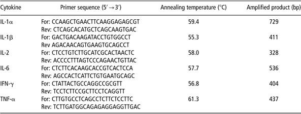
The RNA quantification was carried out by spectrophotometer (Genequant pro®, Amersham Pharmacia, St Albans, UK). RT was carried out using a Ready-to-go™ You-Prime First-Strand Beads kit (Amersham Pharmacia Biotech., Uppsala, Sweden) as described by the manufacturer. Two micrograms of total RNA were used in the RT. Aliquots (5 μl) from the generated cDNA were used for subsequent PCR amplification in the reaction buffer containing 1.5 μl MgCl2 (50 mM), 1 μl dNTPs (12.5 mM) and 1 μl Taq DNA polymerase (1 μg/μl), to a final volume of 50 μl. Amplification was carried out for 27 cycles, when the reaction was in the middle of the linear range (before reaching the amplification plateau). Each cycle consisted of denaturation at 94°C for 1 min, annealing at specific temperature for each primer set for 1 min, extension at 72°C for 1 min; at the end of 27th cycle, an additional extension was carried out for 5 min. Specific primer set used (Giguère and Prescott, Reference Giguère and Prescott1999), their annealing temperature and amplified product weight (expressed in bp) were summarised in Table 1. PCR products were visualised after electrophoresis on 2% agarose gel added with Sybr® Safe DNA gel stain (Invitrogen, Carisbad, CA, USA) and analysed on a computerised densitometry programme (Image J; http://rsb.info.nih.gov/ij/). 18S ribosomal RNA was used as an internal standard according to the instructions of the manufacturers (QuantumRNA™ 18S Internal Standards Kit, Ambion Inc., Austin, TX, USA). The values are represented as the ratio of the band densities of products over the corresponding ribosomial 18 S RT-PCR product and expressed as arbitrary units.
Sequences of PCR primers annealing temperature and PCR product length
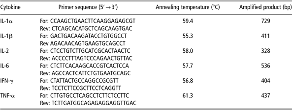
IL = interleukin; IFN = interferon; TNF = tumour necrosis factor; For = forward; Rev = reverse.
Statistical analysis
Data were submitted to ANOVA (SAS, 2003), by applying a mixed model with fatness group (two levels), sampling period (five levels) and interaction as fixed factors and mare, nested within fatness group, as random factor.
Results
The mean fat thickness of the L and H groups resulted 10.33 ± 2.07 mm and 18.00 ± 2.00 mm, respectively (P < 0.001).
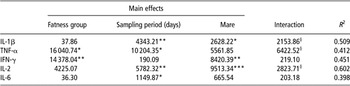
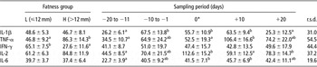
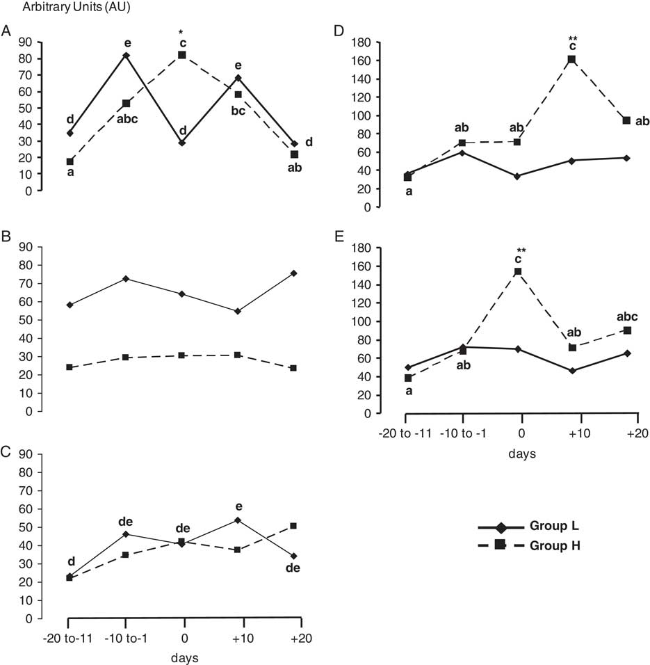
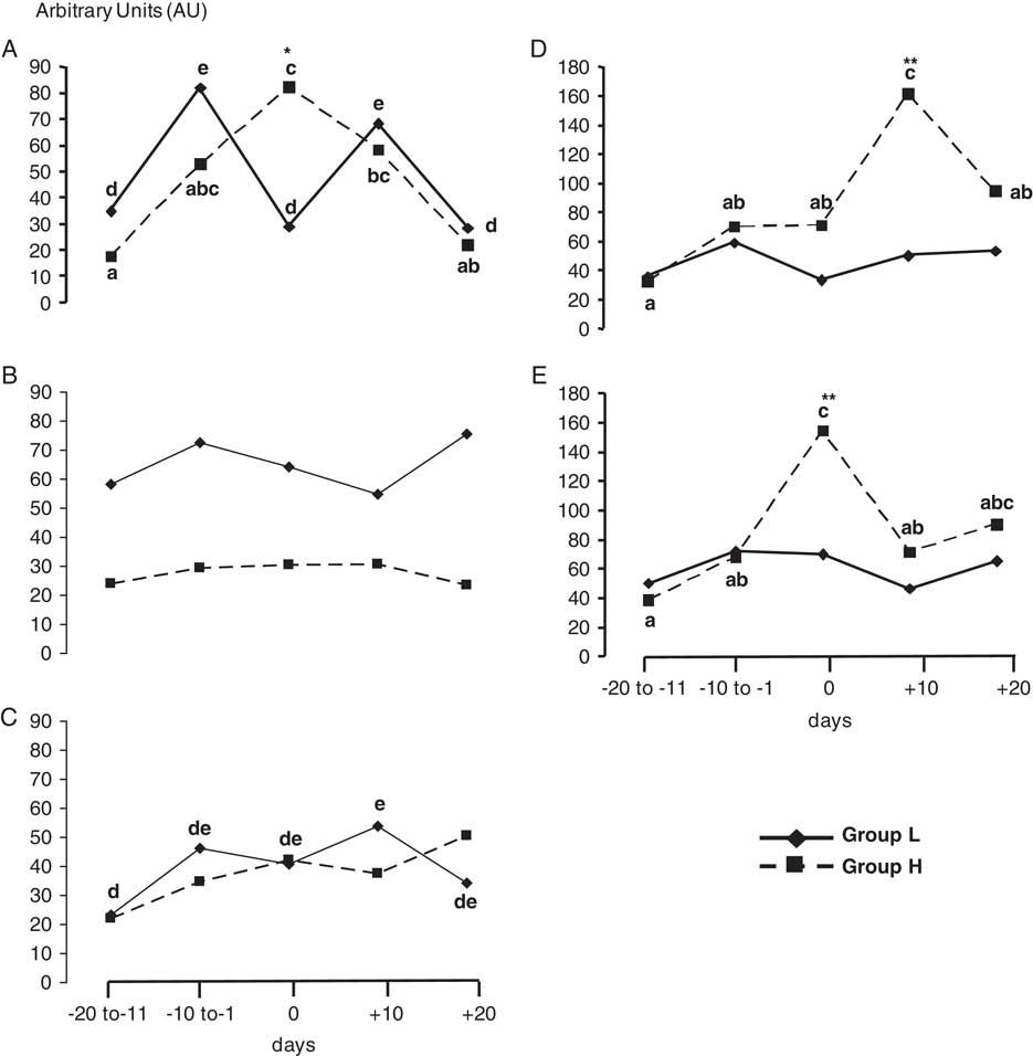
No IL-1α expression was found in mare PBMCs. The expression of peripheral cytokines was affected by the different factors (fatness group, sampling period, mare) as reported in Table 2. The statistical model explained from 41.2% (TNF-α) to 60.2% (IL-2) of total variability. Fatness group significantly influenced TNF-α (P < 0.05) and IFN-γ (P < 0.01) expression while the sampling period significantly affected all parameters but not the IFN-γ levels (P > 0.05). The interaction between fatness group and sampling period tended to be significant (P < 0.10) only for IL-1β, TNF-α and IL-2. Least square means of cytokines as affected by fatness group and sampling period are reported in Table 3. The H group showed higher TNF-α (P < 0.05) and lower IFN-γ (P < 0.01) expression as compared with L group. Regardless to fatness group, cytokines, with the exception of IFN-γ, were influenced by sampling period. However, as shown in Figure 1, different trends were shown in H and L groups. In particular, IL-1β expression rose from −20 days with a diametrically opposite trend (P < 0.05) at parturition: in the L group we observed a decrease, while H group showed a peak (Figure 1A). A similar rise (P < 0.05) was observed at parturition also for IL-2 (Figure 1E) and only in H group. No significant differences (P > 0.05) in IFN-γ and IL-6 were found between groups at different sampling period (Figure 1B and C). The TNF-α peripheral expression showed two different trends in L and H groups (Figure 1D): in L group, no significant variations were spotlighted during the experimental time, whilst in H group we found an increase from parturition to reach the peak at +10 days (P < 0.05).
ANOVA

IL = interleukin; TNF = tumour necrosis factor; IFN = interferon.
§P < 0.10; *P < 0.05; **P < 0.01; ***P < 0.001.
Least squares means (± s.e.) of cytokine expression (AU), as affected by fatness group and sampling period

AU = arbitrary units; L = low; H = high; IL = interleukin; TNF = tumour necrosis factor; IFN = interferon.
a,bP < 0.05.
*0 = delivery.
Cytokine mRNA expression in peripheral blood mononuclear cell during the periparturient period in low (L) and high (H) fat thickness groups (0 = delivery; significant differences between groups are labelled with asterisks: *P < 0.05, **P < 0.01; significant differences (P < 0.05) among sampling periods within groups are labelled with different letters: a, b and c = group H; d and e = group L. A = interleukin (IL)-1β; B = interferon-γ; C = IL-6; D = tumour necrosis factor-α; E = IL-2).
Discussion
To our knowledge, this is the first study evaluating the effect of fatness variations on the expression of cytokines in blood leucocytes from mares during the periparturient period. WAT has been the topic of increasing interest for its role as an endocrine organ, producing a variety of cytokines, mediators and hormones (Trayhurn and Wood, Reference Trayhurn and Wood2004). In particular, WAT has been recognised as an active participant in numerous physiological and path physiological processes as obesity, which is associated with an inflammation status of WAT, resulting from chronic activation of the innate immune system (Nishimura et al., Reference Nishimura, Manabe and Nagai2009). In this study, animals with normal BCS (5) but different instrumental fat thickness were chosen to assess the direct involvement of WAT on the modulation of the PBMCs response. The starting hypothesis was that the increases in adiposity can trigger a low-grade of inflammation and modulate the expression of peripheral cytokines. Group differences in cytokine expression are mainly centred at birth. Production of mRNA or protein for many cytokines has been shown to change during normal labour and parturition (Saeed et al., Reference Saeed, Qureshi, Saleem, Quadri, Sheikh and Waqar2008). These alterations may play a significant role in the processes that culminate in successful delivery. It has been reported previously that IFN-γ is an important modulator of the hypothalamus–pituitary–adrenal (HPA) axis (Zarković et al., Reference Zarković, Ignjatović, Dajak, Cirić, Beleslin, Savić, Stojković, Bulat and Trbojević2008; Borghetti et al., Reference Borghetti, Saleri, Mocchegiani, Corradi and Martelli2009), particularly under stress and/or pathologic conditions. In addition, IL-1β seems strongly involved in the HPA axis and stimulates the production of corticotrophin-releasing hormone by catecholaminergic neurons, ACTH and corticosterone (Berkenbosch et al., Reference Berkenbosch, van Oers, del Rey, Tilders and Besedovsky1987; Sapolsky et al., Reference Sapolsky, Rivier, Yamamoto, Plotsky and Vale1987; Besedovsky and del Rey, Reference Besedovsky and del Rey2000). The onset of parturition starts by foetal glucorticoids that stimulate cortisol production from the foetal adrenal and placenta (Ousey, Reference Ousey2004). Our results (Figure 1A) indicate that IL-1β rises within the last 2 to 3 weeks of gestation both in groups L and H, with a plausible effect on circadian cortisol fluctuations. Nevertheless, in this period, the foetus should be protected from maternal cortisol transplacental transfer because the placenta converts cortisol to inactive cortisone (Kajantie et al., Reference Kajantie, Dunkel, Turpeinen, Stenman, Wood, Nuutila and Andersson2003) and in fact, all our animals have given birth at term. On the contrary, at birth, the expression of IL-1β increases in group H and decreases in group L, as no difference between groups H and L were detected in the delivery progress. These findings could suggest that the role of maternal IL-1β on foetal HPA activation is to read only as part of the redundant mechanisms involved in HPA control.
The absence of IL-1α expression in both groups is similar to what reported for humans, in which it is known that placental IL-1α production plays a pivotal role in the onset of delivery (Bowen et al., Reference Bowen, Chamley, Keelan and Mitchell2002). In addition, in equine we found the IL-1α expression in placenta at term (Saleri R, unpublished results), probably involved in the labour-associated local mechanisms (e.g. membrane rupture).
The significant rise at parturition of IL-2 in H group compared with L group could be explained with an analogous role to IL-1β, even if directed to different targets. IL-2, which is an immunoregulatory cytokine, has been shown to stimulate prostacyclin production by human umbilical vein endothelial cells, through the induction of prostaglandin H synthase (Frasier-Scott et al., Reference Frasier-Scott, Hatzakis, Seong, Jones and Wu1988). Furthermore, IL-2 has been shown to significantly stimulate decidual and chorion prostaglandin E2 (PGE2) production and to enhance the stimulatory actions of IL-1 on PGE2 production by decidual cells (Coulam et al., Reference Coulam, Edwin, LaMarche and Mitchell1993).
With reference to TNF-α we reported a significant rise at day +10 only in H group. Further data relate to the actions of TNF-α on energy metabolism (Coppack, Reference Coppack2001): in in vitro studies it is able to increase lipolysis by stimulating hormone-sensitive lipase (Sumida et al., Reference Sumida, Shiosaka, Nagar, Isshikimasuda, Okuda and Hamada1997) and to reduce the expression of mRNA for glucose transporter 4 (Stephens and Pekala, Reference Stephens and Pekala1991). The net effect is to induce insulin resistance and to mobilise lipids (Ogawa et al., Reference Ogawa, Nielsen and Kawakami1989; Hauner et al., Reference Hauner, Petruschke, Russ and Eckel1995). In mares with higher adiposity, TNF-α may actively modulate glucose and lipid metabolism to warrant a good response to lactation needs.
In conclusion, the study suggests that in mares, the peripheral cytokine expression in time-related changes appears modulated by small fatness variations. Moreover, it indicates that adipose tissue is involved in the modulation of cytokines during the periparturient period, when events strongly linked to energy balance occur. Further investigations should be carried out for a better understanding of the complex relationship between adiposity and immune status in horses.