Introduction
The vast majority of our current understanding of biologic phenomena comes from routine classical cell culture experiments; growing of cells onto flat and rigid two-dimensional (2D) substrates. Even though these efforts have provided the research community with valuable insights into the mechanisms underlying a variety of biologic processes, it is nowadays widely accepted that knowledge obtained from these studies might be too reductionist to accurately translate to the human situation.[Reference Lee, Cuddihy and Kotov1, Reference Edmondson, Broglie, Adcock and Yang2] Growing cells onto 2D substrates deviates significantly from the dynamic three-dimensional (3D) in vivo situation; cells lack tissue-specific polarity, have limited contact with neighboring cells, and are exposed to non-physiologically uniform diffusion kinetics, which together alter how cells perceive and respond to their surrounding microenvironment (Fig. 1).[Reference Pampaloni, Reynaud and Stelzer3–Reference Antoni, Burckel, Josset and Noel5] This discrepancy between traditional in vitro culture conditions and the in vivo environment has been recognized among a variety of research areas, including the area of neuroscience and more specifically neurological disease, the latter presenting a great challenge in modern medicine.[Reference McMurtrey6]
Schematic representation of cells (a) cultured on a stiff 2D tissue culture substrate and (b) embedded in a more physiologically relevant 3D hydrogel environment, which has unique design variables (e.g. degradation sites and crosslinking mechanisms) that direct cell behavior. In vivo, cells reside in a complex and dynamic environment, which provides binding ligands for cell adhesion and directs processes such as cell adhesion, proliferation, migration, and morphology. Cells grown onto classical tissue culture substrates are confined to a planar surface, which leads to abnormal integrin binding and (mechano) signaling cascades, resulting in aberrant phenotypes. In the transition toward the third dimension, hydrogel-based strategies play a pivotal role. Hydrogel matrices can be tuned to meet application-specific requirements (e.g. degradation rate, porosity, and mechanical properties). Adapted by permission from Macmillan Publishers Ltd: [NATURE METHODS][Reference Caliari and Burdick7], copyright (2016).

A glance at our nervous system
Our nervous system encompasses two main cell types, neurons and glial cells, which both have a crucial role in nervous system functioning.[Reference Banerjee and Bhat8, Reference Struzyna, Katiyar and Cullen9] Neurons are highly polarized cells, responsible for the transfer and processing of electrical and chemical signals that regulate body function. According to their function, neurons can be classified into sensory neurons, motor neurons, and interneurons, which can be excitatory, inhibitory, or modulatory in their effect. Typically, neurons consist of a cell body with projections, known as axons and dendrites. These projections can vary in terms of number and position, which is highly linked to neuronal function.[Reference Miller and Jacobs10] The non-neuronal glial cells come in a variety of subtypes and have different roles in the development, maintenance, and functioning of the nervous system.[Reference Araque and Navarrete11] For a long time, glial cells were regarded to have a relatively simple supportive function. However, research over the past years has uncovered their vital role and active participation in the development and functioning of the nervous system. Beyond these cellular components and their intricate communication, appropriate nervous system functioning requires a highly regulated extracellular environment.[Reference Barros, Franco and Müller12] This dynamic mechanical and biochemical interplay plays a key role in many essential processes, including spatiotemporal cell signaling, cell identity, and cell function.[Reference Bradke and Dotti13–Reference Tang-Schomer, White, Tien, Schmitt, Valentin, Graziano, Hopkins, Omenetto, Haydon and Kaplan17] To gain more insights into neurological disorders and advance current health care, it is essential to recapitulate this complex relationship between neurons and their interaction with the surrounding environment, which requires more accurate in vitro nervous systems models.
The rise of the third dimension
Over the years, various strategies aiming at capturing 3D tissue physiology have been developed. Examples range from relatively simple approaches, such as the formation of cellular spheroids, to more sophisticated scaffold-based approaches.[Reference Haycock18–Reference Liu, Gill and Huang21] While scaffold-free approaches mostly rely on the inherent tendency of adherent cells to form aggregates, scaffold-based approaches require a thorough consideration of a plethora of factors. Clearly, the scaffold-based environment should mimic the native niche as closely as possible, which calls for multidisciplinary expertise from biologist, engineers, and material scientists. Unfortunately, accurately recapitulating the complex nature of tissues and/or organs by means of traditional scaffold-based approaches remains challenging, especially when it comes to achieving precise architectural configurations and spatial positioning of (multiple) cells and scaffolding materials. To this end, the growing field of biofabrication offers an exciting toolbox.[Reference Melchels, Domingos, Klein, Malda, Bartolo and Hutmacher22, Reference Malda, Visser, Melchels, Jüngst, Hennink, Dhert, Groll and Hutmacher23] As described by Groll et al., the term biofabrication refers to the automated generation of products with biologic function by means of bioprinting or bioassembly and subsequent maturation processes.[Reference Groll, Boland, Blunk, Burdick, Cho, Dalton, Derby, Forgacs, Li, Mironov, Moroni, Nakamura, Shu, Takeuchi, Vozzi, Woodfield, Xu, Yoo and Malda24] More specifically, biofabrication approaches encompass living cells, cell aggregates, bioactive molecules, and biomaterials to generate biologically functional products in an automated and highly organized manner.
In this review, we focus on bioprinting-based biofabrication approaches and the potential to advance currently available 3D cell culture platforms for nervous system applications, particularly in the context of neurodegenerative diseases using Alzheimer's disease as an example. Noteworthy, here we refer to bioprinting as the use of printing technology to pattern and/or organize biologic entities in 2D or 3D as described by Derby.[Reference Derby25] After a brief description of the general concept of 3D cell culture platforms, an introduction into the field of bioprinting is provided. Subsequently, the potential of bioprinting technology and its emerging applications for the nervous system will be discussed. To conclude, we highlight possible translational bottlenecks, which will need to be addressed as the technology matures. Key aspects include the necessary advances in converging disciplines, the need for standardization in the field, and ethical regulations.
Neural tissue engineering: the need for relevant three-dimensional cell culture platforms
Neurodegenerative disease is a collective term for a group of disorders which primarily affect neurons.[Reference Przedborski, Vila and Jackson-Lewis26] Whereas a variety of disorders (e.g. Alzheimer's disease, Parkinson's disease, and Huntington's disease) are covered by this umbrella term, in this review the focus is put on Alzheimer's disease, which represents the most common cause of dementia among adults and affects millions of people worldwide. As reported by the World Health Organization (WHO), the number of people living with dementia is currently estimated at over 47 million, which is expected to almost triple by 2050.
Alzheimer's disease is clinically characterized by gradual deterioration in cognitive function, including progressive memory loss, impaired judgment, and changes in personality and behavior, which can become incapacitating at later stage of illness.[Reference Yiannopoulou and Papageorgiou27, Reference Korolev28] Besides the impact on patients’ quality of life, dementia is a tremendous social and economic challenge; the disease takes heavy toll on caregivers and has been identified as a major economic burden.[Reference Dharmarajan and Gunturu29–Reference Wimo, Jönsson, Bond, Prince and Winblad31]
At the cellular level, Alzheimer's disease is characterized by damage to cortical neurons, particularly in the associative neocortex and hippocampus.[Reference Korolev28, Reference Donev, Kolev, Millet and Thome32] This neuronal damage is correlated to deposition and accumulation of abnormal proteins; extracellular amyloid-β peptides and intracellular filamentous hyperphosphorylated tau proteins, which are the core hallmarks of Alzheimer's disease and also known as plaques and tangles, respectively.[Reference Deleglise, Magnifico, Duplus, Vaur, Soubeyre, Belle, Vignes, Viovy, Jacotot, Peyrin and Brugg33] Whereas research was initially focused on neuronal cells and protein-mediated neuronal damage, it has become increasingly apparent that Alzheimer's pathogenesis has a more complex nature and is not restricted to these events. Over the years research has evidenced that other factors, such as immunological mechanisms, have an accompanying role in the pathway leading to progression of the disease.[Reference Heneka, Carson, El Khoury, Landreth, Brosseron, Feinstein, Jacobs, Wyss-Coray, Vitorica, Ransohoff, Herrup, Frautschy, Finsen, Brown, Verkhratsky, Yamanaka, Koistinaho, Latz, Halle, Petzold, Town, Morgan, Shinohara, Perry, Holmes, Bazan, Brooks, Hunot, Joseph, Deigendesch, Garaschuk, Boddeke, Dinarello, Breitner, Cole, Golenbock and Kummer34] Despite progress in understanding the underlying mechanisms and aspects of neurodegenerative disease, there are still many unknowns. This is reflected by the lack of effective therapeutic options for the vast majority of these disorders, including Alzheimer's disease, which are nowadays limited to symptomatic relief.[Reference Cuny30, Reference Lunn, Sakowski, Hur and Feldman35] To advance the treatment of these disorders it is critical to gain a deeper understanding of processes such as neural network formation, organization, and functioning.
Classical three-dimensional cell culture strategies
In response to the growing demand for more physiologically relevant models, a number of 3D cell culture platforms have been developed.[Reference Achilli, Meyer and Morgan36, Reference Simian and Bissell37] Examples range from cell-self-assembly-based approaches, to scaffold-based approaches, and on-chip biomimicry. In the next section, we aim to sketch and illustrate the importance of these classical 3D cell culture platforms and capturing the third dimension.
Cell-self-assembly-based approaches
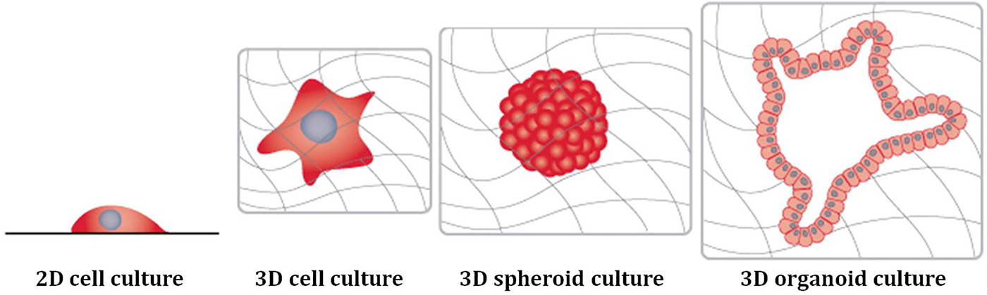
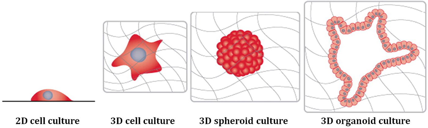
One of the most common cell-self-assembly-based approaches is the formation of cellular spheroids (Fig. 2). Spheroid formation is generally considered to be a relatively simple 3D cell culture method and has shown to be a convenient means toward more accurate tissue models. The main benefit of using spheroids over 2D monolayer cultures is the presence of more physiologically relevant diffusion gradients (e.g. oxygen and nutrients) and cell–cell and cell–extra cellular matrix (ECM) interactions.[Reference Achilli, Meyer and Morgan36, Reference Fang and Eglen38, Reference Mehta, Hsiao, Ingram, Luker and Takayama39] With time, a variety of methods have been developed to produce spheroids, one well-known example being the hanging drop method.[Reference Achilli, Meyer and Morgan36, Reference Fang and Eglen38] In this approach, aliquots of cell suspensions are seeded on an adherent surface, which is subsequently reverted, yet allowing the droplets to stay attached due to surface tension, resulting in gravity-enforced cell aggregation at the bottom of the droplets. Alternative popular approaches are based on dynamic culture conditions (e.g. the spinner flask method), low-adhesive surfaces (e.g. forced-floating technique), and more recently, micro-/nano-patterned surfaces and micromolding, the latter providing defined regions for cell immobilization and subsequent spheroid formation.[Reference Fang and Eglen38] All these different methods are associated with their own advantages and disadvantages, resulting in spheroids of different sizes and shapes, and should be chosen depending on the experimental needs and final application.[Reference Achilli, Meyer and Morgan36, Reference Mehta, Hsiao, Ingram, Luker and Takayama39]
Cell culture platforms: monolayer culture on conventional tissue culture substrates, single cell in 3D matrix, spheroid culture, and organoid culture. Obtained and modified from[Reference Boutin, Kramer, Livi, Brown, Moore and Hoffman-Kim42] with permission from Elsevier.
To date, spheroids have been successfully generated from a variety of cell types, including neuronal cells.[Reference Fennema, Rivron, Rouwkema, van Blitterswijk and de Boer40] For example, Dingle et al. generated neural spheroids from primary rat cortical tissues by means of micromolding, which were reproducible in size and cellular composition.[Reference Dingle, Boutin, Chirila, Livi, Labriola, Jakubek, Morgan, Darling, Kauer and Hoffman-Kim Diane41] Spheroid characterization revealed resemblance to in vivo cortical tissue in terms of stiffness, neuronal electrophysiology, neural cell types, and morphology. Following on from this study, Boutin et al. assessed the potential of this heterogeneous spheroid model to form capillary-like networks, the later playing an important role in protecting the neuronal environment.[Reference Boutin, Kramer, Livi, Brown, Moore and Hoffman-Kim42] Immunohistochemical analysis revealed that within 3 days, cortical endothelial cells seemed to form networks in the spheroids, which were surrounded by basement membrane components and relevant cell types. In future studies, this relatively simple model could offer valuable insights in understanding disorders linked to neurovascular dysfunction.
A more advanced cell-self-assembly-based 3D cell culture platform that has been gaining increasing interest is the organoid model (Fig. 2).[Reference Ranga, Gjorevski and Lutolf43] Whereas various definitions are reported in the literature, here we refer to organoids as self-organizing 3D cell clusters, which can be established from different cell sources (e.g. primary cells, cell lines, and stem cells), resembling the tissue architecture of origin.[Reference Simian and Bissell37, Reference Fatehullah, Tan and Barker44, Reference Schneeberger, Spee, Costa, Sachs, Clevers and Malda45] Over the last decade, organoid technology has made tremendous progress and several in vitro organoids have been established.[Reference Simian and Bissell37] Whereas the relatively scarce amount of literature on neuronal organoids is mainly focusing on neurodevelopmental phenomena, Raja et al. have shown its potential to study neurodegeneration.[Reference Raja, Mungenast, Lin, Ko, Abdurrob, Seo and Tsai46] In this study, neural organoids were formed from induced pluripotent stem cells derived from Alzheimer's disease patients. This organoid model has shown to be robust, able to recapitulate both hallmarks of Alzheimer's disease, and has been shown to be amenable to experimental manipulation in terms of drug treatment.
Biomaterial-aided three-dimensional cell culture platforms
In this section, we only focus on the non-biofabrication biomaterial-aided platforms that involve either pre-fabricated scaffolds for cell cultivation or polymer hydrogels for cell encapsulation. In both cases, the biomaterials do not only provide structural and mechanical support, but also guide the cells via acting as a template for tissue formation.[Reference Pereira, Awatade, Loureiro, Matos, Amaral and Jordan20, Reference O'brien47, Reference Breslin and O'Driscoll48] Intuitively, these biomaterials should resemble the dynamic native microenvironment of the specific target tissue as closely as possible, a task that can be extremely challenging; it does not only require the biologic and functional integration of the cells with the surrounding biomaterial(s), but also careful consideration of biodegradability, mechanical properties, scaffold architecture, and manufacturing technology.[Reference O'brien47, Reference Chan and Leong49]
Over the years, a myriad of materials have been investigated for their potential to support neuronal 3D cell culture.[Reference Murphy, Laslett, O'Brien and Cameron50] These range from natural polymers (e.g. collagen, hyaluronic acid, alginate, Matrigel, and silk) and synthetic polymers [e.g. PLGA [poly(lactide-co-glycolide)], poly-L-lactic acid (PLLA), and polyurethane (PU)] to hybrid materials, metals, ceramics, glass, and carbon nanotubes. For example, Frampton et al. described a 3D culture system based on cell entrapment in peptide-functionalized alginate hydrogels.[Reference Frampton, Hynd, Shuler and Shain51] Cell viability and functional analysis of primary rat glial and neuronal cells demonstrated appropriate cell viability and metabolic function within the constructs over a period of 14 days. Interestingly, cell morphology, particularly of astrocytes differed from cells cultured on 2D substrates. Analysis of the functional activity of primary rat neurons showed formation of functional synaptic elements and electrical activity. Together, the results showed that this system could offer a viable cell culture platform to (co-)culture a variety of relevant cell types for use in neural cell culture, supporting cell-type-specific function. Lai et al. compared neuronal voltage-gated calcium channel functionality in 2D to 3D setting.[Reference Lai, Cheng and Kisaalita52] Primary neuronal cells were harvested from mice and either cultured onto 2D flat surfaces or in 3D synthetic polymer scaffolds, the latter consisting of PLLA and polystyrene. It was found that cells in 3D scaffolds resembled native intact tissue more closely in terms of cell morphology and ion channel functioning. Another striking example that illustrates the importance of recapitulating an in vivo-like 3D cellular environment is described by Choi et al. who studied the pathogenic mechanisms of Alzheimer's disease in a 3D Matrigel-based brain tissue-like environment.[Reference Choi, Kim, Hebisch, Sliwinski, Lee, D'Avanzo, Chen, Hooli, Asselin, Muffat and Klee53] Whereas amyloid-β is thought to diffuse into the culture medium in 2D systems and to be removed during regular media changes, it was found that this closed 3D culture model system promoted amyloid-β aggregation, thereby more closely recapitulating Alzheimer's disease pathology. More recently, Kraus et al. developed a spheroidal co-culture model to examine neurite formation.[Reference Kraus, Boyle, Leibig, Stark and Penna54] In this study, spheroids were formed from glial cells (i.e. Schwann cells) and neurons and subsequently encapsulated in a 3D collagen matrix. Evaluation of neurite lengths demonstrated a significant increase in neurite length after a period of 7 days in the 3D setup compared with 2D culture.
Organ-on-a-chip technology
Organ-on-a-chip technology refers to microfluidic cell culture devices, which are designed to reconstitute critical tissue- and organ-level functions and rely on a sophisticated interplay between engineering and biology.[Reference van der Meer and van den Berg55, Reference Bhatia and Ingber56] These systems are used for and offer control over a variety of key system parameters, including the choice of cells and their spatial positioning. However, the added value of on-chip biomimicry lies within its potential to control parameters that are not easily controlled in 3D static cultures or bioreactors (e.g. fluid flow, physical, and chemical gradients), which makes them promising tools to address fundamental questions in the area of tissue development, organ physiology, and disease etiology.[Reference Bhatia and Ingber56]
Over the years, various microfluidic devices have been developed to provide insights into the pathophysiology of neurodegenerative disorders.[Reference Yi, Park, Lim, Lee and Lee57] For example, Choi et al. and Cho et al. utilized a microfluidic-based approach to investigate the effects of amyloid-β (aggregation) on neurons and microglia, respectively.[Reference Choi, Chae, Kim, Barald, Park and Lee58, Reference Cho, Hashimoto, Wong, Hori, Wood, Zhao, Haigis, Hyman and Irimia59] In their study, Choi et al. used a low-flow microfluidic system to assess the neurotoxicity of time-dependent amyloid-β aggregation and the effect of physiological flow on neuronal cell survival.[Reference Choi, Chae, Kim, Barald, Park and Lee58] With their microfluidic platform, Cho et al. studied the effect of amyloid-β on microglial accumulation, which demonstrated a different role for soluble and bound amyloid-β during microglia recruitment and localization.[Reference Cho, Hashimoto, Wong, Hori, Wood, Zhao, Haigis, Hyman and Irimia59] Another well-reported topic involves amyloid-β and tau protein propagation. Kunze et al. have utilized a microfluidic device to study the neurodegenerative propagation processes of Alzheimer's disease.[Reference Kunze, Meissner, Brando and Renaud60] Primary rat cortical neurons were cultured in two separated cell compartments, one housing diseased neurons (i.e. showing tau-hyperphosporylation) and the other healthy neurons. By designing the microfluidic device in such a manner that connectivity was favored through neurite outgrowth from both compartments over time, a co-pathological state within the same neural cell culture was established. More recent studies regarding amyloid-β and tau protein propagation are presented by Song et al. who demonstrated amyloid-β spreading via neuronal connections along axonal membranes[Reference Song, Shim, Kim, Won, Joo, Kim, Jeon and Yoon61], Deleglise et al. who investigated the distant effects of local amyloid-β stress on neuronal subcompartments and networks[Reference Deleglise, Magnifico, Duplus, Vaur, Soubeyre, Belle, Vignes, Viovy, Jacotot, Peyrin and Brugg33], and Dujardin et al. and Calafate et al. who offered novel insights into tau protein transfer.[Reference Dujardin, Lécolle, Caillierez, Bégard, Zommer, Lachaud, Carrier, Dufour, Aurégan, Winderickx, Hantraye, Déglon, Colin and Buée62, Reference Calafate, Buist, Miskiewicz, Vijayan, Daneels, De Strooper, De Wit, Verstrekenand and Moechars63]
Bioprinting
3D cell culture platforms provide the target tissues with a (temporary) microenvironment and thereby contribute to spatiotemporal cell signaling, cell identity, and cell function. Although these 3D cell culture platforms have been demonstrated to offer a promising route toward more physiologically relevant neuronal models and in-depth understanding of cellular behavior and pathological conditions, there are still a number of limitations. One of the primary general challenges is related to the lack of adequate control. Regardless of the platform, issues such as irregular geometry (e.g. external shape and internal architecture of the scaffolds) and size, poor reproducibility, and difficulties regarding scaling-up impede their widespread use. To this end, bioprinting offers an exciting opportunity. By combining biomaterials, cells, and/or growth factors (composites referred to as bioink) with automated fabrication processes such as additive manufacturing [the generation of 3D constructs in a layer-by-layer manner based on a CAD (computer-aided design)], it facilitates the generation of 3D bio-engineered constructs with superior organization and more closely resembling native tissues.[Reference Melchels, Domingos, Klein, Malda, Bartolo and Hutmacher22, Reference Malda, Visser, Melchels, Jüngst, Hennink, Dhert, Groll and Hutmacher23, Reference Groll, Boland, Blunk, Burdick, Cho, Dalton, Derby, Forgacs, Li, Mironov, Moroni, Nakamura, Shu, Takeuchi, Vozzi, Woodfield, Xu, Yoo and Malda24] This computer-controlled deposition of biologically relevant materials allows the fabrication of (multi)-cellular constructs. Besides, the layer-by-layer architecture allows tailoring beyond the cellular level, namely in terms of materials, and biochemical cues, which could be optimized to meet cell-specific requirements. In this manner, multiple cell types could be contained throughout a single 3D construct, yet in an environment optimized for each of the individual components.
Bioprinting technologies
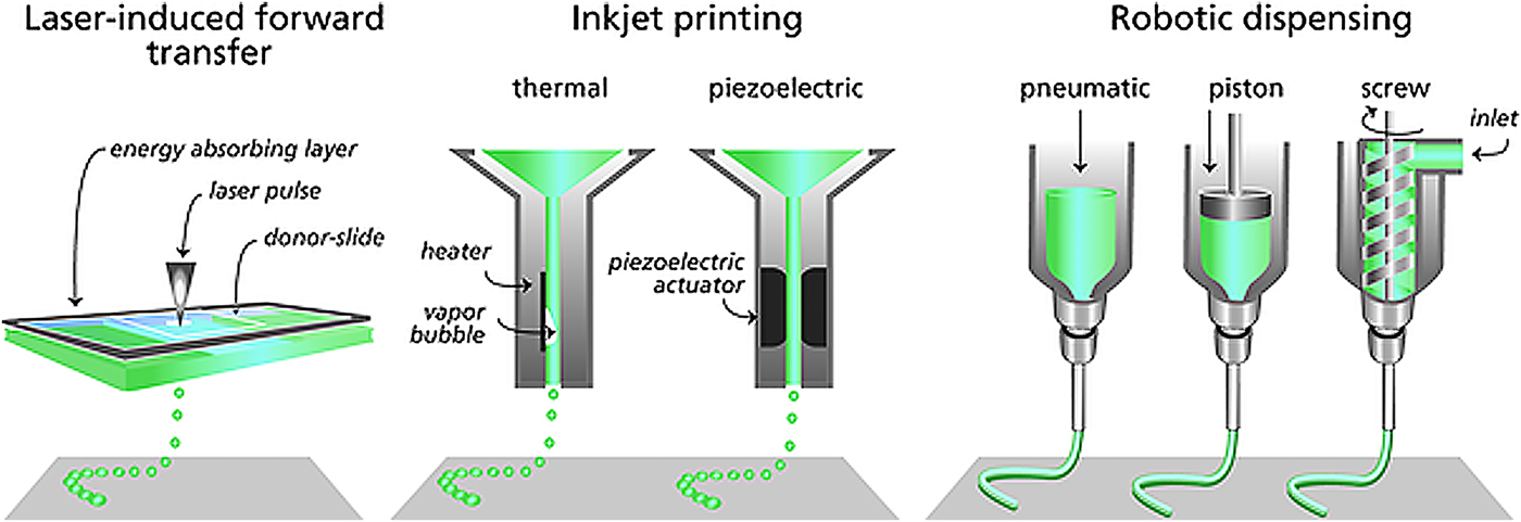
Although a wide variety of bioprinting technologies have been described in the literature, three main categories can be distinguished: inkjet printing, laser-assisted printing, and extrusion-based printing (Fig. 3).[Reference Murphy and Atala64, Reference Hölzl, Lin, Tytgat, Van Vlierberghe, Gu and Ovsianikov65]
Main bioprinting technologies: (a) Inkjet printing, (b) laser-assisted printing, (c) extrusion printing. Figure obtained and modified from[Reference Malda, Visser, Melchels, Jüngst, Hennink, Dhert, Groll and Hutmacher23] with permission from John Wiley & Sons.

Inkjet printing can be described as a non-contact printing process in which controlled volumes of (bio)ink (1–100 picoliters; 10–50 µm diameter) are dispensed on predefined locations, driven by thermal or piezoelectric forces [Fig. 3(a)]. Both in thermal inkjet printing and piezoelectric inkjet printing, (bio)ink droplets are forced from the nozzle by creating a pressure pulse. In thermal inkjet printing, this pressure pulse is generated by heating. This heating results in vaporization of small volumes of the (bio)ink, producing the pulse required to expel material from the nozzle. Piezoelectric inkjet printing involves the application of a direct mechanical pulse to the (bio)ink, which on its turn generates the pressure needed to force material though the nozzle. Regardless of the high resolution, relatively low cost, and wide availability of inkjet printing, several factors impede its successful widespread application.[Reference Malda, Visser, Melchels, Jüngst, Hennink, Dhert, Groll and Hutmacher23, Reference Hölzl, Lin, Tytgat, Van Vlierberghe, Gu and Ovsianikov65] These include the challenges regarding cell viability due to the small orifices, the potential risks of exposing the (bio)ink to thermal and mechanical stress, inconsistency in droplet size, nozzle clogging when using biologically relevant cell densities (>106 cells/mL), and the low upper viscosity limit of the (bio)ink (ideally <10 mPa/s), which although considered to be beneficial for cell migration, proliferation, and cell viability, influences printing fidelity.[Reference Malda, Visser, Melchels, Jüngst, Hennink, Dhert, Groll and Hutmacher23, Reference Hölzl, Lin, Tytgat, Van Vlierberghe, Gu and Ovsianikov65]
As the name indicates, laser-assisted printing encompasses a plethora of manufacturing technologies, which utilize a laser to deposit (biologic) materials on a substrate. Laser-assisted bioprinting stems from the principles of a subclass of laser-assisted printing, namely laser-induced forward transfer (LIFT) [Fig. 3(b)]. In LIFT approaches, a focused laser-source is used to transfer material from a so-called donor slide toward the receiving substrate. Typically the donor slide is covered with a laser energy absorbing layer and the desired (bio)ink. The focused laser pulse causes evaporation of the absorbing layer, leading to the formation a high-pressure bubble, which induces propulsion of the material. As this approach is nozzle free, the clogging issue associated with inkjet-based printing or extrusion-based printing is avoided. In addition, laser-assisted printing is compatible with materials with a broad range of viscosities (1–300 mPa/s) and able to deposit cells at medium cell density (i.e. 108 cells/mL) without detrimental effects on cell viability.[Reference Malda, Visser, Melchels, Jüngst, Hennink, Dhert, Groll and Hutmacher23, Reference Murphy and Atala64, Reference Hölzl, Lin, Tytgat, Van Vlierberghe, Gu and Ovsianikov65] Despite these advantages, the presence of metallic residues in the final construct, the challenging and time-consuming nature of preparing individual ribbons, and relatively high costs are several examples of hurdles yet to clear.
Nowadays, most of the commercially available bioprinters are based on extrusion-based dispensing [Fig. 3(c)]. These systems function by robotically controlled extrusion of the (bio)ink, which is usually driven by mechanical action (piston or screw) or a pneumatic system, each posing their own (dis)advantages. Whereas mechanically driven deposition usually provides more control over the material flow, pneumatic systems have simpler drive-mechanism components, the latter allowing for higher maximum force capabilities. In contrast to inkjet printers, which yield single droplets of material, extrusion printers yield continuous filaments of the (bio)ink, usually with a resolution of the order of 200 µm. Besides, extrusion printing is compatible with materials with a wide range of viscosities (30–6 × 10⁷ mPa/s). Even though the resolution is substantially lower when compared with laser- or inkjet-based systems, the fabrication speed, clinically relevant sizes, and ability to deposit high cell densities (>108 cells/mL) with high cell viability make this technology very promising for tissue engineering and regenerative medicine (TERM) applications.[Reference Malda, Visser, Melchels, Jüngst, Hennink, Dhert, Groll and Hutmacher23, Reference Hölzl, Lin, Tytgat, Van Vlierberghe, Gu and Ovsianikov65]
Bioprinting in three-dimensional neuronal cell culture
Given that the field of bioprinting in general, and even more for this specific application, is still in its infancy, early studies of bioprinting in 3D neuronal culture have been focused mostly on healthy tissue. However, even though these studies do not directly concern diseased tissue(-like) constructs, these could be used as tool for a variety of applications, ranging from cell behavior studies to understanding pathological conditions and drug testing, which is related to and relevant for neurodegenerative disease.
Various studies relating to this topic have been reported. Among these, several have been focusing on the effect of printing processes on cell viability. For example, Xu et al. studied the delivery of neuronal cells by a modified thermal inkjet printer, and demonstrated no significant effect on cell survival.[Reference Xu, Jin, Gregory, Hickman and Boland66, Reference Xu, Gregory, Molnar, Cui, Jalota, Bhaduri and Boland67] More recently, Lorber et al. studied the potential of inkjet printing of adult rat retinal ganglion cells and retinal glia, demonstrating no significant difference in cell survival compared with non-printed controls.[Reference Lorber, Hsiao, Hutchings and Martin68] In a similar approach, Tse et al. printed primary porcine Schwann cells and neuronal analog NG108-15 cells with a piezoelectric inkjet printer.[Reference Tse, Whiteley, Yu, Stringer, MacNeil, Haycock and Smith69] This study showed that a higher range of experimental voltages has no adverse effects over a period of 7 days. Whereas these studies could be considered relatively simplistic in nature (i.e. printing of cells to explore compatibility with the printing modality) these efforts have illustrated that relevant cells can be delivered and positioned by means of inkjet printing approaches without substantial damage.
A major advantage of bioprinting approaches is the ability to deliver and distribute cells and other biologic factors in a very precisely controlled manner. An example is given by Lee et al. who fabricated cell–hydrogel composites for the purpose of in vitro neural culture.[Reference Lee, Polio, Lee, Dai, Menon, Carroll and Yoo70] Murine neural stem cells were deposited on top of a layer of printed collagen, subsequently crosslinked to immobilize the cells, and covered with an additional layer of collagen. Standard cell viability and proliferation assays showed that the printing technique did not affect cell viability. To explore the effect of growth factor release, a vascular endothelial growth factor-releasing fibrin gel was added to the system. Whereas further studies should be conducted, initial experiments have shown that cellular proliferation and migration was supported over time. In a more advanced approach, Hsieh et al. explored bioprinting of cell-laden thermo-responsive biodegradable PU hydrogels.[Reference Hsieh, Lin and Hsu71] Murine neural stem cells were harvested from adult mouse brain, encapsulated in different PU hydrogels, and subsequently bioprinted, forming a grid-like structure. Cell viability, proliferation, and gene expression analysis demonstrated its potential as a suitable niche for neuronal stem cell proliferation and differentiation. Furthermore, the potential of this strategy in central nervous repair was evaluated in zebrafish and embryo neural injury models, which demonstrated its potential to rescue central nervous system functioning. Whereas the above-mentioned approaches illustrate the potential of bioprinting technology for neuronal applications, these fabricated constructs are mainly constricted to relatively thin layers of cells within cell culture media or on top of a hydrogel.
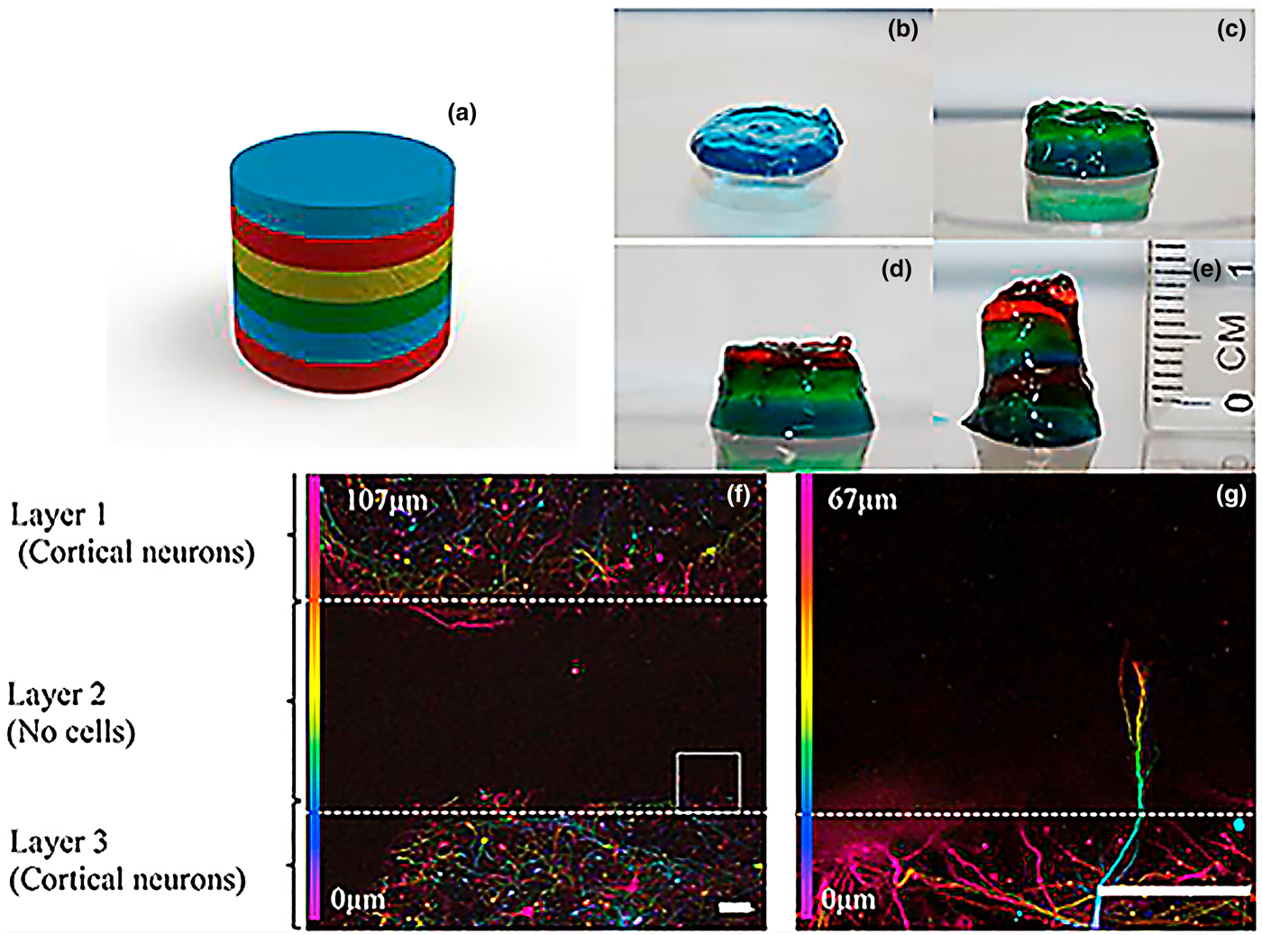
Given that the utility of actual bioprinting platforms for this particular application is at early stage, only a few studies have been reported in literature. One example is presented by our group and involves the development of a novel bioprinting-based method to recapitulate the layered structure of the brain and thereby fabricate 3D brain-like structures.[Reference Lozano, Stevens, Thompson, Gilmore, Gorkin, Stewart, in het Panhuis, Romero-Ortega and Wallace72] This study was based on the previous work by Tang-Schomer et al., who developed compartmentalized 3D brain-like cortical tissue with silk fibroin-based biomaterials.[Reference Tang-Schomer, White, Tien, Schmitt, Valentin, Graziano, Hopkins, Omenetto, Haydon and Kaplan17] Our group developed an Arg–Gly–Asp (RGD) peptide-modified gellan gum-based bioink (RGD-GG), which was assessed in terms of neural cytocompatibility, network formation, and printability. Discrete layers of cell-encapsulating (i.e. mouse primary neurons) RGD-GG were deposited using a simple hand-held printing device, resulting in layered constructs [Figs. 4(a)–4(e)]. Standard calcein/propodium iodide assays revealed acceptable cortical neuron and glial cell viability, both after encapsulation and printing RGD-GG. Immuno-staining 7 days after encapsulation demonstrated neuronal network formation throughout the RGD-GG structures. To assess the potential of this strategy to create more complex multi-layered brain-like structures, six-layer structures were printed. Visual analysis revealed presence of distinct layers, within the solid structure. To assess the neural cytocompatibility of these larger structures, a three-layer construct was printed, consisting of two cell-laden RGD-GG layers, separated by an acellular RGD-GG layer. Neuronal network formation was observed after 5 days of culture, with axons starting to penetrate into the acellular middle layer [Figs. 4(f) and 4(g)]. These multi-layer brain-like structures have shown to provide an environment in which the dynamic process of neural growth can be observed and quantified in 3D and thereby offer a promising tool for a variety of applications ranging from cell behavior studies to understanding pathological conditions and drug testing.
Layered brain-like structure: (a) SolidWorks schematic representation of the envisioned six-layered construct, (b–e) construct fabrication process, in which each color represents a distinct layer, (f) confocal microscope images of the neurons colored for their z-axis distribution after 5 days of culture, (g) showing neurite penetration through the gel into the acellular RGD-GG layer. Scale bars represent 100 µm. Figure obtained and adapted from[Reference Lozano, Stevens, Thompson, Gilmore, Gorkin, Stewart, in het Panhuis, Romero-Ortega and Wallace72] with permission from Elsevier.

In another approach toward establishing more accurate neuronal tissue-like structures, our group investigated in situ differentiation of human neural stem cells in bioprinted constructs.[Reference Gu, Tomaskovic-Crook, Lozano, Chen, Kapsa, Zhou, Wallace and Crook73] Frontal cortical human neural stem cells were encapsulated in an optimized polysaccharide bioink consisting of alginate, carboxymethyl-chitosan, and agarose, and followed by a micro-extrusion approach. Characterization of the cells revealed homogenous cell distribution and high viability throughout the printed construct. Immunophenotyping and reverse-transcription quantitative PCR (RT–qPCR) analysis demonstrated successful differentiation into functional neurons and supporting neuroglia, illustrating the potential of this platform to be used in translational studies.
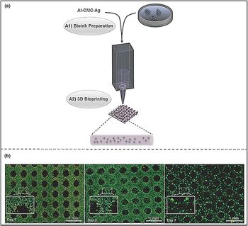
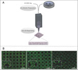
Following on from this study, Gu et al. described the first example of bioprinted human induced pluripotent stem cell constructs, which were successfully differentiated into homogenous neural tissues.[Reference Gu, Tomaskovic-Crook, Wallace and Crook74] Human-induced pluripotent stem cells were encapsulated in a bioink consisting of alginate (5% w/v), carboxymethyl-chitosan (5% w/v), and agarose (1.5% w/v) and subsequently bioprinted [Fig. 5(a)]. Extrusion printing of the cell-laden bioink resulted in homogenous cell distribution throughout the constructs, with negligible cell death over a period of 7 days, which was determined by standard calcein/propodium iodide assays [Fig. 5(b)].[Reference Gu, Tomaskovic-Crook, Wallace and Crook74] Flow cytometry studies, performed after ten days in culture, revealed expression of pluripotency markers, which was further confirmed by induction of embryoid bodies. The bioprinted stem cells were then differentiated into cells with phenotypes that represent neuronal subtypes and microglia, as confirmed by immunophenotyping and RT–qPCR. This study has successfully demonstrated the ability to produce human-induced pluripotent stem cell-laden constructs and differentiate these into relevant cell types, which is an important step towards bioprinting-based neuronal tissue models.
Bioprinting of human-induced pluripotent stem cells: (a) schematic representation of the approach, in which the bioink is prepared by adding the cells to an optimized alginate, carboxymethyl-chitosan, and agarose (Al–CMC–Ag) mixture and subsequently bioprinted. Cell viability studies by means of calcein/propidium iodide staining have shown (b) homogenous cell distributions and high cell viability over a period of 7 days post-printing. Scale bars represent 1 mm. Figure obtained and adapted from[Reference Gu, Tomaskovic-Crook, Wallace and Crook74] with permission from John Wiley & Sons.

Bioprinting: a complementary approach
The true power of bioprinting technology lies in its superior potential to offer spatial and temporal control in a reproducible manner. However, it is important to realize that different applications may require a different approach, which could be offered by other 3D cell culture platforms. Therefore, rather than being considered as superior, it is noteworthy to mention the complementary role of bioprinting technology, when being integrated with other existing 3D cell culture platforms.[Reference Mironov, Visconti, Kasyanov, Forgacs, Drake and Markwald75] Several papers have been reported regarding the integration of scaffold-free 3D cell culture platforms in combination with 3D printing technology. For example, Mironov et al. have demonstrated the potential of bioprinting to precisely deposit tissue spheroids formed from human dermal fibroblasts according to predefined patterns.[Reference Mironov, Khesuani, Bulanova, Koudan, Parfenov, Knyazeva, Mitryashkin, Replyanski, Kasyanov and DAS76] Very recently, Schneeberger et al. have reviewed the convergence of organoids and bioprinting technologies, highlighting the benefits bioprinting could have in terms of increasing the (clinical) application potential of organoids.[Reference Schneeberger, Spee, Costa, Sachs, Clevers and Malda45]
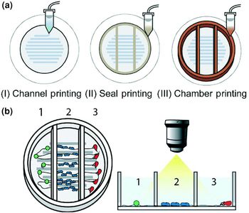
Besides its added value for scaffold fabrication, additive manufacturing could facilitate the development of custom-made microfluidics devices.[Reference Yi, Lee and Cho77] For example Johnson et al. used additive manufacturing technology to fabricate a nervous system-on-a-chip.[Reference Johnson, Lancaster, Hogue, Meng, Kong, Enquist and McAlpine78] This biomimetic chip consisted of three chambers with separate fluid environments and microchannels, the latter providing axonal guidance and alignment (Fig. 6). The different compartments were loaded with appropriate cell types and the complete chip was studied over time in terms of cell organization and its potential to be applied in viral infection assays. Whereas spatial separation was indeed achieved, axonal networks were able to penetrate into the chambers, allowing axon-to-cell integration between compartments. In line with this, analysis of axon-to-cell spreading of viral particles from one compartment to another demonstrated biologic connectivity between the compartments. In this approach, relevant cell types were successfully integrated on their chip, illustrating that additive manufacturing could be an effective fabrication approach for the development of tailor-made on-a-chip biomimicry devices.
3D printed nervous-system-on-chip: (a) schematic representation of the printing approach, which consisted of three consecutive steps. First, microchannels providing axonal guidance were printed; subsequently, a sealant layer was printed, after which the individual chambers were fabricated. The chip was completed by (b) functionalizing the chambers with the relevant cell types. Obtained and modified from[Reference Johnson, Lancaster, Hogue, Meng, Kong, Enquist and McAlpine78] with permission of The Royal Society of Chemistry.

Future perspectives: challenges and opportunities
Over the years, bioprinting technology has shown to offer great potential to establish 3D tissue-like constructs and thereby approximate in vivo-like conditions. Even though bioprinting technology and its implications for the nervous system are still in its infancy, several important challenges that remain to be addressed are already identified: (1) systematic studies of cell–material and cell–ECM interactions in 3D to enable optimization of bioink formulations and scaffold design; (2) improved characterization methods to monitor the dynamic process of neural growth in 3D, such as tracking of cell migration and differentiation; (3) coupling of high throughput screening methodologies with 3D printed neural systems for fundamental studies and pharmacological tests.
Given its potential, research is increasingly turning to biofabrication technology to fabricate 3D functional constructs. However, it is essential to bear in mind that biofabrication represents an integrated group of enabling technologies and disciplines, which together have as a common goal to develop biologically functional products.[Reference Groll, Boland, Blunk, Burdick, Cho, Dalton, Derby, Forgacs, Li, Mironov, Moroni, Nakamura, Shu, Takeuchi, Vozzi, Woodfield, Xu, Yoo and Malda24, Reference Rezende, Selishchev, Kasyanov, da Silva and Mironov79] As the biofabrication field matures, developments within all these converging technologies and disciplines become equally important and will need to be streamlined in order for the field to advance.
The importance of converging disciplines
Central to the biofabrication approach is the software, which is the starting point for construct design and drives the transition from this design phase to actual printing.[Reference Atala and Yoo80] The software forms the basis for the quality and usability of the final construct, and should cover construct design and machine control in a user-friendly manner. It is essential to develop the software as well as the hardware, which can keep up with our enhanced understanding and appreciation of biofabrication-based tissue engineering and associated disciplines (e.g. the need for multiscale geometric complexity and multimaterial management).
Another crucial pillar for the development of the biofabrication arena is the field of materials science. Key in the biofabrication discipline is the choice of the bioink. Regardless of the final application, this bioink should meet various general requirements, two crucial ones being its biocompatibility and printability. On the one hand, the bioink should resemble the native in vivo niche as closely as possible, which requires careful consideration of cellular behavior, cellular functions, and—depending on the application—host integration. On the other hand, the bioink should exhibit appropriate viscous and mechanical properties, which enable accurate deposition and shape-maintenance post-printing. Even though the scope of printable materials is expanding, to date, meeting both the requirements is hard to achieve, and usually involves a suboptimal, yet tolerable balance between both factors.[Reference Chimene, Lennox, Kaunas and Gaharwar81] In order to advance the field, it is essential to develop more advanced biomaterials, which meet application-specific requirements. A relevant and exciting development is the emerging field of four-dimensional (4D) bioprinting, which is based on 3D bioprinting, but incorporates an extra fourth dimension in the form of ‘time’ to the system. 4D bioprinting goes hand-in-hand with developments in the area of smart stimuli-responsive materials.[Reference Gao, Yang, Zhao, Jin, Ma and Xu82, Reference Li, Zhang, Akpek, Shin and Khademhosseini83] These materials can be defined as materials that undergo a change in shape and/or function in response to an external stimulus such as temperature. Together, this facilitates the fabrication of constructs that can evolve over time after being printed, which makes it possible to add another level of complexity to the fabricated constructs, more closely resembling dynamic tissue conformations.
While the above-mentioned challenges are mostly centerd on the pre-printing and printing phase, it is important to anticipate on post-printing issues. One important, yet limiting aspect is related to the lack of optimized methods to analyze and assess the more complex 3D structures. For decades, research was predominantly focusing on thin, optically transparent 2D culture platforms, not only in terms of experimental design, but also in terms of subsequent data analysis. Whereas the increasing in vivo relevance that is offered by 3D cell culture platforms has its benefits, data interpretation becomes more challenging and requires developments in imaging technologies and establishing standard protocols.
To bring biofabrication-based innovation from the bench to the clinic, it is not only necessary to look into the scientific and technical aspects, but also to look beyond. Key aspects to consider are the need for standardization in the field and the ethical considerations.
The need for standardization
Among the abundant literature describing biofabrication and biofabrication approaches, a variety of ambiguous terms exist.[Reference Hourd, Medcalf, Segal and Williams84, Reference Chhaya, Poh, Balmayor, van Griensven, Schantz and Hutmacher85] For example, terminology to describe the technology ranges from 3D printing, to bioprinting, additive manufacturing, additive biomanufacturing, and biofabrication, which are used interchangeably, often without being appropriately defined. This way of reporting could lead to potential confusion and makes it challenging to find relevant literature and compare results, which hinders knowledge transfer.
If the field is to move forward, it is not only essential to establish standards in terms of terminology, but also in terms of experimental design, procedures, and performance. In this context, it is essential to develop a regulatory framework, which accounts for the potential of biofabrication technology to create tailor-made products that are integrated with living cells. As described in detail by Hourd et al. points of consideration do not only arise around testing and validation of these customizable products, but already at the stage of experimental design.[Reference Hourd, Medcalf, Segal and Williams84] For example, successful implementation requires sufficient knowledge of the materials in terms of cytocompatibility, which indeed influences performance, but also on its reaction when subjected to the printing process and to its post-printing conditions/environment. One should take into account that it is not only about a relatively novel manner of manufacturing, but also about a complex product, with the potential to be placed in a dynamic environment. The true challenge lies in developing a regulatory environment in which products pass a set of standards and regulations, while retaining the customizable nature and ability to fabricate patient-specific products.
Ethical considerations
Whereas the biofabrication arena has substantial similarities with the fields of regenerative medicine and tissue engineering, the unique character of biofabrication technology and its potential applications necessitates thorough biofabrication-specific ethical considerations.[Reference Baker, McQuilling and King86–Reference Gilbert, O'Connell, Mladenovska and Dodds88] These involve questions related to the novelty of the technology itself, but also regarding its integration with biologic products (e.g. stem cell technology). In addition, seen from a translational-application point of view, the great benefits that 3D biofabricated tissue products could offer in terms of personalized treatments raise their own ethical questions. From a preclinical ‘bench-side’ perspective, questions mainly arise around the use of animal and human materials. Several topics of debate are the choice and use of animals, use of cells sourced from embryonic and fetal tissues, and the privacy-related concerns associated with biobanking.[Reference Otto, Breugem, Malda and Bredenoord87, Reference Gilbert, O'Connell, Mladenovska and Dodds88] Other factors worth to consider include data integrity and appropriate study design, which could prevent premature transition from the bench to the bedside stage.[Reference Baker, McQuilling and King86] This bedside stage, which involves clinical trials, predominantly addresses questions related to the participants; selection of participants, and most importantly, the risks for the participant, which should be appropriate compared with the potential benefits on the larger scale. Lastly, there are society-related ethical considerations, which include the public's perception of the biofabrication field. As with any emerging technology, this perception is highly influenced by the public media. To make scientific developments more accessible to the general audience, complex findings are usually presented with excessive enthusiasm, giving weight to particular aspects of the topic. Given the multidisciplinary character and unknown risks of biofabrication technology, biofabricated products, and their possible applications, it may be difficult to look beyond the hype. However, it is crucial to realize that the manner of reporting affects the public's view on science. Overselling could easily lead to false expectations, disappointment, and eventually distrust in researchers and the technology. Sufficiently informing the public is essential for appropriate general understanding of science and could help when leading discussions on related topics (e.g. the role of biofabrication on human enhancement).
Conclusion
The vast majority of our understanding of neurological disorders is based on simplified 2D cell culture experiments; an environment that profoundly differs from the dynamic native in vivo situation. To gain a better understanding of our nervous system, the associated disorders, and to develop effective therapeutics, more accurate and meaningful models are imperative. As discussed, bioprinting-based biofabrication approaches offer the potential to more accurately recapitulate tissue features. These models are envisioned to shed new light on the causes and the mechanisms underlying (neurodegenerative) diseases and improve and accelerate the translation of in vitro findings to clinically relevant applications. However, one should realize that biofabrication encompasses a variety of disciplines, requiring constructive collaboration between engineers, scientists, and clinicians. Successful implementation of a novel biotechnology such as biofabrication requires scientists to look beyond experimental design, keep the final application in mind, and consider the interplay between their research and associated (potential) ethical and regulatory issues.
Acknowledgments
The authors wish to acknowledge funding from the Australian Research Council (ARC) Centre of Excellence Scheme (CE140100012), the use of facilities at the University of Wollongong Electron Microscopy Centre, support of the Australian National Fabrication Facility (ANFF)—Materials Node. Professor Gordon Wallace acknowledges the support of the ARC through an ARC Laureate Fellowship (FL110100196). Funding from the Australian Government through the International Education and Training program in relation to project BIOFABrication for Future Manufacturing and the EU ICI ECP international joint program biofabrication mobility grant are gratefully acknowledged.