The primary extracardiac inferior cavopulmonary connection, coined the “Southern Glenn”, was first performed at our institution on a patient with hypoplastic left heart syndrome, 6 months after an uneventful Norwood stage I operation, but with unfavourable upper-body systemic venous anatomy at the time of assessment for a planned standard bi-directional Glenn operation.Reference Dodge-Khatami, Aggarwal, Taylor, Maposa and Salazar 1 The superior caval vein was small, the innominate vein thrombosed, with multiple inadequate veno-venous collateral tributaries.Reference Dodge-Khatami and Salazar 2 We chose to perform a primary extracardiac inferior cavopulmonary connection as a better alternative to leaving our patient with shunt-dependent single-ventricle physiology, although the future of this new circulation was unpredictable, as was its eventual role in any type of staging process. In our discussion section, we acknowledged the unknown outcome: “Longer follow-up is certainly warranted … whether it leads to an unplanned inter-stage reoperation, will be an ideal set-up for future Fontan completion with the superior caval vein, or results in a final palliative stage not needing any further intervention …”.Reference Dodge-Khatami, Aggarwal, Taylor, Maposa and Salazar 1
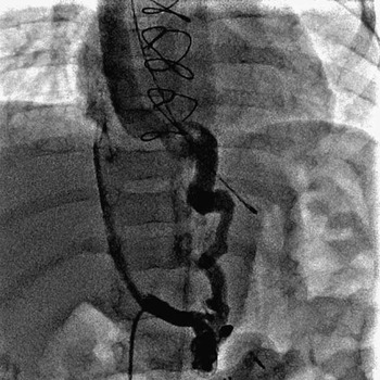
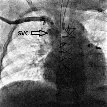
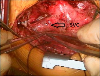
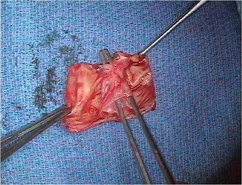
We report the follow-up surgery of our patient 16 months after the Southern Glenn, who was 22 months old at the time of completion Fontan, consisting of a standard “Northern” bi-directional cavopulmonary anastomosis with the superior caval vein, and extracardiac fenestration. Briefly, the primary extracardiac inferior cavopulmonary connection had been performed using an extracardiac roll fashioned from photofixed bovine pericardium to a diameter of 14 mm, rather than a ready-to-use Goretex conduit. We thought the roll could be tailored to an asymmetrical shape that would better fit behind an undivided superior caval vein–right atrial junction. The infant had an uneventful postoperative course, went home on postoperative day 8 on oral Aspirin, and maintained oxygen saturations equal to those expected after a standard bi-directional Glenn procedure. The patient gained weight, and showed no abdominal distension or ascites. After a period of 14 months postoperatively, the child had intermittent desaturations down to the 60s, leading to diagnostic catheterisation. A large veno-venous collateral from the iliac system decompressed the inferior caval vein (Fig 1), and drained back into an enlarged superior caval vein. The collateral vessel was successfully occluded with a device, and what was seen as left pulmonary artery stenosis at the inflow of the extracardiac conduit–pulmonary artery junction ballooned. Surprisingly, the previously diminutive superior caval vein had grown to larger-than-normal dimensions (Fig 2). Pressure measurements were deemed suitable for proceeding with Fontan completion with an inferior caval vein (pulmonary artery) pressure of 15 mmHg, left pulmonary artery of 15 mmHg, and pulmonary vascular resistance at 1.47 Wood Units/m2. Given the favourable catheter data yet persisting cyanosis despite coiling of the major veno-venous collateral, Fontan completion was planned at 22 months of age with a standard superior cavopulmonary anastomosis. Through redo-sternotomy, routine aortic and bicaval cannulation, the procedure was performed with normothermic beating heart cardiopulmonary bypass. A very large superior caval vein was visualised (Fig 3), transected from the common atrium, and a standard “Northern” bi-directional Glenn performed with a generous connection to the pulmonary arteries. The extracardiac photofixed bovine pericardium roll was opened in the middle to check for residual left pulmonary artery stenosis. An extensive intraluminal peel formed a true and false lumen, thereby reducing the diameter and effective pulmonary blood flow (Fig 4). The extant conduit was completely excised, and replaced with a 16 mm Goretex graft from the inferior caval vein to the underside of the pulmonary artery confluence. Weaning from cardiopulmonary bypass was uneventful. A fenestration was added secondary to high Fontan pressures (22–25 mmHg) and soft systemic pressures with an under-filled systemic ventricle. The hospital course was uneventful, with a 10-day intensive care stay and home discharge on day 17, with 1-litre supplemental oxygen and empirical sildenafil. He has been thriving at home since last follow-up with appropriate pulse oximetry saturations corresponding to a small residual fenestration (88–90%), allowing discontinuation from oxygen. No hepatic congestion or dysfunction occurred in the interim after the primary extracardiac inferior cavopulmonary connection, and has not been documented since Fontan completion to date.
Large veno-venous collateral decompressing the inferior caval vein (pulmonary artery system) via the iliac veins, and connecting to upper-body systemic tributaries draining into the superior caval vein.

Pre-Fontan angiogram depicting the superior caval vein (SVC) of larger-than-normal calibre.

Intraoperative picture before cannulation showing the superior caval vein (SVC), which is of larger-than-normal calibre.

The extracardiac roll once excised and opened on the back table, better demonstrating the intraluminal peel leading to stenosis and diminished pulmonary blood flow.

Discussion
Since abandoning one-stage Fontan completion because of higher operative mortality, the sequence of staged palliations for single-ventricle physiology has progressively involved interim palliation with a superior (Northern) caval vein–pulmonary artery anastomosis, or the bi-directional Glenn procedure, followed by completion FontanReference Bridges, Jonas, Mayer, Flanagan, Keane and Castaneda 3 – Reference Jacobs and Norwood 5 with the primary extracardiac inferior cavopulmonary anastomosis. Although initially attempted by Dr Glenn in dogs, no animal survived the inferior cavopulmonary connection as an isolated first palliation, leading to successful attempts with the azygousReference Glenn and Patino 6 and then superior caval vein,Reference Glenn 7 initially in the animal model, and then in humans. Based on the historical failure of an animal model to stage the right heart bypass concept in a different order, namely first from below (“South”), and then from above (“North”), we have inherited the very successful staging sequence of superior followed by inferior, which is used in a routine fashion in everyone’s daily practice.
We performed the primary extracardiac inferior cavopulmonary connection in an infant whose upper-body systemic venous anatomy was unsuitable for a standard bi-directional Glenn.Reference Dodge-Khatami, Aggarwal, Taylor, Maposa and Salazar 1 At the time, a very diminutive superior caval vein, thrombosed innominate vein, and insufficient collateral tributaries made us deviate from the normal staging sequence, short of leaving our patient with a shunt-dependent physiology. The type of material chosen for the extracardiac conduit was photofixed bovine pericardium, which we hoped could be fashioned into a shape better suited to lie behind an intact superior caval vein–right atrial junction. In retrospect, this extra caution proved unnecessary, as a standard Goretex tube could have fit easily. Furthermore, the poor choice in material may have even been detrimental to and instrumental in our patient’s health status, as the intimal peel that formed inside the pericardial roll despite Aspirin therapy led to conduit stenosis, and thereby to increased cyanosis from diminished pulmonary blood flow, the need for a large pop-off veno-venous collateral, and a premature reoperation. Although speculative and based on limited experience, it is questionable whether photofixed bovine pericardium is suited for low-pressure systems with laminary flow and only anti-aggregant therapy. If faced with the same situation in the future with the need for a primary inferior cavopulmonary connection, a standard Goretex conduit, as used for extracardiac Fontan completions, will be chosen, as its performance in the same setting and response to Aspirin are more predictable. A valuable lesson was learnt in this process, namely that a diminutive superior caval vein in small infancy may grow to larger-than-normal size by childhood, when faced with presumed enhanced flow, as it represents the only systemic venous blood return to the heart. Whether this is directly due to altered flow patterns created by the primary extracardiac inferior cavopulmonary conduit, or secondarily from the decompressing veno-venous collateral from the inferior caval vein, is speculative. The connections of the bi-directional cavopulmonary anastomosis we were able to perform to complete the “Northern” Fontan were wide open, owing to a very generously sized superior caval vein.
In conclusion, our experience in one patient with reverse geographical staging, namely initial partial right heart bypass from below, followed by complete right heart bypass from above, demonstrates the feasibility of such an approach. If faced with unfavourable anatomical conditions for a bi-directional Glenn in future patients, we suggest repeating the reversed staging procedure from South to North, which leads to earlier ventricular unloading, and seems intuitively preferable to leaving a volume-loaded heart with shunt-dependent physiology indefinitely. It is unknown whether an early primary extracardiac inferior cavopulmonary connection may lead to long-term hepatic sequelae.
Acknowledgement
None.
Financial Support
This research received no specific grant from any funding agency, commercial, or not-for-profit sectors.
Conflicts of Interest
None.