FOCUS POINTS
• Dyskinesia is a complication of long-term treatment with levodopa for Parkinson's disease (LID) and antipsychotics for schizophrenia (TD).
• Dyskinesia may be related to a degeneration or malfunctioning of indirect extrapyramidal pathway medium spiny neuron (MSN) within the striatum.
• Blocking MSN neurotoxicity induced by corticostriatal glutamatergic synapses may play an important role in preventing dyskinesia in HD and LID.
• Blocking MSN neurotoxicity induced by oxidative stress may play an important role in preventing TD.
The extrapyramidal system is a circuit
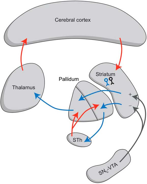
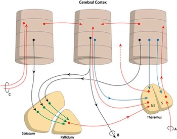
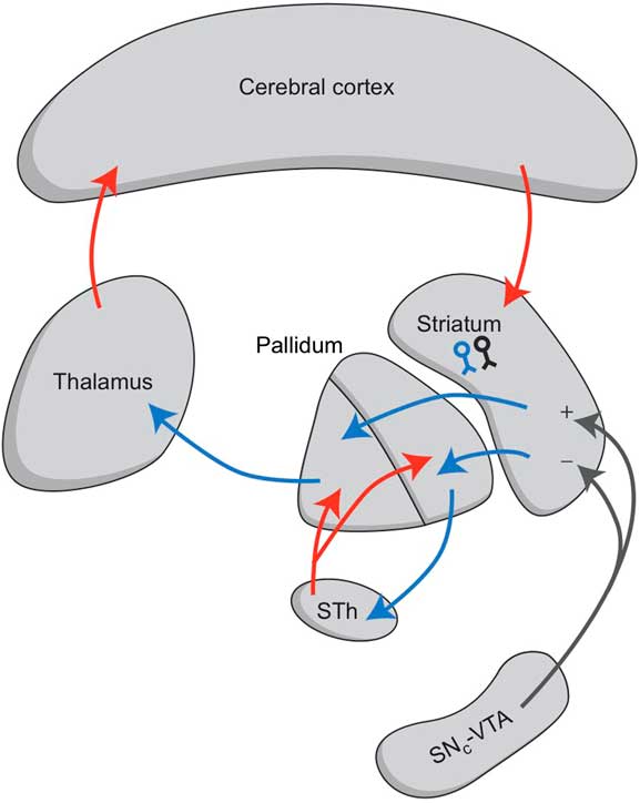
Drug-induced parkinsonism and dyskinesia are classified as extrapyramidal movement disorders. This term was derived from the original belief that all motions were controlled by two parallel systems: (1) a pyramidal tract running from the cerebral cortex to the brain stem and spinal cord; (2) the other was thought to be a parallel tract, therefore called the extra-pyramidal system.Reference Lance and McLeod1 Currently, it is known that the extrapyramidal system is not a parallel pathway, but a circuit that starts and ends in the cerebral cortex (Figure 1).Reference Groenewegen2 It plays an important role in adjusting the amplitude and velocity of muscle contractions that are generated by the motor areas of the frontal cerebral cortex. By regulating and adjusting the contractions of different muscle groups during kinesis, the extrapyramidal circuit allows a proper collaboration between these groups while executing a specific movement. The system also plays an important role during learning how these movements are best executed. It memorizes the exact interplay of muscle contractions during the execution of specific actions. Within the extrapyramidal system, information from different parts of the cerebral cortex converges on specific parts of the frontal cortex. For example, the premotor cortex receives information from the sensorimotor cortex, the prefrontal cortex, and the supplementary motor cortex. This information is also transferred directly through intracortical projections (Figure 2). Therefore, the motor cortex receives its commands through connections within the cerebral cortex itself, but also in a parallel way through the extrapyramidal circuit. This construct makes it possible to optimally adjust the executed movement.
The so-called cortico-striato-pallido-thalamo-cortical or extrapyramidal circuit. Striatal medium spiny neurons (MSNs) receive information from glutamatergic corticostriatal neurons and are influenced by dopaminergic fibers from the midbrain substantia nigra, pars compacta (SNc), and ventral tegmental area (VTA). STh = subthalamic nucleus.
Simplified representation of the cortico-striatal processing unit in which cortical information leading to a movement is processed in an intracortical and (parallel) extrapyramidal fashion: (A) sensory input, (B) projections to brain stem and spinal cord, (C) projection to and from ipsilateral and contralateral cortical areas.

The extrapyramidal system is not limited to the motor system. Within the extrapyramidal system, several parallel divisions can be distinguished that adjust different forms of output, eg, cognitive output or behavioral motivation.Reference Groenewegen and Trimble3 However, these aspects will not be discussed further in this article.
Medium spiny neurons
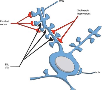
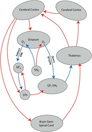
Within the striatum, the projections of glutamatergic pyramidal cells synapse on a specific type of GABAergic neurons, called medium spiny neurons (MSNs; Figure 3). Two types of MSNs are distinguished, and these constitute the starting points of two different pathways.Reference Groenewegen2 MSNs of the direct pathway carry dopamine D1 receptors and use substance P and dynorphin as co-transmitters. These fibers run directly to the outlet structures of the basal ganglia, including the internal segment of the pallidum (GPi) and the reticular part of the substantia nigra (SNr). The other type of MSN is the starting point of the indirect pathway. It carries dopamine D2 receptors and contains enkephalin as a co-transmitter. These indirect pathway MSNs project to the outer segment of the pallidum (GPe) and, from there, the signal runs to the subthalamic nucleus (STh) before ending in the outlet structures (Figure 4). Several other types of GABAergic neurons are found in the striatum. A minor fraction (about 5%) of striatal interneurons is cholinergic in nature. The latter nerve cells also carry dopamine D2 receptors and synapse with MSNs through muscarinic (AChM) receptors.
Medium spiny neuron. SNc: substantia nigra, pars compacta; VTA: ventral tegmental area; MSN: other medium spiny neuron (from ref. 6; adapted from ref. 2).

The cortico-striato-thalamo-cortical circuits, including the indirect and direct pathways: ENK = enkephalin; GPe = globus pallidus, external segment; GPi = globus pallidus, internal segment; SNc = substantia nigra, pars compacta; SNr = substantia nigra, pars reticulata; SP/DYN = substance P/dynorphin; STh = subthalamic nucleus; D1, D2: medium spiny neurons with D1 or D2 receptors.

Function of the direct and indirect extrapyramidal pathways
Functionally, the direct and indirect pathways have opposite influences. Activation of the direct pathway increases the amplitude and velocity of movements; conversely, activation of the indirect pathway reduces the magnitude and speed of movements. Because the corresponding direct and indirect pathways run to the same spot on the cerebral cortex, this organization allows for the exact learning of the amplitude and speed of the muscle contractions necessary for a proper execution of a certain movement. However, global dominance of the direct pathway would be expected to result in hyperkinesia/dyskinesia, and dominance of the indirect pathway would be expected to result in hypokinesia/bradykinesia.
Dopaminergic and serotonergic influence
Dopaminergic terminals from the midbrain synapse with both types of MSNs and several types of interneurons (Figures 1–4). Stimulation of D1 receptors on MSNs in the direct pathway causes activation of this pathway and results in hyperkinesia. Stimulation of D2 receptors on MSNs of the indirect pathway reduces the activity of the indirect pathway, and this also results in hyperkinesia by disinhibiting the activity of the cerebral cortex. Serotonergic terminals also influence the activity of cortico-striato-pallido thalamo-cortical (CSTC) circuits, but probably in an indirect manner.Reference Stahl4 Serotonergic terminals inhibit dopaminergic neurons in the midbrain by stimulating serotonin 5-HT2A/C-receptors on GABAergic interneurons in the compact part of the substantia nigra (SNc). The same mechanism may mediate the inhibitory influence of serotonergic terminals on the release of dopamine in the striatum. Thus, atypical antipsychotics that antagonize these 5-HT2A/C receptors would disinhibit the release of dopamine, and consequently, increase motor activity. This effect is often postulated to explain why atypical antipsychotics, usually 5-HT2A-antagonists, have fewer extrapyramidal side effects than classical neuroleptics. However, the affinity of these antipsychotics for D2 (and usually also D1) receptors is often far higher than the affinity of dopamine itself. It is difficult to understand how dopamine could displace these drugs from their firm binding to D1 or D2 receptors in order to stimulate these receptors. So, there exists room for debate about the validity of this hypothesis.
Glutamatergic neurotransmission
The cortico-striatal synapse is glutamatergic in nature; this confers the ability to learn by means of long-term potentiation (LTP) or long-term depression (LTD).Reference Bashir5, Reference Loonen6 These learning processes provide the potential to memorize the precise sequences and magnitude of augmenting and diminishing activation patterns during the execution of complex movement patterns. LTP was initially described for glutamatergic neurons of the hippocampus, but it can also occur at cortico-striatal synapses. LTP results from activation of a specific subtype of the ionotropic glutamatergic receptors—the NMDA receptor. During normal glutamatergic neurotransmission, AMPA receptors are activated and induce depolarization of the postsynaptic membrane. However, when the postsynaptic membrane is already in a partially depolarized state, or when an additional factor simultaneously activates NMDA receptors, the heightened depolarization can also activate NMDA receptors. This results in intracellular release of calcium ions, which serve as a second messenger to induce long-term changes in the sensitivity of the synapse to activation. Thus, the synapse is more readily activated with subsequent neurotransmission; in this manner, activation patterns can be memorized. The process of LTD is somewhat more complex; it involves retrograde signaling mediated by endocannabinoids. LTD can diminish once LTP has occurred, but it can also arise de novo.
LTP and LTD occurring at cortico-striatal synapses allows the extrapyramidal system to memorize the exact activation level of a set of mutually corresponding direct and indirect pathway MSNs during training in how to execute specific movements.
Dyskinesia
Dyskinesia is a collective name for a variety of involuntary hyperkinetic movements.Reference Loonen and van Praag7 The movements are irregular, repetitive, and typically include motionless intervals. Dyskinesia is reduced when the muscles are used voluntarily and when attention is focused on it. Dyskinesia increases during activity and with anxiety.
The most widely described phenomenon is the facio-bucco-linguo-masticatory (BLM), or orofacial, syndrome, which consists of movements of the lips, jaw, tongue, and eyelids. In addition, dyskinesia may present with choreiform movements of the extremities and distal athetosis. These movements consist of twisting movements of the fingers, the feet, and the toes. Also, the neck and trunk may exhibit rocking movements, and even the throat and diaphragm may exhibit vocalizations and disturbed swallowing. These movements comprise limb-truncal dyskinesia.
Dyskinesia may result from long-term treatment with antipsychotic drugs. This involuntary movement syndrome is termed tardive dyskinesia (TD).Reference Kane8, Reference Margolese, Chouinard, Kolivakis, Beauclair and Miller9 Dyskinesia may also occur spontaneously in Huntington's disease.Reference Novak and Tabrizi10 Another type of drug-induced dyskinesia results from long-term treatment with levodopa in Parkinson's disease; this is termed levodopa-induced dyskinesia (LID).Reference Thanvi, Lo and Robinson11 Slight differences are observed in the phenomenology of these three different types of dyskinesia. In TD, the orofacial syndrome is more prevalent than limb-truncal movements. In contrast, in HD and LID, limb-truncal dyskinesia is more prevalent than the orofacial syndrome.
The mechanism of drug-induced dyskinesia: a new idea based on the results of a Dutch-Siberian Collaborative Research Project
Dyskinesia is considered to be caused by disinhibition of the CSTC circuit, which finally results in overstimulation of the motor cortex. Initially, this disinhibition was believed to be caused by overactivation of dopamine receptors. In TD, long-term treatment with antipsychotics induced supersensitivity of dopamine receptors; this was thought to result in intense activation of these receptors when the concentration of antipsychotics dropped. In LID, the therapeutic administration of the dopamine precursor, levodopa, results in the presence of a relatively high extracellular concentration of dopamine. This was believed to result in a temporarily high activation of dopamine receptors. However, these models failed to explain why both TD and LID required significant treatment durations for development. Supersensitivity of dopamine D2 receptors occurs within weeks after initiation of treatment with antipsychotic drugs; in contrast, the development of TD often requires years of treatment. A similar time frame is required for the development of LID. Moreover, the model does not explain the rapid switch from a clinical condition of hyperkinesias to a condition of hypokinesias, which often occurs in LID (“on-off” phenomenon). This behavior is indicative of deregulation in a neuronal circuit, comparable with an electronic toggle switch. It has been suggested that LID might result from supersensitivity of the direct pathway MSNs to stimulation by glutamatergic cortico-striatal pyramidal neurons.Reference Thanvi, Lo and Robinson11 In this hypothesis, the relative surplus of dopamine shortly after the administration of a levodopa immediate release tablet could result in overstimulation of dopamine D1 receptors on direct pathway MSNs. In turn, this might promote the facilitation of LTP, which would then result in hypersensitivity of these cortico-striatal synapses.
Dyskinesia is also a primary symptom of Huntington's disease. This heritable progressive neurological disorder is associated with a pathological variant of the gene that encodes the Huntington protein. In patients with Huntington's disease, a slow, progressive degeneration occurs in indirect pathway MSNs.Reference Estrada Sanchez, Mejia-Toiber and Massieu12–Reference Kumar, Kalonia and Kumar14 Two independent research groups have reported that the age of onset of dyskinesia in patients with Huntington's disease was correlated with the presence of a variant of the gene that encodes one subunit of the glutamatergic NMDA receptor (GRIN2A).Reference Arning, Kraus and Valentin15–Reference Arning, Saft and Wieczorek17 This variant probably increases the sensitivity of the indirect pathway MSNs to excitotoxicity induced by the stimulation of glutamatergic NMDA receptors from cortico-striatal synapses. We recently found a strong association between the presence of the same GRIN2A variant and the occurrence of LID in patients with Parkinson's disease.Reference Ivanova, Loonen and Pechlivanoglou18 This suggests that the primary defect in LID may also involve degeneration of indirect pathway MSNs, rather than hypersensitivity of direct pathway MSNs, as was formerly believed.Reference Thanvi, Lo and Robinson11 Indirect pathway MSNs carry dopamine D2 receptors; therefore, they are also affected by antipsychotic drugs, which, currently, are all dopamine D2 antagonists. However, the development of TD was not associated with the presence of GRIN2A variants. Thus, the same type of indirect pathway MSNs appear to be involved in the pathogenesis of Huntington's disease, LID, and TD, but the mechanisms may differ slightly. This is consistent with the observation that the clinical picture also differs for these three conditions.
The hypothesis that both HD and LID are related to the degeneration of indirect pathway MSNs corresponds to an entirely different hypothesis for the pathogenesis of TD: the neurotoxicity theory.Reference Lohr, Kuczenski and Niculescu19 This theory states that TD is the result of neurotoxic effects of the free radicals produced by excessive metabolism of dopamine. Thus, drugs that block dopamine D2 receptors trigger a compensatory release of excess dopamine, and this excess requires increased metabolism of the spilled neurotransmitter. Increased dopamine metabolism releases high levels of hydrogen peroxide, which results in the production of free radicals, which then cause cell damage. Thus, excessive dopamine metabolism results in the production of more free radicals than the cell can handle. This hypothesis is consistent with the reported association between the incidence of TD and the presence of variants in the gene that encodes manganese superoxidedismutase (MnSOD), an enzyme that scavenges free radicals.Reference Al Hadithy, Ivanova and Pechlivanoglou20 A reduction in MnSOD activity would increase the likelihood of neurotoxic effects. This mechanism may also contribute to the pathogenesis of LID, which arises after prolonged levodopa treatment for Parkinson's disease. The externally applied levodopa is metabolized to dopamine, which in turn, is broken down by the mitochondrial enzyme monoamine oxidase (MAO). In fact, patients with Parkinson's disease may be excessively susceptible to the formation of neurotoxic free radicals, because this same mechanism explains the neurotoxic degeneration of dopaminergic neurons in Parkinson's disease. To date, it remains unclear why indirect pathway MSNs are particularly vulnerable to this neurotoxicity.
Conclusion
These findings have indicated that the selective damage of indirect pathway MSNs could represent a primary pathogenetic factor in Huntington's disease, LID, and TD. However, the exact mechanisms involved may differ slightly between these three conditions. The differences may result from slight differences in the exact location in the striatum of the MSNs that are primarily damaged. The different locations of damage may determine the occurrence of primarily peripheral or orofacial manifestations. In both LID and HD, glutamatergic neurotoxicity seems to play an important role in increasing the vulnerability of MSNs. However, this does not appear to be the case in TD. It remains to be determined whether TD is related to another form of neurotoxicity and/or whether the different mechanisms of action give rise to dissimilarities in the manifestations.
Disclosures
Dr. Loonen served as a consultant to Dutch Courts and Solicitors. He received speaker's fees from Bristol Myers Squibb and Servier. He received grants of AstraZeneca and Servier. Dr. Ivanova received grants from RHF (Russian Human Foundation) and Ministry of Health and Social Development of the Russian Federation.