Introduction
Acoustic neuromas account for about 5–10 per cent of all primary intracranial tumours and 85 per cent of cerebellopontine angle and internal auditory canal tumours.Reference Tong, Harnsberger, Swartz, Swartz and Harnsberger1 They originate from a vestibular branch of the VIIIth cranial nerve and spread into the internal auditory canal to reach the cerebellopontine angle.
The advent of magnetic resonance imaging (MRI) has enabled an increasing number of schwannomas to be diagnosed early. In some series, up to 28 per cent of acoustic neuromas were reported as still being completely within the internal auditory canal.Reference Casselman2
In the past, hearing preservation was considered an accessory goal of acoustic neuroma removal. However, it is now gaining increasing importance, especially if the tumour has affected the better ear of a patient with asymmetrical bilateral hearing loss or the healthy ear of a patient with unilateral hearing loss.Reference Domb and Chole3–Reference Larson12
To achieve the goal of hearing preservation, the middle fossa approach and the retrosigmoid approach are indicated.Reference House, Gardner and Hughes13–Reference Driscoll, Jackler, Pitts and Banthia16 These techniques have strict indications, specific advantages and well defined limitations.
The middle fossa approach can easily control small acoustic neuromas involving the fundus of the internal auditory canal. However, this route is contraindicated for tumours protruding more than 5 mm into the cerebellopontine angle.
The opposite effect is seen with the retrosigmoid approach. This approach ensures adequate exposure of the cerebellopontine angle but fails to reach the distal portion of the internal canal when hearing preservation is attempted.
In order to overcome these disadvantages, McElveen et al. proposed in 1991 a modification of the traditional translabyrinthine approach, by sealing the vestibule with bone wax.Reference McElveen, Wilkins, Erwin and Wolford17–Reference Hirsch, Cass, Sekhar and Wright18 These authors aimed to preserve hearing function, although all three semicircular canals had to be sacrificed. This aim was achieved in one patient. Unfortunately, this outcome could not be reproduced, either by these authors or by other investigators.
In 2004, Magliulo et al. introduced another modified translabyrinthine approach and reported encouraging hearing outcomes. They emphasised the need to completely close off access to the vestibule of the semicircular canals in order to preserve hearing function.Reference Magliulo, Parrotto, Stasolla and Marini19
The aim of our study was to evaluate the effectiveness of this technique, comparing the hearing results with imaging data.
Materials and methods
Our study group consisted of 16 prospectively selected patients with vestibular schwannoma (10 women and six men) aged between 23 and 65 years (average age, 47.4 years). Each patient was informed of the possibility of preserving their hearing function and was given detailed information on the indications, results, complications and consequences of the traditional hearing preservation techniques used in cerebellopontine angle surgery.
Each patient underwent puretone and speech audiometry to evaluate their degree of hearing loss. Patients with a speech discrimination score of 50 per cent or less were excluded from the study. The pre-operative hearing of all patients was graded as class one or two according to the Gardner–Robertson scale.Reference Gardner and Robertson20
The possibility of attempting to preserve hearing function via adopting an alternative method, namely, the modified translabyrinthine approach, was then presented to the patients: All patients accepted to be operated on via modified translabyrinthine approach.
The size of the schwannoma was calculated according to the final pre-operative axial and coronal MRI scans. Measurements were taken and the schwannoma size was calculated, assessing separately the canalicular part and the part outside the acoustic porus of the cerebellopontine angle. In seven patients, the intracanalicular portion of the schwannoma involved the fundus of the internal auditory canal. In the remaining nine cases, the schwannoma had spread to involve two-thirds of the meatus, sparing its lateral third.
The size of the cerebellopontine angle portion of the schwannoma was calculated from the axial MRI by measuring the medial distance at the level of the acoustic porus and the perpendicular distance at the petrous bone. The schwannoma, including the intracanalicular segment, was less than 2 cm along its greatest diameter in size in all patients undergoing surgery.
The modified translabyrinthine approach has been widely described elsewhere.Reference Magliulo, Parrotto, Stasolla and Marini19 In brief, a total mastoidectomy is performed and the semicircular canals are resected (taking care to avoid damage to the membranous labyrinth) and sealed with bone wax. The removal of the posterior bony labyrinth is completed by occluding the canals with bone wax, maintaining the anatomical integrity of the vestibule. The internal auditory canal is then exposed and the tumour removed. After endoscopy of the internal auditory canal, the wound is closed with abdominal fat.
Computed tomography imaging
Computed tomography (CT) examinations were performed with a multislice scanner (Somatom Plus 4 Volume Zoom, Siemens, Erlangen, Germany), using the following parameters: 140 kV; 110–180 mAs; 4×0.5 mm slice thickness; and 0.75 seconds rotation time.
Magnetic resonance imaging
Magnetic resonance imaging was performed using a 1.5 Tesla unit (Magnetom Vision, Siemens). Both axial and coronal T2 turbo spin echo – thin slice (3 mm) images were obtained (2735 ms/102 ms/15=time repetition/time echo/echotrain length). A standard T1-weighted spin-echo sequence (539 ms/12 ms/2=time repetition/time echo/number of excitation) was used in the transverse plane prior to and following contrast medium administration (Omniscan, Nycomed Amersham Sorin, Saluggia [VC], Italy). High resolution, T2-weighted three-dimensional images (constructive interference at steady state) were obtained using the following parameters: time repetition=12.25; time echo=5.90; flip angle=65°; matrix=230×512; field of view=165×220; section thickness=0.7 mm; acquisition time=8 minutes 40 seconds.
Image analysis
The CT and MRI images were examined by two independent readers, blinded to the clinical status of the patients, in order to establish the integrity of the bony vestibule and the presence of a regular, hyperintense signal on the T2-weighted images at vestibular level. The readers agreed in every case.
The MRI scans were studied to exclude the possibility of neoplastic residue and to identify possible complications (e.g. haemorrhage or cerebral ischaemia).
Results
The schwannoma was completely removed in all patients. None showed any signs of persistence or relapse of the tumour on the post-operative MRI.
The pre-operative hearing of all patients was graded as class one or two according to the Gardner–Robertson scale. The final post-operative follow up showed that seven patients had maintained their hearing function (four patients had class one hearing and three had class two).
Magnetic resonance imaging sequences using constructive interference at steady state were employed to evaluate the membranous labyrinth, in all but one case. This one patient's MRI scans had contained artefacts due to magnetic susceptibility and were consequently considered unreliable. Therefore, this patient was evaluated using turbo spin echo T2-weighted MRI scans.
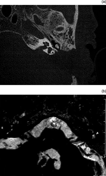
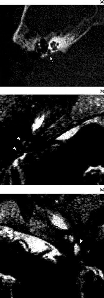
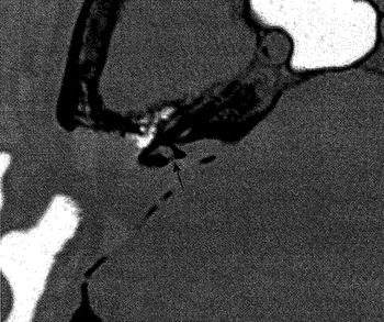
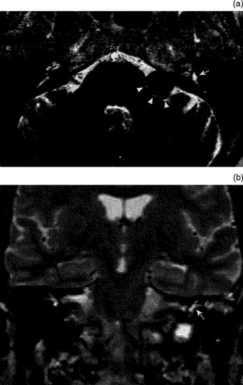
The MRI vestibular signal on T2-weighted images was well depicted only in patients with hearing preservation. Bony vestibular CT integrity was observed in all cases with hearing preservation, but also in three cases with no socially useful hearing (i.e. hearing loss classes three to five) (Figures 1 to 4; Table I).
Case 1; hearing preservation. (a) Axial computed tomography scan showing preservation of vestibular bony structure (arrow). (b) Axial constructive interference at steady state magnetic resonace image (time repetition/time echo/flip angle=12.25/5.90/65°) showing normal vestibular signal.

Case 4; hearing loss. (a) Axial computed tomography scan showing a regular-shaped vestibule on the right (arrow). (b) Axial constructive interference at steady state magnetic resonance image (time repetition/time echo/flip angle=12.25/5.90/65°) showing only a faintly depicted vestibulo-cochlear signal (arrowheads). (c) Normal signal on the contralateral side (arrowhead).

Case 5; hearing preservation. Axial computed tomography scan showing integrity of vestibular bony structure. (The constructive interference at steady state magnetic resonance images showed a normal cochlear and vestibular signal; image not shown.)

Case 9; hearing preservation. (a) Axial constructive interference at steady state magnetic resonance image (time repetition/time echo/flip angle=12.25/5.90/65°) showing a normal vestibular signal (arrow), prior to eradication of an internal auditory canal vestibular schwannoma extending into the cerebellopontine angle (arrowheads). (b) After eradication by modified translabyrinthine approach, the coronal turbo spin echo T2-weighted magnetic resonance image (time repetition/time echo/echotrain length=2735 ms/102 ms/15) shows a preserved vestibular signal (arrow).

imaging appearance of bony and membranous vestibule after modified translabyrinthine approach

* Classes 1–2; †classes 3–4. MRI=magnetic resonance imaging; CISS=constructive interference at steady state; SE=spin echo; CT=computed tomography
Discussion
For small tumours in the internal auditory canal with no or minimal extrameatal spread, the modified translabyrinthine approach may be considered a positive development of the classical translabyrinthine approach; it retains the advantages of the classical approach, and it may potentially preserve hearing function.
After the bony labyrinth has been exposed, the modified translabyrinthine approach entails rapid closure of the semicircular canal openings with bone wax, being careful to preserve the membranous labyrinth. According to Magliulo et al., this modification prevents endolymph leakage and consequently preserves hearing, despite the destruction of all three semicircular canals.Reference Magliulo, Parrotto, Stasolla and Marini19
Like all fluids, the endolymph and perilymph in the membranous labyrinth produce a high signal on the spin echo T2-weighted images normally indicated for study of the encephalus. However, considering the small size of the structure under examination, high resolution sequences are usually preferable in order to study the membranous labyrinth in detail. It is thus possible to obtain thin slices and, at the same time, a good signal/noise ratio. Three-dimensional, gradient echo or turbo spin echo sequences are aimed specifically at the region of interest and subsequently allow reconstruction of slices less than 1 mm thick. Three-dimensional T constructive interference at steady state sequences were the first to be used for this purpose,Reference Casselman, Kuhweide, Deimling, Ampe, Dehaene and Meeus21 and our department uses them routinely to study the internal auditory ducts and the membranous labyrinth. These sequences are also adopted for ‘magnetic resonance cisternography’ in order to study the nervous branches of the cerebellopontine angle cistern and the internal auditory canal in detail. These sequences can also enable morphological evaluation of the membranous labyrinth, since endolabyrinthic fluids (e.g. cerebrospinal fluid) remain stable and maintain a high signal compared with surrounding tissues.
However, when various materials are used to fill the surgical wound the disadvantages is the possibility of distortion due to magnetic susceptibility, which may invalidate the diagnostic capacity of the images. In theory, these adulterated images are more likely to occur in areas that have been subjected to surgical bone milling or packing of autological substances, etc. In one of our cases, the quality of the constructive interference in steady state images was worse than that of the conventional turbo spin echo T2 sequences (which had produced a valid signal for the vestibule) (Figure 4b).
Our results demonstrate that the CT appearance of the vestibule does not correlate with successful hearing preservation. In fact, we observed post-operative hearing loss in six out of six cases which had shown discontinuity of the vestibule on CT, and in three out of 10 cases in which the vestibule appeared intact on CT. This confirms the view that the surgical opening must be obliterated in order to preserve hearing function, and that this must be performed sufficiently quickly to prevent endolymphatic leak.
Computed tomography does not seem to be the most reliable method of judging the outcome of the operation. On the contrary, we found the MRI appearance to be closely connected with the outcome of surgery. Of our seven cases with an endolabyrinthic fluid signal clearly depicted on post-operative T2-weighted images, all had preserved hearing. Of our nine cases with no or low endolabyrinthic fluid signal post-operatively, none had any socially useful hearing preservation.
• This study investigated whether, following removal of vestibular schwannoma via a modified translabyrinthine approach, the computed tomography (CT) and magnetic resonance imaging (MRI) appearances of the vestibule correlated with a successful outcome, defined as hearing preservation
• The study group consisted of 16 patients with vestibular schwannoma. The state of the labyrinth, in particular the integrity of the vestibule, was evaluated by CT and MRI prior to and following surgery
• The results of this study suggest that complete isolation and maintenance of the anatomical integrity of the vestibule, as depicted by MRI examination, is fundamental to successful hearing preservation after surgery for vestibular schwannoma
Conclusion
Our study confirms the hypothesis that the maintenance of endolabyrinthic fluids is fundamental to the preservation of hearing function. This is shown by the observed relationship between the preservation of socially useful hearing and the demonstration of constant, well depicted post-operative T2-weighted vestibule signals.
Obviously the methods of choice to evalute the degree of hearing preservation remain pure-tone and speech audiometry. In our opinion, MRI should be considered a valid method of establishing a positive or negative outcome of surgery with regard to the anatomical preservation of the membranous labyrinth.
In those cases in which MRI has failed to give no signal or has produced a low signal in the vestibular area, CT may be used to attribute the bad outcome to insufficient or untimely obliteration of the semicircular canals.