INTRODUCTION
NG2-glia are a distinct class of CNS glial cells that are identified by their specific expression of the NG2 chondroitin sulphate proteoglycan (CSPG) and alpha receptors for platelet-derived growth factor (PDGFαR). NG2-glia are phenotypically distinct from astrocytes and oligodendrocytes (Butt et al., Reference Butt, Kiff, Hubbard and Berry2002), and generally classed as oligodendrocyte progenitor cells (OPCs) (Nishiyama et al., Reference Nishiyama, Lin, Giese, Heldin and Stallcup1996). It has been shown by genetic fate mapping in transgenic mice that NG2/PDGFαR-expressing cells generate oligodendrocytes during development (Zhu et al., Reference Zhu, Bergles and Nishiyama2008) and in the adult (Rivers et al., Reference Rivers, Young, Rizzi, Jamen, Psachoulia and Wade2008). In addition, the same authors showed that in the forebrain at least, NG2/PDGFαR-expressing cells generate astrocytes during development (Zhu et al., Reference Zhu, Bergles and Nishiyama2008), but not apparently in the adult (Rivers et al., Reference Rivers, Young, Rizzi, Jamen, Psachoulia and Wade2008), and that NG2-glia generate a restricted population of neurons throughout life (Rivers et al., Reference Rivers, Young, Rizzi, Jamen, Psachoulia and Wade2008). However, there are regional differences indicated in the literature. The astrogenic potential of NG2-glia is indicated in the pons, where NG2-glia express the astroglial reporter gene enhanced green fluorescent protein (EGFP) driven by glial fibrillary acidic protein (GFAP) in transgenic mice (Grass et al., Reference Grass, Pawlowski, Hirrlinger, Papadopoulos, Richter and Kirchhoff2004). We have confirmed this is the case in the pons and provide evidence that NG2-glia ‘turn on’ the GFAP reporter in cerebellar slices ex vivo (Leoni et al., Reference Leoni, Rattray and Butt2009). The neurogenic potential of NG2-glia has been indicated in the hippocampus (Belachew et al., Reference Belachew, Chittajallu, Aguirre, Yuan, Kirby and Anderson2003; Aguirre et al., Reference Aguirre, Chittajallu, Belachew and Gallo2004), olfactory bulb (Aguirre et al., Reference Aguirre, Chittajallu, Belachew and Gallo2004) and cortex (Dayer et al., Reference Dayer, Cleaver, Abouantoun and Cameron2005). Nonetheless, NG2-glia are equally numerous outside areas of neurogenesis and in areas where oligodendrocytes are not found, such as the molecular layer of the cerebellum (Butt et al., Reference Butt, Hamilton, Hubbard, Pugh and Ibrahim2005). Moreover, NG2-glia are highly populous throughout the adult CNS, making up 5–7% of cells, and 50% of these cells and do not appear to be normally involved in generating oligodendrocytes, astrocytes or neurons (Rivers et al., Reference Rivers, Young, Rizzi, Jamen, Psachoulia and Wade2008; Psachoulia et al., Reference Psachoulia, Young and Richardson2009). It is ambiguous to consider NG2-glia as OPCs, because this implies the fate of all NG2-glia ultimately is to turn into oligodendrocytes. The functions of non-proliferating NG2-glia in the adult CNS are unknown and we have asked the question whether they represent a new class of glia, which we called synantocytes (Butt et al., Reference Butt, Kiff, Hubbard and Berry2002, Reference Butt, Hamilton, Hubbard, Pugh and Ibrahim2005). These novel glial cells form exquisite associations with neurons at synapses (Bergles et al., Reference Bergles, Roberts, Somogyi and Jahr2000; Lin and Bergles, Reference Lin and Bergles2004; Lin et al., Reference Lin, Huck, Roberts, Macklin, Somogyi and Bergles2005; Ge et al., Reference Ge, Zhou, Luo, Jan and Jan2009), with unmyelinated axons at ‘synapse-like’ sites (Ziskin et al., Reference Ziskin, Nishiyama, Rubio, Fukaya and Bergles2007), and with myelinated axons at nodes of Ranvier (Butt et al., Reference Butt, Duncan, Hornby, Kirvell, Hunter and Levine1999, Reference Butt, Hamilton, Hubbard, Pugh and Ibrahim2005; Hamilton et al., Reference Hamilton, Vayro, Wigley, Robbins and Butt2009). In addition, NG2-glia express a range of ion channels and neurotransmitter receptors, which enable them to respond to neuronal and glial signals (Bergles et al., Reference Bergles, Roberts, Somogyi and Jahr2000; Lin and Bergles, Reference Lin and Bergles2004; Lin et al., Reference Lin, Huck, Roberts, Macklin, Somogyi and Bergles2005; Karadottir et al., Reference Káradóttir, Cavelier, Bergersen and Attwell2005, Reference Káradóttir, Hamilton, Bakiri and Attwell2008; Ge et al., Reference Ge, Yang, Zhang, Wang, Shen and Deng2006, Reference Ge, Zhou, Luo, Jan and Jan2009; Hamilton et al., Reference Hamilton, Vayro, Wigley, Robbins and Butt2009; Bakiri et al., 2009). In this review, we examine how NG2-glia are integrated with neurons, astrocytes and the vasculature of the CNS.
NG2-GLIA AND ASTROCYTES ARE SEPARATE POPULATIONS WITH OVERLAPPING DOMAINS
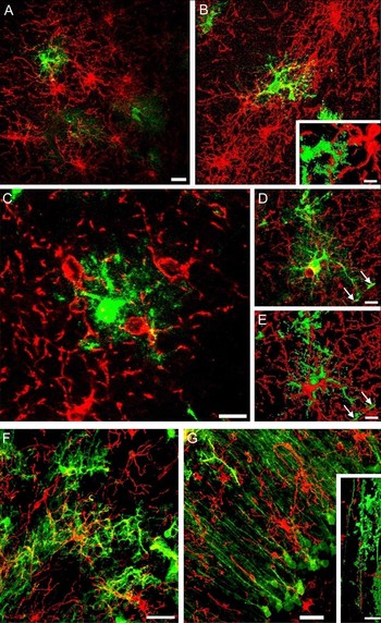
We have examined NG2-glia and astrocytes in the brain using immunohistochemistry in transgenic mice in which the expression of EGFP is driven by the human promoter for GFAP (Nolte et al., Reference Nolte, Matyash, Pivneva, Schipke, Ohlemeyer and Hanisch2001). We have previously reported that outside the pons we do not normally find co-expression of NG2 immunolabelling and the GFAP-EGFP reporter (Wigley et al., Reference Wigley, Hamilton, Nishiyama, Kirchhoff and Butt2007; Leoni et al., Reference Leoni, Rattray and Butt2009). EGFP identifies the true morphological complexity of astrocytes, but it is important to recognise that not all astrocytes express the reporter (Nolte et al., Reference Nolte, Matyash, Pivneva, Schipke, Ohlemeyer and Hanisch2001; Wigley et al., Reference Wigley, Hamilton, Nishiyama, Kirchhoff and Butt2007). NG2-glia and astrocytes have a similar distribution throughout grey matter, but differ in their antigenic and morphological phenotype (Fig. 1). NG2-glia have a characteristic stellate morphology, with a central cell body and fine processes that extend outwards radially to distances of up to 50 µm, as illustrated in the hippocampus (Fig. 1A–E), and the cerebellar granule cell layer (Fig. 1F) and molecular layer (Fig. 1G). Astrocytes have five or more thick primary processes from which emanate a multitude of fine processes, giving the cell a ‘furry’ appearance (Fig. 1A–F). NG2-glia and astrocytes define overlapping three-dimensional domains, in which the domains of the same cell type overlap only slightly at the edges, but those of different cell types overlap considerably (Fig. 1A–F). NG2-glial domains are significantly larger than those of astrocytes (P < 0.05, unpaired t-test), with an average diameter in the hippocampus of 99±1 µm (n = 17) in NG2-glia and 78±2 µm (n = 13) in astrocytes. The domains of individual astrocytes overlap with those of three or more NG2-glia (Fig. 1A–C), whereas those of NG2-glia overlap with the domains of more than six astrocytes (Fig. 1F), bearing in mind that this number is likely to be greater because not all astrocytes express GFAP or the reporter gene. Within overlapping domains, the processes of NG2-glia and astrocytes interdigitate and display bipartite ensheathment, whereby short segments of NG2-glial and astroglial processes ensheath each other (Fig. 1B, inset). The somata of some NG2-glia and astrocytes are directly apposed to one another, with the appearance of sister cells (Fig. 1D). In these cases, the domains of the NG2-glial cells and astrocyte are almost identical and there is a large surface area of contact between the somata and their processes, which interdigitate extensively within the overlapping process fields of the two adjoining cells (Fig. 1E). Both NG2-glia and astrocytes extend processes to blood vessels, but those of astrocytes expand to form perivascular end-feet (Fig. 1D,E, arrows). In the cerebellum, astrocytes display functional and morphological specialisation, with velate astrocytes in the granule cell layer (Fig. 1F), and Bergmann glia with cell somata in the Purkinje cell layer and primary processes extending through the molecular layer to the pia (Fig. 1G). In contrast, NG2-glia do not display morphological heterogeneity, and individual cells extend processes to traverse the different layers, to form overlapping domains with velate astrocytes (Fig. 1F) and Bergmann glia (Fig. 1G). NG2-glial cell processes are directly apposed to the primary radial processes of Bergmann glia and they ensheath each other regularly along their lengths (Fig. 1G, inset). The domain organisation of astrocytes is considered to be important for integrated activity at the synapses in their domain (Bushong et al., Reference Bushong, Martone, Jones and Ellisman2002; Nedergaard et al., Reference Nedergaard, Ransom and Goldman2003; Oberheim et al., Reference Oberheim, Wang, Goldman and Nedergaard2006), and presumably this is also true for NG2-glia.
Domain structure of NG2-glia and astroglia. Immunolabelling for NG2 (red) in brain sections from GFAP-EGFP mice. Confocal images are shown as two-dimensional snapshots (A,B,D,F,G), single z-section (C) and with isosurface rendering (B inset, E,G inset). NG2-glia and astrocytes are arranged in overlapping radial domains mice in the molecular layer of the hippocampus (A,C,D), the dentate gyrus (B), the granule cell layer of the cerebellum (F) and the molecular layer of the cerebellum (G). NG2-glial cell and astrocyte processes ensheath each other at numerous points other along their length (B, inset). (A–E) Domains of individual astrocytes interact with those of three or more NG2-glia (C), whereas individual NG2-glia interact with six or more astroglia (F,G). (D,E) NG2-glial cell and astrocyte somata are often directly apposed and have completely overlapping domains, with processes that interdigitate extensively. NG2-glia and astrocytes extend end feet to blood vessels, but those of astrocytes expand to form perivascular end feet (D,E, arrows). (F,G) NG2-glia traverse the different layers of the cerebellum to associate with multiple velate astrocytes and Bergmann glia; the process of NG2-glia and primary process of Bergmann glia are extensively apposed along their length (G, inset). Scale bars = 20 µm in (A,F,G) and 10 µm in (B,C,D,E) and 15 µm (G inset).

INTEGRATION OF NG2-GLIA INTO THE NEUROGLIAL NETWORK
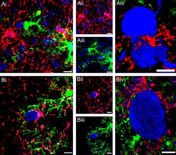
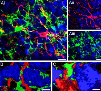
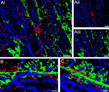
We have examined the interrelationships between NG2-glia, astrocytes and neurons in the cortex and cerebellum. Double immunolabelling for NG2 and calretinin or calbindin for neurons in the cortex of GFAP-EGFP mice illustrates the integration of the three elements (Fig. 2). The processes of NG2-glia, like those of astrocytes, are directly apposed to most, if not all, neuronal somata within their domains (Fig. 2A). It is important to note that our results demonstrate the close associations that NG2-glia form with other elements in the CNS, but it is not possible at the confocal microscopic level to distinguish whether these contacts are actually functional. Nonetheless, using a combination of electron microscopy and patch-clamp electrophysiology, it has been estimated that only a few of the neuronal contacts formed by NG2-glia are electrophysiologically active ‘synapses’ (Lin et al., Reference Lin, Huck, Roberts, Macklin, Somogyi and Bergles2005; Ge et al., Reference Ge, Zhou, Luo, Jan and Jan2009). In contrast, astrocytes form many hundreds of active contacts with neuronal synapses (Araque et al., Reference Araque, Parpura, Sanzgiri and Haydon1999; Grosche et al., Reference Grosche, Matyash, Moller, Verkhratsky, Reichenbach and Kettenmann1999; Bushong et al., Reference Bushong, Martone, Jones and Ellisman2002). NG2-glia and astrocytes form intimate contacts with the same neurons, extending processes that almost completely circumnavigate neuronal somata (Fig. 2B), and perisynaptic processes from both cells types form a latticework (reticulation) on the cell body (Fig. 2Biv). The close apposition of NG2-glia and astrocytes to the same neurons suggests they may contact the same synapses, but they subserve neurons in different ways. Electron microscopy studies demonstrate the different associations made by NG2-glia and astrocytes with neurons; NG2-glia form specific points of synaptic like contacts with neurons (Bergles et al., Reference Bergles, Roberts, Somogyi and Jahr2000), whereas astrocytes ensheath pre- and post-synaptic boutons (Nedergaard et al., Reference Nedergaard, Ransom and Goldman2003). This close relationship between NG2-glia, astrocytes and neurons is also reflected in the cerebellum (Fig. 3). In the granule cell layer, individual neurons are contacted by astrocytes and NG2-glial cell processes (Fig. 3A). However, whilst velate astrocyte processes circumnavigate neuronal cell somata, the processes of NG2-glia extend radially to form en passant associations with both neurons and astrocytes as they weave around the somata (Fig. 3B,C); as noted above it is not currently known whether these contacts have any function in communication. In the molecular layer, NG2-glia are closely integrated with Bergmann glia and Purkinje cells (Fig. 4). NG2-glial cell somata often lie along the primary process of Bergmann glial cells and Purkinje cell primary dendrites (Fig. 4A). NG2-glia extend processes both parallel to and perpendicular to Purkinje cell dendritic trees (Fig. 4A), and their processes interdigitate with those of Bergmann glia and Purkinje cell dendrites (Fig. 4B,C). A single NG2-glial cell subserves multiple Bergmann glia and Purkinje cells, and many NG2-glia extend processes into both the molecular and granule cell layers, to form additional contacts with velate astrocytes and granule neurons. NG2-glia contact synapses between climbing fibres and Purkinje neurons, but they appear to form functional contacts with only a small number of synapses within their domain (Lin et al., Reference Lin, Huck, Roberts, Macklin, Somogyi and Bergles2005), whereas Bergmann glia form many hundreds of microdomains that interact with the synapses they ensheath (Grosche et al., Reference Grosche, Matyash, Moller, Verkhratsky, Reichenbach and Kettenmann1999, Reference Grosche, Kettenmann and Reichenbach2002). Similarly, a single hippocampal astrocyte can contact in excess of 100,000 synapses (Bushong et al., Reference Bushong, Martone, Jones and Ellisman2002), and cortical astrocytes wrap on average four neuronal cell bodies and hundreds of dendrites (Halassa et al., Reference Halassa, Fellin, Takano, Dong and Haydon2007b). In contrast, NG2-glia do not have the profuse fine processes of protoplasmic astrocytes, and on average may form electrophysiologically active contacts with less than 20 synapses (Bergles et al., Reference Bergles, Roberts, Somogyi and Jahr2000; Lin and Bergles, Reference Lin and Bergles2004; Ge et al., Reference Ge, Zhou, Luo, Jan and Jan2009). However, the intimate associations between NG2-glia and astrocytes independent of neurons, both on their somata and along their processes, are sites of neurotransmitter mediated glia-to-glia calcium signalling (Hamilton et al., Reference Hamilton, Vayro, Wigley, Robbins and Butt2009). The overlapping domain organisation of NG2-glia and astrocytes may therefore be important for integrating glial and neuronal activity.
NG2-glia and astrocytes contact the same neurons in the cortex. Immunolabelling for NG2 (red) and calretinin (A, blue) or calbindin (B, blue) in 100 µm mid-sagital brain sections from the cortex of GFAP-EGFP mice. Confocal images are shown in three channels (Ai,Bi), and two channels (Aii,Aiii,Bii,Biii), and with isosurface rendering (Aiv,Biv). (A) NG2-glia and astrocytes are associated with 10 or more neurons within their overlapping domains. (B) NG2-glia and astroglial processes almost circumnavigate the same neuronal somata. Scale bars = 10 µm in (Ai–Aiii) and (Bi–Biii), and 5 µm in (Aiv) and (Biv).

Interactions of NG2-glia with astrocytes and neurons in the granule cell layer of the cerebellum. Immunolabelling for NG2 (red) and calretinin (blue) in the cerebellum of GFAP-EGFP mice. Confocal images are shown in three channels (Ai) and two channels (Aii,Aiii), and with isosurface rendering (B,C). Astrocytes and NG2-glia contact 20 or more neurons within their overlapping domains. Velate astrocyte processes encompass neuronal somata (Aii), whereas NG2-glial cell processes from en passant associations (Aiii). The processes of Ng2-glia and astrocytes are directly apposed at the same contact sites with neuronal somata, suggesting they subserve the same synapses (B,C). Scale bars = 10 µm in (A), 5 µm in (B) and 2 µm in (C).

Integration of NG2-glia with Purkinje neurons and Bergmann glia in the molecular layer of the cerebellum. Immunolabelling for NG2 (red) and calbindin (blue) in the cerebellum of P15 GFAP-EGFP mice. Confocal images are shown in three channels (Ai,Bi) and two channels (Aii,Aiii) and with isosurface rendering (B,C). NG2-glial cell somata lie along the primary processes of Bergmann glia and the Purkinje neuron dendrites, and extend processes parallel and perpendicular to the primary processes of Bergmann glia. The processes of NG2-glia and Bergmann glia are intimately associated with Purkinje neuron dendrites (C,D). Scale bars = 20 µm in (A) and 2 µm in (B,C).

COMMUNICATION BETWEEN NG2-GLIA AND THE NEUROGLIAL NETWORK
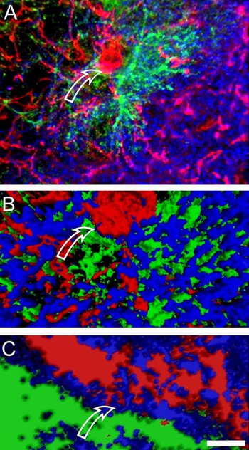
Electrophysiological studies show that NG2-glia express glutamate and GABA receptors and respond to neuronally released neurotransmitters (Bergles et al., Reference Bergles, Roberts, Somogyi and Jahr2000; Lin and Bergles, Reference Lin and Bergles2004; Lin et al., Reference Lin, Huck, Roberts, Macklin, Somogyi and Bergles2005). Calcium imaging and immunocytochemistry show that NG2-glia express functional AMPA-type glutamate receptors and metabotropic P2Y and ionotropic P2X receptors, including the P2X7 subtype (Ge et al., Reference Ge, Yang, Zhang, Wang, Shen and Deng2006; Hamilton et al., Reference Hamilton, Vayro, Wigley, Robbins and Butt2009; Wang et al., Reference Wang, Cai, Chen, Deng and Zhao2009). Cerebellar NG2-glia can display both GABAergic and glutamatergic inputs (Lin and Bergles, Reference Lin and Bergles2004; Karadottir et al., 2005), consistent with our observations that individual NG2-glia traverse the cell layers of the cerebellum to contact glutamatergic and GABAergic elements. In addition, we have used immunolabelling for synaptophysin to examine the relations of NG2-glia with synapses (Fig. 5). The density of synaptophysin immunolabelling in the grey matter precludes the quantification of specific synapses contacted by NG2-glia (Fig. 5A). Nevertheless, our results provide evidence that NG2-glia and astrocytes contact the same neurons (Figs 3–5), and the immediate apposition of synaptophysin to points of contact between NG2-glia and astrocytes suggests they may be integrated at the same synapses (Fig. 5). Moreover, synaptophysin is localised to sites of immediate apposition between astrocytes and NG2-glia, and a region of particular interest is the expression of synaptophysin between the NG2-glial cell body and an astrocyte process (Fig. 5B). Isoforming at the integral communication site between the NG2-glial cell and astrocyte process provides further evidence of the localisation of synaptophysin to the NG2-glial cell body and to the interface between NG2-glia and astrocytes (Fig. 5C). The results presented above show that the cell somata of astrocytes and NG2-glia are often directly apposed, and that their processes exhibit regions where they tightly ensheath each other (Figs 1B inset, 1G inset, 3B,C and 4B,C). The localisation of synaptophysin to these areas indicates these are potential sits of vesicular communication between the two cell types. Astrocytes have mechanisms to release neurotransmitters via vesicles (Parpura et al., Reference Parpura, Basarsky, Liu, Jeftinija, Jeftinija and Haydon1994; Araque et al., Reference Araque, Parpura, Sanzgiri and Haydon1999). We have provided evidence that astrocytes also signal onto NG2-glia via glutamate and ATP (Hamilton et al., Reference Hamilton, Vayro, Kirchhoff, Verkhratsky, Robbins and Gorecki2008, Reference Hamilton, Vayro, Wigley, Robbins and Butt2009). Moreover, the evidence that NG2-glia express synaptophysin, indicates they may be capable of neurotransmitter release and bidirectional communication with astrocytes and neurons.
Synaptophysin is localised to sites of close apposition between NG2-glia and astrocytes. Immunolabelling for NG2 (red) and synaptophysin (blue) in the hippocampus of GFAP-EGFP mice. Confocal images are shown in three channels (A) and with isosurface rendering (B,C). The processes of NG2-glia and astrocytes are associated with a large density of synapses (A,B). There is immediate apposition of the NG2-glial cell and astrocyte cell bodies and processes, and synaptophysin is localised to these sites of contact (arrows), indicating sites of synaptic communication onto both NG2-glia and astrocytes. In addition, synaptophysin was localised within the soma of the NG2-glia cell, confirming our recent report (Hamilton et al., Reference Hamilton, Vayro, Wigley, Robbins and Butt2009), and suggesting NG2-glia are capable of bidirectional communication with astrocytes and neurons. Scale bar = 20 µm (A), 5 µm (B) and 1 µm (C).

RELATIONS BETWEEN NG2-GLIA AND THE CEREBRAL VASCULATURE
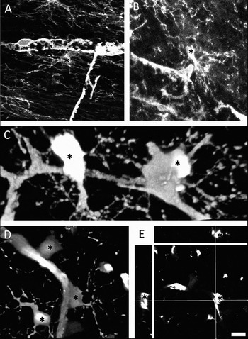
Astrocytes are able to ‘sense’ changes in neuronal activity and transmit signals to the vasculature to regulate local blood flow and provide metabolic support to neurons (Halassa et al., Reference Halassa, Fellin and Haydon2007a). Astrocytes may use glutamate signalling mechanisms to communicate with pericytes on blood vessels that are able to regulate cerebral blood flow (Peppiatt et al., Reference Peppiatt, Howarth, Mobbs and Attwell2006). NG2-glia also contact pericytes on cerebral blood vessels (Fig. 6), and they contact astrocytes and neurons within their domains (Figs 1–5), and express synaptophysin (Hamilton et al., Reference Hamilton, Vayro, Wigley, Robbins and Butt2009), and may therefore play a role in regulating blood flow. NG2-glia and pericytes both express the NG2 CSPG (Ozerdem et al., Reference Ozerdem, Monosov and Stallcup2002), and the two cell types are distinguished by their morphology, NG2-glia as stellate process-bearing cells and pericytes as perivascular cells with two or more primary processes extending along blood vessels. According to these distinguishing features, it can be seen that NG2-glia and pericytes lie in very close proximity to each other (Fig. 6A,B). The processes of NG2-glia contact pericyte processes on the blood vessel (Fig. 6A). In some cases, it appears that the pericyte cell soma extends out from the capillary to almost contact that of a neighbouring NG2-glial cell (Fig. 6B), and in many cases NG2-glial cell somata are directly apposed to blood vessels, identified by immunolabelling (Fig. 6C) and in the NG2-DsRed transgenic mouse (Fig. 6D,E). Pericytes have a neurogenic potential in vitro (Dore-Duffy et al., Reference Dore-Duffy, Katychev, Wang and Van Buren2006) and proliferate in response to injury (Gerhardt and Betsholtz, Reference Gerhardt and Betsholtz2003), and the intimate associations between NG2-glia and pericytes is suggestive of a lineage relationship between the two cell types (Wigley and Butt, unpublished observations). It remains to be seen whether NG2-glia communicate with pericytes and help regulate blood flow, but this is a possibility since they express synaptophysin and contact synapses, astrocytes and pericytes.
NG2-glia are in direct contact with pericytes. NG2-glia are marked by an asterisk in each image. Images show immunolabelling for NG2 in optic nerve of non-transgenic mice (A,B) and NG2-glia and pericytes in sections of NG2-dsRed mouse brain (C–E). NG2-glia contact multiple pericytes and in many cases their cell somata appear to be in direct contact (C,D) and this is confirmed in cross sectional images (E). Scale = 10 µm (A), 12 µm (B), 5 µm (C), 8 µm (D) and 10 µm (E).

CONCLUSIONS
NG2-glia and astrocytes form overlapping neuron–glial–vascular domains. Within these domains, NG2-glia and astrocytes contact the same neurons and most likely the same synapses, but they perform different functions. Astrocyte processes envelop synapses and one of their main functions is the removal of excess neurotransmitters. NG2-glia form fine connections with synapses and do not express transporters, and their function at synapses is unknown. The large domains of NG2-glia enable them to ‘sense’ neuronal activity from many neurons over a large area that overlaps with the domains of multiple astrocytes. Their relation with pericytes indicates that NG2-glia and astrocytes form parallel communications pathways between neurons and blood vessels which may be important in the regulation of blood flow. The evidence that NG2-glia also receive input from astrocytes and the possibility that NG2-glia express mechanisms for vesicular release of gliotransmitters raises the possibility that NG2-glia may also have an output and be able to feed back to neurons or astrocytes. The fact that NG2-glia and astrocytes form parallel communications pathways suggests they have different functions, since otherwise this level of redundancy is difficult to explain. Astrocyte domain organisation segregates intercellular communication within neuronal circuits (Houades et al., Reference Houades, Koulakoff, Ezan, Seif and Giaume2008), and astrocytes are coupled by gap junctions that provide an activity-dependent intercellular pathway for transport between blood vessels and neurons (Rouach et al., Reference Rouach, Koulakoff, Abudara, Willecke and Giaume2008). NG2-glia are not coupled by gap junctions, but by forming ‘synapses’ with neurons, astrocytes and pericytes, they connect otherwise segregated astroglial domains. An important question for future studies is to determine how these multiple inputs are integrated in individual NG2-glia, and whether they have a physiological output.
ACKNOWLEDGEMENTS
Supported by the BBSRC and INTERRIG.
Statement of interest
None.