Disease
Bovine respiratory disease (BRD) is a polymicrobial infection involving several viruses and bacteria as well as the predisposing factor of stress. The bacterial pathogens most often include Mannheimia haemolytica, Pasteurella multocida and Histophilus somni (Angen et al., Reference Angen, Ahrens, Kuhnert, Christenesen and Mutters2003), also called Haemophilus somnus. In addition to BRD, H. somni induces respiratory disease in sheep, bison and big horn sheep as well as infertility, septicemia, abortion, myocarditis, arthritis and meningoencephalitis in cattle (Griner et al., Reference Griner, Jensen and Brown1956; Biberstein Reference Biberstein, Kilian, Frederiksen and Biberstein1981; Stephens et al., Reference Stephens, Little, Wilkie and Barnum1981; Humphrey and Stephens, Reference Humphrey and Stephens1983; Miller et al., Reference Miller, Lein, McEntee, Hall and Shaw1983; Widders et al., Reference Widders, Paisley, Goglewski, Evermann, Smith and Corbeil1986; Corbeil et al., Reference Corbeil, Arthur, Widders, Smith and Barbet1987, Reference Corbeil, Gogolewski, Stephens, Inzana, Donachie, Lainson and Hodgson1995; Gogolewski et al., Reference Gogolewski, Kania, Inzana, Widders, Liggitt and Corbeil1987a, Reference Gogolewski, Leathers, Liggitt and Corbeilb, Reference Gogolewski, Schaefer, Wasson, Corbeil and Corbeil1989; Corbeil, Reference Corbeil1990; Haritani et al., Reference Haritani, Nakazawa, Hashimoto, Narita, Tagawa and Nakagawa1990; Kwiecien and Little, Reference Kwiecien and Little1991; Lees et al., Reference Lees, Yates and Corbeil1994; Ward et al., Reference Ward, Jaworski, Eddow and Corbeil1995, Reference Ward, Dyer and Corbeil1999, Reference Ward, Weiser, Anderson, Cummings, Arnold and Corbeil2006; Dyer, Reference Dyer2001). Not only is H. somni the etiologic agent in all of these syndromes but also it commonly exists in an asymptomatic carrier state on the reproductive and respiratory mucosa (Humphrey and Stephens, Reference Humphrey and Stephens1983; Humphrey et al., Reference Humphrey, Little, Stephens, Barnum, Doig and Thorsen1985). Several reviews of the naturally occurring disease are available (Humphrey and Stephens, Reference Humphrey and Stephens1983; Miller et al., Reference Miller, Lein, McEntee, Hall and Shaw1983; Harris and Janzen, Reference Harris and Janzen1989; Kwiecien and Little, Reference Kwiecien and Little1991). BRD due to H. somni is sometimes seen in young calves (enzootic pneumonia) but most often in feedlot cattle (Humphrey and Stephens, Reference Humphrey and Stephens1983; Harris and Janzen, Reference Harris and Janzen1989). A study of hemophilosis in feedlot calves showed that H. somni was a significant cause of death even though calves were immunized with a commercial H. somni vaccine on arrival (Van Donkersgoed et al., Reference Van Donkersgoed, Janzen and Harland1990). In that study, the median onset of fatal pneumonia was day 12 after arrival in the feedlot and death due to pneumonia was during the first five weeks. Although metaphylaxis (with antibiotics given before clinical signs are evident) has become popular for the prevention of BRD (Vogel et al., Reference Vogel, Laudert, Zimmermann, Guthrie, Mechor and Moore1998; Step et al., Reference Step, Engelken, Romano, Holland, Krehbiel, Johnson, Bryson, Tucker and Robb2007), the long-term results in selecting for antibiotic resistant bacteria is not known. Characteristics of the pathology of naturally occurring H. somni pneumonia have been defined in several studies (Andrews et al., Reference Andrews, Anderson, Slife and Stevenson1985; Bryson et al., Reference Bryson, Ball, Mcaliskey, McConnell and McCullough1990; Tegtmeier et al., Reference Tegtmeier, Uttenthal, Friis, Jensen and Jensen1999b). Much information on the definition of H. somnus disease also has been gleaned from experimental reproduction of the spectrum of syndromes caused by this organism. Several groups have reproduced thrombotic meningoencephalitis (Stephens et al., Reference Stephens, Little, Wilkie and Barnum1981), reproductive failure (Miller et al., Reference Miller, Lein, McEntee, Hall and Shaw1983) and pneumonia (Andrews et al., Reference Andrews, Anderson, Slife and Stevenson1985; Jackson et al., Reference Jackson, Andrews and Hargis1987; Groom et al., Reference Groom, Little and Rosendal1988; Potgieter et al., Reference Potgieter, Helman, Greene, Breider, Thurber and Peetz1988; Tegtmeier et al., Reference Tegtmeier, Bloch, Jensen and Jensen1999a; Tagawa et al., Reference Tagawa, Sanders, Uchida, Bastida-Corcuera, Kwashima and Corbeil2005). Interestingly, H. somni also is found commonly in an asymptomatic carrier state (Humphrey and Stephens, Reference Humphrey and Stephens1983). The pathogenesis of H. somni infection was studied by our group by reproducing both abortion and pneumonia in cattle. To simulate hematogenous infection, 19 pregnant cows were inoculated intravenously or intrabronchially (Widders et al., Reference Widders, Paisley, Goglewski, Evermann, Smith and Corbeil1986; Corbeil et al., Reference Corbeil, Arthur, Widders, Smith and Barbet1987). Reproductive failure occurred in six cows. Cultures showed massive infection of the uterus and placenta (Widders et al., Reference Widders, Paisley, Goglewski, Evermann, Smith and Corbeil1986; Corbeil et al., Reference Corbeil, Arthur, Widders, Smith and Barbet1987). Placentitis was characterized by thrombosis and vasculitis (Widders et al., Reference Widders, Paisley, Goglewski, Evermann, Smith and Corbeil1986), as in thrombotic meningoencephalitis (Stephens et al., Reference Stephens, Little, Wilkie and Barnum1981) and pneumonia (Gogolewski et al., Reference Gogolewski, Kania, Inzana, Widders, Liggitt and Corbeil1987a, Reference Gogolewski, Leathers, Liggitt and Corbeilb, Reference Gogolewski, Kania, Liggitt and Corbeil1988).
In other studies, pneumonia was reproduced in 6–12- week old calves (Gogolewski et al., Reference Gogolewski, Kania, Inzana, Widders, Liggitt and Corbeil1987a, Reference Gogolewski, Leathers, Liggitt and Corbeilb, Reference Gogolewski, Kania, Liggitt and Corbeil1988), since our earlier studies indicated that calves are most susceptible to pneumonia at this time (Corbeil et al., Reference Corbeil, Watt, Corbeil, Betzen, Brownson and Morrill1984). At 24 h after intrabronchial inoculation of 107H. somni, pneumonia was characterized by neutrophilic to fibrinoid vasculitis, degeneration of alveolar macrophages, necrotizing bronchiolitis, suppurative bronchiolitis, lobular necrosis and dilation and thrombosis of lymphatics (Gogolewski et al., Reference Gogolewski, Leathers, Liggitt and Corbeil1987b). Infection lasted for 6–10 weeks in studies of chronic infection (Gogolewski et al., Reference Gogolewski, Schaefer, Wasson, Corbeil and Corbeil1989). Interestingly, H. somni was isolated more often from the bronchioalveolar lavage fluid than from nasal swabs in weekly samples during these studies on chronic disease. This suggested that H. somni may preferentially colonize the lower respiratory tract rather than the nasal mucosa. Severe clinical signs in these chronically infected calves were only obvious for a few days. After that only an occasional cough was detected, indicating that chronic H. somni infection of the lower respiratory tract can be almost subclinical for weeks.
Immunodiagnostic assays have proven to be useful for diagnosis of H. somni infection. Culture of bacteria has usually been the gold standard for diagnosis of bacterial infection. This is also true for H. somni when large numbers are obtained in pure culture from lesions. However, a common genital asymptomatic carrier state and a less common respiratory carrier state complicate diagnosis by culture. In experimental disease, Gogolewski et al. (Reference Gogolewski, Leathers, Liggitt and Corbeil1987b) showed that organisms could be detected in pneumonic lesions by immunohistochemistry. This assay has been developed for diagnosis and used very successfully by Haines and Clark (Reference Haines and Clark1991). Microagglutination assays have been used widely for diagnosis but many cattle have high titers, even with no history of H. somni disease and no current clinical signs (Widders et al., Reference Widders, Paisley, Goglewski, Evermann, Smith and Corbeil1986). This can be explained by several antigens of H. somni which are cross-reactive with other members of the family Pasteurellaceae (Kania et al., Reference Kania, Gogolewski and Corbeil1990; Corbeil et al., Reference Corbeil, Kania and Gogolewski1991). In addition, the microagglutination test preferentially detects IgM antibodies and natural cross-reactive antibodies are often of this Ig class. Thus the high microagglutination titers in normal cattle with no history of H. somni infection are probably due to antibodies induced by other Pasteurellaceae. Alternatively, some of these antibodies could be due to induction by H. somni in the asymptomatic carrier state, especially in the genital tract. Our laboratory showed that ELISA tests using second antibody conjugates for bovine IgG2 were more specific than secondary antibodies to other immunoglobulin classes (Widders et al., Reference Widders, Paisley, Goglewski, Evermann, Smith and Corbeil1986) and IgG2 antibody assays against the 270 kDa FcR (now called IbpA or IgBPs) were most specific compared with other antigens in immunodiagnostic assays (Yarnall and Corbeil, Reference Yarnall and Corbeil1989).
Virulence factors
We and others have identified and characterized several H. somni antigens or virulence factors that are likely to be important in host–parasite relationships. The 40 kDa outer membrane protein (OMP; p40) is surface exposed and is surely important in protection since convalescent phase serum recognizing p40 and monospecific bovine antibodies against p40 are passively protective against experimental bovine pneumonia (Gogolewski et al., Reference Gogolewski, Kania, Inzana, Widders, Liggitt and Corbeil1987a, Reference Gogolewski, Kania, Liggitt and Corbeil1988; Corbeil et al., Reference Corbeil, Kania and Gogolewski1991). Others have cloned and sequenced a 40 kDa lipoprotein OMP of H. somni (Theisen et al., Reference Theisen, Rioux and Potter1992) which may be the p40 protective antigen. The 40 kDa OMP recognized by protective antibodies is different from the 41 kDa major outer membrane protein (MOMP) (Gogolewski et al., Reference Gogolewski, Kania, Inzana, Widders, Liggitt and Corbeil1987a; Yarnall et al., Reference Yarnall, Gogolewski and Corbeil1988a, Reference Yarnall, Widders and Corbeilb). We showed that p40 was immunodominant (Corbeil et al., Reference Corbeil, Kania and Gogolewski1991) but that the MOMP was not recognized by bovine convalescent phase serum at dilutions of 1:500–1:1000 or more; the dilutions which we usually use in Western blots (Gogolewski et al., Reference Gogolewski, Kania, Inzana, Widders, Liggitt and Corbeil1987a, Reference Gogolewski, Leathers, Liggitt and Corbeilb; Ward et al., Reference Ward, Jaworski, Eddow and Corbeil1995). Tagawa and colleagues purified and characterized the MOMP (Tagawa et al., Reference Tagawa, Haritani, Ishikawa and Yuasa1993b, Reference Tagawa, Ishikawa and Yuasad; Khan et al., Reference Khan, Tanaka, Ide, Hoshinoo, Hanafusa and Tagawa2005), showing it to be similar to porins of other Gram-negative bacteria. Tagawa also studied immunologic aspects of the MOMP in Corbeil's laboratory (Tagawa et al., Reference Tagawa, Bastida-Corcuera and Corbeil2000). Low dilutions (1:50 or 1:100) of bovine convalescent serum did recognize the MOMP but higher dilutions did not. Also, the MOMP was shown to be antigenically variable (Tagawa et al., Reference Tagawa, Bastida-Corcuera and Corbeil2000) and it is the predominant antigen recognized by IgE (Corbeil et al., Reference Corbeil, Arnold, Kimball, Berghaus and Gershwin2006), which was associated with enhanced respiratory disease. Thus the MOMP is not likely to be a very good vaccine candidate.
Tagawa et al. (Reference Tagawa, Haritani, Ishikawa and Yuasa1993a) also characterized a 37 kDa heat modifiable OMP with N-terminal sequence homology and immunologic cross-reactivity with OmpA of other Gram-negative bacteria. Studies of surface exposure gave contradictory results. This OMP was recognized by convalescent phase serum at 1:800 in Western blots. It is not clear whether it is an important virulence factor. Similarly, a 17.5 kDa OMP has been characterized but its role in virulence is unknown (Tagawa et al., Reference Tagawa, Haritani and Yuasa1993c). Lastly, iron regulated OMPs may be important in host–parasite relationships. Schryvers' group demonstrated transferrin binding proteins (Tbps; Ogunnariwo et al., Reference Ogunnariwo, Cheng, Ford and Schryvers1990) which bind bovine transferrin, but not other ruminant transferrins (Yu et al., Reference Yu, Gray-Owen, Ogunnariwo and Schryvers1992). Later, Ekins and co-workers (Ekins and Niven, Reference Ekins and Niven2001; Ekins et al., Reference Ekins, Bahrami, Sijercic, Mare and Niven2004) showed that some strains of H. somni have two genes for Tbps. Whether antigenic differences among strains result from differences in Tbps is unclear. These Tbps are probably involved in our studies of the role of bovine serum proteins in H. somni virulence in a mouse septicemia model (Geertsema et al., Reference Geertsema, Kimbal and Corbeil2007). That study demonstrated enhanced virulence for mice by preincubation of H. somni in Fetal Calf Serum (FCS) for 5 min before inoculation. To identify the virulence enhancing factor, H. somni was preincubated in each of the main components of FCS. Both fibrinogen and transferrin bound to H. somni but only bovine transferrin enhanced virulence. Mouse transferrin did not enhance virulence but bovine lactoferrin did enhance virulence for mice. Iron chelation experiments indicated that iron uptake from transferrin (but not lactoferrin) was critical for increased virulence in the mouse septicemia model.
In addition to protein virulence factors, endotoxin or lipooligosaccharide (LOS) is important in pathogenesis (Inzana et al., Reference Inzana, Iritani, Gogolewski, Kania and Corbeil1988). Collaborative studies with Inzana (Inzana et al., Reference Inzana, Gogolewski and Corbeil1992), showed that H. somni LOS undergoes phase and antigenic variation. Since then, Inzana's group has contributed much to the understanding of H. somni LOS, including LOS structural analysis of several strains (Cox et al., Reference Cox, Howard, Brisson, Van Der Zwan, Thibault, Perry and Inzana1998, Reference Cox, Howard and Inzana2003; Michael et al., Reference Michael, Howard, Li, Duncan, Inzana and Cox2004, Reference Michael, Li, Howard, Duncan, Inzana and Cox2005, Reference Michael, Inzana and Cox2006), sequences involved in biosynthesis of LOS (McQuiston et al., Reference McQuiston, McQuiston, Cox, Wu, Boyle and Inzana2000; Wu et al., Reference Wu, McQuiston, Cox, Pack and Inzana2000), the mechanism of phase variation (Howard et al., Reference Howard, Cox, Weiser, Schurig and Inzana2000; McQuiston et al., Reference McQuiston, McQuiston, Cox, Wu, Boyle and Inzana2000) and the role of LOS sialylation in resistance to complement mediated killing (Inzana et al., Reference Inzana, Gogolewski and Corbeil1992, Reference Inzana, Glindemann, Cox, Wakarchuk and Howard2002). This work was reviewed recently (Siddaramppa and Inzana, Reference Siddaramappa and Inzana2004), so will not be further discussed here. The antigenic variation in vivo suggests that LOS may not be a good vaccine candidate but this may make it a more effective virulence factor. Czuprynski's group (Sylte et al., Reference Sylte, Corbeil, Inzana and Czuprynski2001, Reference Sylte, Kuckleburg, Inzana, Bertics and Czuprynski2005a, Reference Sylte, Kuckleburg, Atapattu, Leite, McClenahan, Inzana and Czuprynskib, Reference Sylte, Kuckleburg, Leite, Inzana and Czuprynski2006; Kuckleburg et al., Reference Kuckleburg, Sylte, Inzana, Corbeil, Darien and Czuprynski2005) showed that LOS mediated apoptosis of bovine endothelial cells. Later, the same group found that sulfated glycans are involved in adherence to bovine endothelial cells (Behling-Kelly et al., Reference Behling-Kelly, Vonderheid, Kim, Corbeil and Czuprynski2006), similar to our observations on the role of heparin binding motifs in studies of Immunoglobulin Binding Proteins (IgBPs) (Tagawa et al., Reference Tagawa, Sanders, Uchida, Bastida-Corcuera, Kwashima and Corbeil2005). Whether similar mechanisms of apoptosis are involved in phagocyte dysfunction caused by H. somni is not clear but Howard et al. (Reference Howard, Boone, Buechner-Maxwell, Schurig and Inzana2004) demonstrated that LOS did not contribute to H. somni inhibition of superoxide production by phagocytes.
Much of the work from Corbeil's laboratory in the last several years has focused on the surface IgBPs of H. somni. Initially, we showed that a high molecular weight (HMW) antigen, comprised of several bands in SDS–PAGE (∼120–350 kDa; called p120) as well as a 76 kDa antigen (p76), bound bovine IgG2 by the Fc domain (Yarnall et al., Reference Yarnall, Gogolewski and Corbeil1988a, Reference Yarnall, Widders and Corbeilb; Widders et al., Reference Widders, Smith, Yarnall, McGuire and Corbeil1988, Reference Widders, Dorrance, Yarnall and Corbeil1989; Corbeil et al., Reference Corbeil, Bastida-Corcuera and Beveridge1997a, Reference Corbeil, Gogolewski, Kacskovics, Nielsen, Corbeil, Morrill, Greenwood and Butlerb; Sanders et al., Reference Sanders, Bastida-Corcuera, Arnold, Wunderlich and Corbeil2003), meeting the definition of IgBPs. These surface proteins consisted of a fibrillar network on the surface of H. somni which was Sarkosyl soluble, unlike integral membrane proteins (Corbeil et al., Reference Corbeil, Blau, Prieur and Ward1985) and is easily shed from the surface (Yarnall et al., Reference Yarnall, Gogolewski and Corbeil1988a, Reference Yarnall, Widders and Corbeilb). The role of IgBPs in virulence of H. somni is not yet well explained, but it is clear that they are associated with serum resistance (Corbeil et al., Reference Corbeil, Blau, Prieur and Ward1985; Widders et al., Reference Widders, Dorrance, Yarnall and Corbeil1989). All disease isolates of H. somni had both HMW and p76 IgBPs (Yarnall et al., Reference Yarnall, Widders and Corbeil1988b; Cole et al., Reference Cole, Guiney and Corbeil1992) and five serum sensitive preputial strains from asymptomatic carriers lacked Ig Fc binding activity as well as both groups of IgBPs (Widders et al., Reference Widders, Dorrance, Yarnall and Corbeil1989). It should be noted that not all isolates from asymptomatic carriers were serum sensitive (Corbeil et al., Reference Corbeil, Blau, Prieur and Ward1985) and not all serum sensitive isolates lacked IgBPs (Widders et al., Reference Widders, Dorrance, Yarnall and Corbeil1989). Furthermore, surface proteins p76 and p120 are absent in four preputial serum sensitive strains of H. somni (Cole et al., Reference Cole, Guiney and Corbeil1992), which lacked the HMW IgBPs and had a truncated MOMP (Widders et al., Reference Widders, Dorrance, Yarnall and Corbeil1989). Southern hybridization showed that a 13.4 kb segment (including the entire sequence encoding p76 and p120) was absent in these four serum sensitive strains including strains 129Pt and 1P (Cole et al., Reference Cole, Guiney and Corbeil1992). This further supports the hypothesis that IgBPs contribute to serum resistance.
To confirm the role of H. somnus IgBPs in serum resistance, our goal was to knock out the genes for IgBPs, requiring development of genetic exchange systems for H. somnus. This had been a difficult problem for many of the Pasteurellaceae (Frey and MacInnes, Reference Frey, MacInnes, Donachie, Lainson and Hodgson1995), except for Haemophilus influenzae. Jerry Sanders, a former postdoctoral fellow in our laboratory, with previous experience on H. influenzae genetics (Sanders et al., Reference Sanders, Cope, Jarosik, Maciver, Latimer, Toews and Hansen1993, Reference Sanders, Cope and Hansen1994) succeeded in transfer of DNA from H. somni to Escherichia coli and back into H. somni (Sanders et al., Reference Sanders, Tagawa, Briggs and Corbeil1997, Reference Sanders, Bastida-Corcuera, Arnold, Wunderlich and Corbeil2003) using the broad host range vector, pLS88, methylated in H. influenzae. The p76 gene was electroporated into H. somni serum sensitive strain 129Pt, which lacks the gene for p76 (Cole et al., Reference Cole, Guiney and Corbeil1992). Expression of p76 was detected by Western blotting (Cole et al., Reference Cole, Guiney and Corbeil1992; Sanders et al., Reference Sanders, Tagawa, Briggs and Corbeil1997). Later, we found that the HMW IgBPs but not p76 could be partially purified from H. somni culture supernatants by gel filtration (Corbeil et al., Reference Corbeil, Bastida-Corcuera and Beveridge1997a). Extraction of cells and supernatants with Sarkosyl showed that p76 is in the Sarkosyl soluble fraction of the cell pellet, suggesting that it is a peripheral OMP rather than an integral OMP (Corbeil et al., Reference Corbeil, Bastida-Corcuera and Beveridge1997a, Reference Corbeil, Gogolewski, Kacskovics, Nielsen, Corbeil, Morrill, Greenwood and Butlerb). HMW IgBPs predominated in the Sarkosyl-soluble fraction of the culture supernatant, providing a method for purification (Corbeil et al., Reference Corbeil, Bastida-Corcuera and Beveridge1997a).
Ultrastructural studies showed that virulent IgBP+ strains (2336 and 649) were covered with a fibrillar network but IgBP negative strains (129Pt and 1P) were not (Corbeil et al., Reference Corbeil, Bastida-Corcuera and Beveridge1997a, Reference Corbeil, Gogolewski, Kacskovics, Nielsen, Corbeil, Morrill, Greenwood and Butlerb). Fibrils on the surface bound gold-labeled bovine IgG2 anti-dinitrophenol (DNP), indicating that fibrils are IgBPs (Corbeil et al., Reference Corbeil, Bastida-Corcuera and Beveridge1997a). Since anti-DNP did not react with H. somni in competitive inhibition assays with DNP-albumin or DNP (Widders et al., Reference Widders, Smith, Yarnall, McGuire and Corbeil1988; Corbeil et al., Reference Corbeil, Bastida-Corcuera and Beveridge1997a), this binding was not antigen specific, so was due to Fc binding. Furthermore, Fc fragments bound much more efficiently than Fab fragments, confirming that IgG2 bound to IgBPs by the Fc portion (Widders et al., Reference Widders, Smith, Yarnall, McGuire and Corbeil1988). Binding of the IgG2 allotypes to IgBPs was investigated in a separate study. Cattle have two IgG2 allotypes, IgG2a and IgG2b (formerly IgG2A1 and IgG2A2), encoded by codominant alleles. In our studies, IgG2b bound to IgBPs and IgG2a did not (Bastida-Corcuera et al., Reference Bastida-Corcuera, Nielsen and Corbeil1999b). Also, since binding of IgG2 to IgBPs and complement activation (or lack thereof) may both be related to serum resistance, it is noteworthy that IgG2b activates C better than IgG2a (Bastida-Corcuera et al., Reference Bastida-Corcuera, Butler, Yahiro, Yahiro and Corbeil1999a). These findings may partially explain differential susceptibility of cattle to H. somni.
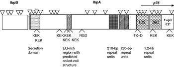
In order to clone genes for the IgBPs, we made a cosmid library of H. somni DNA. Four clones expressed IgBPs and one clone expressed a 60 kDa surface antigen of H. somni (Corbeil et al., Reference Corbeil, Chikami, Yarnall, Smith and Guiney1988). We addressed the IgBPs by subcloning linked genes encoding HMW and p76 IgBPs in an approximately 12 kb PvuII fragment excised from the original cosmid clone. First Cole et al. (Reference Cole, Guiney and Corbeil1993) sequenced and defined motifs in p76, including tandem 1.2 kb direct repeats (DR1 and DR2) with insertion sequence structure. Then Tagawa et al. (Reference Tagawa, Sanders, Uchida, Bastida-Corcuera, Kwashima and Corbeil2005) sequenced the DNA encoding HMW IgBPs ranging from above 200 kDa to 120 kDa (p120). The series of bands in SDS-PAGE and Western blotting is characteristic of not only H. somni HMW IgBPs (Yarnall, Reference Yarnall, Gogolewski and Corbeil1988a, Reference Yarnall, Widders and Corbeilb), but also Staphylococcal Protein A (Bjorck and Kronvall, Reference Bjorck and Kronvall1984) and Streptococcal Protein G (Fahnestock, Reference Fahnestock, Alexander, Nagle and Filpula1986; Fahnestock et al., Reference Fahnestock1987; Akerstrom et al., Reference Akerstrom, Nielsen and Bjorck1987). The sequence of the H. somni ORF encoding HMW IgBPs indicated that it was contiguous with the ORF encoding p76 (Tagawa et al., Reference Tagawa, Sanders, Uchida, Bastida-Corcuera, Kwashima and Corbeil2005). So, one ORF of >12 kb encoded both p76 and the HMW IgBPs (Fig. 1). This very large gene (ibpA, for IgBP A) contained 18 translational start sites (ATG codons), many of which had potential Shine–Delgarno sequences and putative promoters (−35 and −10 consensus sequences). Deletion analysis of the p76 sequence had shown that several start sites were used to express p76 and its truncated peptides (Cole et al., Reference Cole, Guiney and Corbeil1993), perhaps explaining the many bands in SDS–PAGE.
Sequence diagram for H. somni IbpB and IbpA. Multiple ATG translational start sites are shown by small triangles, the p76 gene with a horizontal arrow (labeled p76) and RGD, TKXXD and KEK motifs by open arrows. The EQ-rich region may have Ig binding activity. Sets of repeats are shaded similarly. The 1.2 kb repeat units (DR1 and DR2) are upstream from the cysteine proteinase catalytic domain (YopT) near the C terminus.

A protein sequence homology search of this very large IbpA sequence (both HMW IgBPs and p76) demonstrated similarity to Bordetella pertussis filamentous hemagglutinin (FHA) (which is a surface fibrillar network (Blom et al., Reference Blom, Hansen and Poulsen1983) similar to IgBPs), and other large exoproteins (see below). Tagawa et al. (Reference Tagawa, Sanders, Uchida, Bastida-Corcuera, Kwashima and Corbeil2005) also sequenced an additional upstream ORF of 1758 bp, designated ibpB. The sequence suggested that IbpB is an integral OMP which is likely involved in secretion of IbpA (see below). At the 3′ end of ibpA is another open reading frame, ORF7, with a predicted homology of 64% identity to P. multocida thiamin binding protein A (May et al., Reference May, Zhang, Li, Paustian, Whittam and Kapur2001; Tagawa et al., Reference Tagawa, Sanders, Uchida, Bastida-Corcuera, Kwashima and Corbeil2005). The sequences of the predicted IbpA and IbpB proteins demonstrated homology to large exoproteins and the transporter proteins of Gram-negative bacteria that belong to the two-partner secretion pathway family (Jacob-Dubuisson et al., Reference Jacob-Dubuisson, Locht and Antoine2001). The large predicted or confirmed exoproteins related to IbpA include P. multocida PfhB1 (2615 aa) and PfhB2 (3919 aa) (May et al., Reference May, Zhang, Li, Paustian, Whittam and Kapur2001), Haemophilus ducreyi LspA1 (4152 aa) and LspA2 (4919 aa) (Ward et al., Reference Ward, Lumbley, Latimer, Cope and Hansen1998), B. FhaB (3591 aa) (Domenighini et al., Reference Domenighini, Relman, Capiau, Falkow, Prugnola, Scarlato and Rappuoli1990; Relman et al., Reference Relman, Tuomanen, Falkow, Golenbock, Saukkonen and Wright1990) and others. The greatest identity and similarity was observed with PfhB1 and PfhB2 (53.6 and 61.4%, respectively). The N-terminal half of the whole IbpA sequence overlapped in part with FhaB hemagglutination domains (Blom et al., Reference Blom, Hansen and Poulsen1983).
Low-level homology regions were detected for several known functional domains such as a heparin binding domain at aa 525–975 of IbpA (aa 442–863 of FhaB) (Hannah et al., Reference Hannah, Menozzi, Renauld, Locht and Brennan1994) and a carbohydrate recognition domain at aa 1307–1471 of IbpA (aa 1141–1279 of FhaB) (Prasad et al., Reference Prasad, Yin, Rodzinski, Tuomanen and Masure1993) at 40.8 and 39.3% similarity, respectively (Tagawa et al., Reference Tagawa, Sanders, Uchida, Bastida-Corcuera, Kwashima and Corbeil2005). The integrin binding motif, RGD, was unique in IbpA and FhaB but the TK–D sequence, with a possible role in integrin recognition, was found in all related large exoproteins listed above. Another common motif found in all except FhaB was a putative ATP/GTP binding consensus sequence. Interestingly, multiple KEK motifs were found in IbpA as well as in PfhB1, PfhB2 and LspA2. The KEK motifs in malaria antigens are thought to be involved in binding to erythrocyte membranes (Calvo et al., Reference Calvo, Guzman, Perez, Segura, Molano and Patarroyo1991). A cysteine proteinase catalytic domain with homology to Yersinia YopT as well as PfhB1 and PfhB2 has also been demonstrated (Shao et al., Reference Shao, Merritt, Bao, Innes and Dixon2002). Just upstream from the invariant C/H/D residues of the cysteine proteinase catalytic domain of p76 are the previously described direct repeats, DR1 and DR2 (Cole et al., Reference Cole, Guiney and Corbeil1993).
Several virulence factors have been described above, including LOS, the MOMP, the 40 kDa OMP, Tbps and IgBPs. Other surface proteins such as a 37 kDa OMP, a 31 kDa protein (Won and Griffith, Reference Won and Griffith1993), a 78 kDa OMP (Kania et al., Reference Kania, Gogolewski and Corbeil1990), a 60 kDa protein (Corbeil et al., Reference Corbeil, Chikami, Yarnall, Smith and Guiney1988) and a 17 kDa OMP (Tagawa et al., Reference Tagawa, Haritani and Yuasa1993c) are potential virulence factors. In addition to LOS and surface proteins, H. somni has been shown to produce histamine (Ruby et al., Reference Ruby, Griffith and Kaeberle2002) and exopolysaccharide (Siddaramappa and Inzana, Reference Siddaramappa and Inzana2004). The exopolysaccharide is shed from the surface of H. somni, so may be more like a slime layer than like a capsule. Passive immunization of mice with antibodies to the purified exopolysaccharide was not protective (Siddaramappa and Inzana, Reference Siddaramappa and Inzana2004), consistent with it not being a capsule. Lastly, H. somni has been shown to form biofilms in vitro (Sandal et al., Reference Sandal, Hong, Swords and Inzana2007) and may be capable of quorum sensing (Siddaramappa and Inzana, Reference Siddaramappa and Inzana2004). Since virulence of bacterial pathogens is usually multifactorial, many of the above factors may be involved in H. somni pathogenesis. Probably more virulence factors will be identified by comparative genomics now that the compete genome sequence of serum sensitive strain 129Pt, from an asymptomatic carrier (Corbeil et al., Reference Corbeil, Blau, Prieur and Ward1985) is published (Challacombe et al., Reference Challacombe, Duncan, Brettin, Bruce, Chertkov, Detter, Han, Misra, Richardson, Tapia, Thayer and Xie2007) and the sequence of serum resistant strain 2336, demonstrated to be pathogenic (Gogolewski et al., Reference Gogolewski, Kania, Inzana, Widders, Liggitt and Corbeil1987a, Reference Gogolewski, Leathers, Liggitt and Corbeilb, Reference Gogolewski, Kania, Liggitt and Corbeil1988, Reference Gogolewski, Schaefer, Wasson, Corbeil and Corbeil1989), is expected to be published soon.
Protective immune responses
Antibodies are likely to be important in protection for two reasons. Firstly, convalescent phase serum was passively protective (Gogolewski et al., Reference Gogolewski, Kania, Inzana, Widders, Liggitt and Corbeil1987a). Secondly, although H. somni survives phagocytosis by bovine macrophages (Lederer et al., Reference Lederer, Brown and Czuprynski1987; Gomis et al., Reference Gomis, Godson, Beskorwayne, Wobeser and Potter1997a, Reference Gomis, Godson, Wobeser and Potterb), it destroys the macrophage within hours in vivo (Gogolewski et al., Reference Gogolewski, Leathers, Liggitt and Corbeil1987b). Therefore, it is more like an extracellular parasite than a facultative intracellular parasite which multiplies over an extended time inside normal macrophages. Classically, antibodies are most important in protection against extracellular pathogens. Other studies showed that IgG2 antibodies were more associated with protection against H. somni (Corbeil et al., Reference Corbeil, Bastida-Corcuera and Beveridge1997a, Reference Corbeil, Gogolewski, Kacskovics, Nielsen, Corbeil, Morrill, Greenwood and Butlerb). In chronic H. somni BRD study, infection was maintained for 6–10 weeks with clearance occurring as IgG2 titers to H. somni increased in bronchioalveolar lavage fluids (Gogolewski et al., Reference Gogolewski, Schaefer, Wasson, Corbeil and Corbeil1989). Immune responses of infected calves protected rechallenged convalescent calves against reinfection (Gogolewski et al., Reference Gogolewski, Schaefer, Wasson, Corbeil and Corbeil1989). Convalescent phase serum from these calves passively protected a second group of calves against experimental pneumonia (Gogolewski et al., Reference Gogolewski, Kania, Inzana, Widders, Liggitt and Corbeil1987a). The protective serum recognized several antigens in Western blots, including 37, 40, 60, 78 kDa proteins (Gogolewski et al., Reference Gogolewski, Kania, Inzana, Widders, Liggitt and Corbeil1987a; Kania et al., Reference Kania, Gogolewski and Corbeil1990; Corbeil et al., Reference Corbeil, Gogolewski, Stephens, Inzana, Donachie, Lainson and Hodgson1995; Tagawa et al., Reference Tagawa, Bastida-Corcuera and Corbeil2000) and HMW or p76 IgBPs (Corbeil et al., Reference Corbeil, Arthur, Widders, Smith and Barbet1987; Yarnall and Corbeil, Reference Yarnall and Corbeil1989) as well as LOS (Gogolewski et al., Reference Gogolewski, Kania, Inzana, Widders, Liggitt and Corbeil1987a; Inzana et al., Reference Inzana, Gogolewski and Corbeil1992). Passive protection studies with monospecific bovine antibodies to 78 and 40 kDa antigens showed that antibodies to p40 protected but antibodies purified from monospecific bovine antiserum to p78 resulted in enhanced pneumonia (Gogolewski et al., Reference Gogolewski, Kania, Liggitt and Corbeil1988). Interestingly, the antibodies to p78 consisted of only IgG1 whereas the antibodies to p40 were both IgG1 and IgG2 (Gogolewski et al., Reference Gogolewski, Kania, Liggitt and Corbeil1988). Therefore, passive protection experiments were done with purified IgG1 or IgG2 monospecific bovine antibodies to p40. This experiment showed a trend toward better passive protection with IgG2 anti p40 than IgG1 anti p40 (Corbeil et al., Reference Corbeil, Gogolewski, Kacskovics, Nielsen, Corbeil, Morrill, Greenwood and Butler1997b).
The above studies of passive protection by monospecific antibodies against immunodominant antigens of H. somni did not include antibodies to IbpA. At the time the passive transfer experiments were done, we used OMP preparations in SDS–PAGE and Western Blots to define immundominant antigens because we thought only OMPs and LOS were surface exposed. However, Yarnall and Corbeil (Reference Yarnall and Corbeil1989) found that cattle with experimental or natural H. somni infection had strong IgG2 antibody responses to IgBPs/IbpA (then called FcRs). Later, we showed the IgBPs consisted of surface exposed fibrils (Corbeil et al., Reference Corbeil, Bastida-Corcuera and Beveridge1997a). The surface exposure and strong IgG2 responses to IbpA suggest that it may be a protective antigen also. Since IgG2 antibodies appeared to be important in protection, we then investigated the role of IgG2 allotypes. We found that IgG2 allotype responses develop at different ages, with IgG2a (formerly IgG2A1) being expressed early but IgG2b (IgG2A2) not being expressed until 3 or 4 months of age (Corbeil et al., Reference Corbeil, Gogolewski, Kacskovics, Nielsen, Corbeil, Morrill, Greenwood and Butler1997b). Functional studies indicated that IgG2b but not IgG2a bound to HMW IgBPs of H. somni (Bastida-Corcuera et al., Reference Bastida-Corcuera, Nielsen and Corbeil1999b), perhaps accounting for one mechanism of genetic susceptibility to H. somni infection.
IgE responses also appear to be important in H. somni induced BRD. Ellis and Jong (Reference Ellis and Jong1997) reported systemic adverse reactions similar to anaphylaxis after vaccination with products containing H. somni whole cells. Others demonstrated IgE antibodies to H. somni in calves immunized with commercially available H. somni bacterins (Ruby et al., Reference Ruby, Griffith, Gershwin and Caeberle2000). In studies with Laurel Gershwin, we found that calves given bovine respiratory syncytial virus (BRSV) six days before H. somni had greater clinical scores, longer duration of respiratory disease and higher IgE anti-H. somni levels than calves given either pathogen alone (Gershwin et al., Reference Gershwin, Berghaus, Arnold, Anderson and Corbeil2005). The antigenic specificity of IgE and IgG antibodies in the serum of these calves was studied by Western blotting (Corbeil et al., Reference Corbeil, Arnold, Kimball, Berghaus and Gershwin2006). Although both isotypes recognized some antigens to a similar extent, the predominant antigen recognized by IgE was the 41 kDa MOMP. The predominant antigen recognized by IgG antibodies was the 40 kDa protective OMP, consistent with our earlier studies (Gogolewski et al., Reference Gogolewski, Kania, Inzana, Widders, Liggitt and Corbeil1987a). Since the MOMP is antigenically variable (Tagawa et al., Reference Tagawa, Bastida-Corcuera and Corbeil2000) and IgE antibodies are associated with worse pneumonia of extended duration (Gershwin et al., Reference Gershwin, Berghaus, Arnold, Anderson and Corbeil2005), a vaccine without the MOMP epitopes recognized by IgE might be indicated. With this in mind, we determined the reactivity of these IgE antibodies with H. somni strain 129Pt, which has a truncated MOMP of 33 kDa (Widders et al., Reference Widders, Dorrance, Yarnall and Corbeil1989). The IgE antibodies, which reacted strongly with the 41 kDa MOMP of virulent strains, did not react with the truncated MOMP of strain 129Pt (Corbeil et al., Reference Corbeil, Arnold, Kimball, Berghaus and Gershwin2006). Since this strain does have the protective 40 kDa immunodominant OMP, it may be a good vaccine candidate. These studies of H. somni and BRSV infection alone or together were then extended to investigate the effect of vaccination on disease and cytokine profiles (Berghaus et al., Reference Berghaus, Corbeil, Berghaus, Kalina, Kimbal and Gershwin2006). These aspects of the dual infection studies will be covered in the paper on BRSV by Laurel Gershwin in this volume.
Our studies and those of others have shown that antibody specificity as well as isotype and allotype all contribute to protection versus immune evasion or immunopathogenesis. The Fc binding of IgG2 to HMW and p76 IgBPs could be considered to be a mechanism of immune evasion, but specific antibody binding of IgG2 is at least 16 times more avid. Therefore, the demonstrated specific antibody responses to IbpA/IgBPs are likely to be protective rather than primarily immuno-evasive. A vaccination challenge study of IbpA subunits is underway in our laboratory to test this hypothesis. Our previous studies have made it clear that IgG2 antibodies to p40 are protective in a BRD H. somni challenge model (Gogolewski et al., Reference Gogolewski, Kania, Liggitt and Corbeil1988; Corbeil et al., Reference Corbeil, Gogolewski, Kacskovics, Nielsen, Corbeil, Morrill, Greenwood and Butler1997b). Other observations confirmed the importance of IgG2 in protection since cattle with low IgG2 anti-H. somni titers were more susceptible to experimental H. somni disease and IgG2 antibodies increased the most after infection (Widders et al., Reference Widders, Paisley, Goglewski, Evermann, Smith and Corbeil1986; Gogolewski et al., Reference Gogolewski, Kania, Liggitt and Corbeil1988, Reference Gogolewski, Schaefer, Wasson, Corbeil and Corbeil1989; Corbeil et al., Reference Corbeil, Gogolewski, Kacskovics, Nielsen, Corbeil, Morrill, Greenwood and Butler1997b). Thus adjuvants and antigens which stimulate IgG2 responses are critical. The bovine IgG2 antibody response and cell-mediated immunity are both Th1 responses (Estes and Brown, Reference Estes and Brown2002), so T cell effectors may also play a role. IgG1 and IgE responses are usually considered to be Th2 responses. Our studies suggest that IgG1 antibodies are less critical than IgG2 antibodies and that IgE antibodies are associated with poor outcomes in BRD. Therefore we have concluded that Th1 responses are to be desired in protection against H. somni BRD. The role of IgA is relatively unexplored. Both the 40 kDa OMP and the IgBPs are among critical antigens in eliciting protective host responses. Clearly both the arm of the immune response and the specificity are important in protection.
Conclusions
Virulence factors and immune responses involved in the host–parasite relationships during H. somni-induced bovine pneumonia have been described. Several of these interactions are likely to account for the pathology after intrabronchial inoculation of H. somni into 6–12-week old calves. Histamine production by H. somni (and later histamine release by IgE cross-linking of receptors on mast cells) may account for early lesions of edema, increased mucus secretion, bronchoconstriction and vascular constriction. Complement activation by LOS and perhaps Fc binding of IgG2 to the surface fibrillar network (IgBPs or IbpA) would result in chemotaxis of inflammatory cells characteristic of H. somni pneumonia. LOS has been shown to result in apoptosis of endothelial cells although it does not account for all the apoptosis detected. LOS also activates platelets. Surely both functions are important factors in the etiology of the vasculitis and thrombosis so characteristic of H. somni infection. Vasculitis and histamine release by H. somni would both contribute to vascular leakage and edema. Since fibrinogen attaches to H. somni it would be interesting to know if fibrinogen binding contributes to pathogenesis of fibrinopurulent bronchopneumonia. The mechanism of the damage to macrophages detected in histopathology at 24 h after inoculation of H. somni, is not clear. However, it could also be related to IbpA, similar to the role of the YopT homologous protein in Yersinia infection. Of course the large influx of inflammatory cells, due to the above mechanisms, contributes to the necrotic lesions seen in severe cases.
In early natural infection, exopolysaccharide release and biofilm production are thought to contribute to colonization. Iron acquisition after binding bovine transferrin to Tbps enhances growth in vitro and perhaps in vivo. Histamine release with resulting vascular permeability would increase the plasma transferrin concentration in secretions. Although we did not show that lactoferrin provided iron to H. somni, it did increase virulence for mice. Since lactoferrin is present in secretions and is released by neutrophils, it may contribute to survival of H. somnus in vivo. Unlike several other Gram negative pathogens, the MOMP of H. somni does not induce much of an immune response in cattle and it also undergoes antigenic variation, so it is probably a factor in evasion of host responses, contributing to survival. H. somnus LOS also undergoes antigenic variation, contributing to evasion of immune responses.
Protective immune responses are complex. The critical antigenic proteins identified so far include the 40 kDa OMP and IgBPs. Other antigens may also induce protective responses. Since H. somnus kills macrophages, rather than surviving and multiplying in them for long periods, it is more like an extracellular pathogen than a facultative intracellular pathogen. For this reason and because antibody is passively protective, the antibody response is thought to be most important. Several studies have shown that IgG2 antibodies are most protective. IgE responses, conversely, are associated with more severe disease of longer duration. This accounts for at least part of the synergy of BRSV and H. somni in the etiology of BRD. The dynamic balance between immunoprotection and immunopathogenesis is critical in determining the outcome of infection.
Acknowledgments
This work was supported in part by USDA NRI grant numbers 2005-35204-16290 and 2005-35204-16257. Thanks are also due to the many colleagues and co-authors listed in the ‘references’ section.