Introduction
Partial anomalous pulmonary venous connection is a congenital anomaly characterised by the failure of one or more, but not all, pulmonary veins to connect to the left atrium. This condition is observed in approximately 0.4% to 0.7% of post-mortem examinations. Reference Dillman, Yarram and Hernandez1 With advancements in surgical techniques and postoperative care, the mortality and morbidity associated with the correction of partial anomalous pulmonary venous connection have significantly improved. Therefore, increasing attention is being paid to minimising surgical trauma and reducing postoperative stay. Reference Nicholson, Chard, Nunn and Cartmill2-Reference Amirghofran, Nirooei and Edraki6 Although median sternotomy is a conventional approach that provides optimal exposure to mediastinal structure, it has several disadvantages, including longer and more visible scars, increased invasiveness, and the potential for long-term sternal deformity. Right anterolateral thoracotomy or left anterolateral thoracotomy has already been reported to obtain cosmetic and less invasive outcomes in partial anomalous pulmonary venous connection correction. Reference ElBardissi, Dearani, Suri and Danielson3,Reference Dodge-Khatami and Dodge-Khatami7 However, the studies rarely compare different approaches between minimally invasive techniques and median sternotomy approach. The application of different approaches is still confusing. In our single-centre study, we would compare the perioperative and mid-term outcomes to seek the indication of different approaches and provide guidance for the surgical treatment of partial anomalous pulmonary venous connection.
Patients and methods
From July 2019 to August 2023, a total of 51 patients diagnosed with partial anomalous pulmonary venous connection were reviewed at our centre. Excluding six patients with other concomitant complex intracardiac abnormalities and one patient who received conservative treatment, a total of 44 patients underwent surgical correction of partial anomalous pulmonary venous connection through three kinds of approaches. According to the three approaches, median sternotomy, right anterolateral thoracotomy, and left anterolateral thoracotomy, we categorised patients into three groups: the Median (M) group including 13 (29.6%) patients, the Right (R) group including 28 (63.6%) patients, and the Left (L) group including three (6.8%) patients. Patients’ records were reviewed for the clinical data, such as demographics, cardiac medical history, surgical records, and preoperative and postoperative clinical data. Follow-up information was obtained from subsequent clinic visits. The cardiac anatomic data were evaluated by using an ultrasonic cardiogram or CT. All of the procedures performed in this study were in accordance with the ethical standards of the institutional research committee, and the Institutional Review Board at Beijing Children’s Hospital approved the use of clinical data for this study (Figure 1, Figure 2, Figure 3, Figure 4, and Figure 5).
Flow of patients in our study.

Anatomic types and surgical methods of PAPVC in different group. (a) Proportion of anatomic types. (b) Proportion of surgical methods. PAPVC = partial anomalous pulmonary venous connection; PV = pulmonary vein; SVC = superior vena cava.

Preoperative and follow-up echocardiographic characteristics. (a) Proportion of different degrees of right atrial enlargement. (b) Proportion of different degrees of tricuspid regurgitation.

CT reconstruction of chest wall and heart. (a) Coronal structure of chest and heart. (b) Sagittal structure of chest and heart. The green area represent the third intercostal space.

Left anterolateral thoracotomy applied for PAPVC correction. (a) The incision of left anterolateral thoracotomy (LALT) at preoperation. (b) The incision of LALT at postoperation. (c) Operative field exposure.

As a conventional approach, median sternotomy is widely applied for different kinds of cardiac defects. Considering this point, we would emphatically introduce the surgical technique of right anterolateral thoracotomy and left anterolateral thoracotomy. As previously described by Liu Ying-long et al. Reference Liu, Zhang, Sun, Li, Su and Yu8 regarding right anterolateral thoracotomy, we partially modify the technique. Patients would be placed in lateral decubitus position with the right side elevated at an angle of 60 to 80 degrees. The skin incision, measuring approximately 4 to 6 cm in length and shorter than reported previously, Reference Liu, Zhang, Sun, Li, Su and Yu8 is made obliquely between the midaxillary line and the anterior axillary line. The upper border is the third rib and the lower border is the sixth rib. This incision, which is far from breast, would avoid influencing the development of breast, especially for female patients. The subcutaneous tissue is carefully dissected along the incision, and pectoralis muscle is subsequently dissected along the fourth or third intercostal space from which we enter the chest cavity. On the other hand, the left anterolateral thoracotomy is approximately equivalent to that of the right. The left and right incisions are symmetrical. Before the operation, we typically perform a CT reconstruction of chest wall and left vertical vein to decide to open the left third or fourth intercostal space, as illustrated in Figure 4. The M and R groups always need to establish cardiopulmonary bypass to complete the correction of partial anomalous pulmonary venous connection. The L group adopts an off-pump approach, as previously described by Dearani and colleagues for adult patients. Reference ElBardissi, Dearani, Suri and Danielson3 The vertical vein would be transected at the distal end, and the innominate vein would be repaired using 5-0 Prolene sutures. A bulldog clamp is placed at the proximal end of vertical vein and a side-biting clamp is placed at the base of the left atrial appendage. An end-to-end anastomosis would be performed between the distal end of vertical vein and the opened appendage. Intra-atrial rerouting was employed for partial anomalous pulmonary venous connection connected to right atrium or coronary sinus. Single patch technique, double patch technique, and Warden procedure were employed for partial anomalous pulmonary venous connection connected to superior vena cava. For the majority of cases, single patch technique was employed for pulmonary vein connected to the orifice of superior vena cava, Warden was employed for pulmonary vein connected to superior vena cava around azygos vein, and double patch technique was employed for pulmonary vein connected to superior vena cava between the orifice and azygos vein. Direct anastomosis was employed for scimitar syndrome, left partial anomalous pulmonary venous connection connected to innominate vein, and a part of partial anomalous pulmonary venous connection connected to superior vena cava.
All the data were analysed by SPSS version 18.0 statistical software. Continuous variables, such as age and weight, were presented as mean ± standard deviation. Categorical variables, such as gender and surgical approach, were presented as frequencies accompanied by percentage. The Student t tests were performed to compare continuous variables, while Pearson χ2 and Fisher exact tests were performed to compare categorical variables. Statistical significance was considered at a p value less than 0.05.
Results
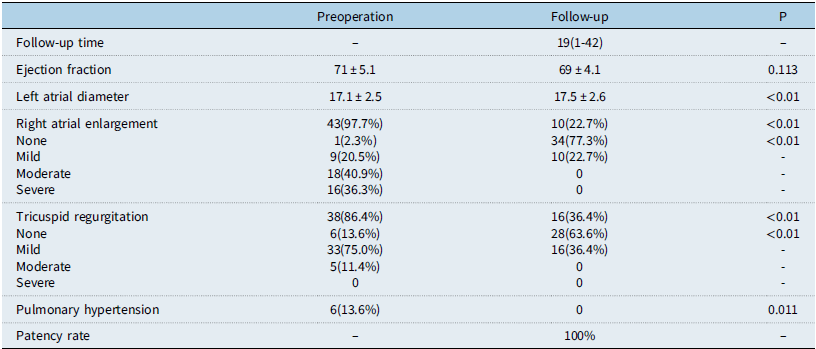
The demographic and perioperative data of the patients are presented in Table 1. The overall mean age was 49.3 months, mean weight was 16.5 kg, and male patients accounted for 43.2%. Pneumonia (24, 54.5%) was the most common presenting symptom of all the patients, which implied the left-to-right shunt has a potential influence on lung. Other presenting symptoms included fatigue (2, 4.5%), dyspnoea (2, 4.5%), and arrhythmia 1(2.3%). Fifteen patients (34.2%) diagnoses were made incidentally without any symptoms. The anatomic types of partial anomalous pulmonary venous connection included 11 (25.0%) right pulmonary vein connected to superior vena cava, 24 (54.5%) pulmonary vein connected to right atrium or coronary sinus, seven (16.0%) left pulmonary vein connected to innominate vein, and two (4.5%) scimitar syndrome. In the aspect of operative and early postoperative clinical data, three patients (6.8%) in the L group adopted off-pump approach. Other patients received the operation on cardiopulmonary bypass with mean cardiopulmonary bypass time of 87.8 mins and mean cross-clamping time of 48.8 mins. Thirteen patients (29.5%) underwent median sternotomy, 28 patients (63.7%) underwent right anterolateral thoracotomy, and three patients (6.8%) underwent left anterolateral thoracotomy. The surgical methods included intra-atrial rerouting (24, 54.6%), single-patch (6, 13.6%), double-patch (2, 4.5%), Warden procedure (1, 2.3%), and direct anastomosis (11, 25%). The mean mechanical ventilation time was 6.4 hours, ICU stay was 1.3 days, and postoperative stay was 6.6 days. Two early postoperative complications happened in this study. One patient whose right pulmonary vein connected to superior vena cava in R group received direct anastomosis developed superior vena cava obstruction with a gradient of 15 mmHg and one patient whose right pulmonary vein connected to superior vena cava in M group received Warden procedure developed superior vena cava obstruction with a gradient of 13 mmHg at early postoperation. There was no operative death. In the aspect of data comparisons between different surgical approaches, the age and weight of M group were significantly higher compared with R group (p<0.05). Because of the limited patients of L group, there was no statistical difference between L group and M group. However, the age and weight of L group trended lower compared with M group. M group included all the four types of partial anomalous pulmonary venous connection mentioned above. R group included two types of partial anomalous pulmonary venous connection, and pulmonary vein connected to the right atrium or coronary sinus accounted for the most. L group only included one type of partial anomalous pulmonary venous connection which was left pulmonary vein connected to innominate vein. The detailed proportion is presented in Table 1 and Figure 2. The proportion of concomitant atrial septal defect showed no significant difference between M group and R group, while all the patients in L group had no atrial septal defect. In addition, proportion of surgical methods is presented in Table 1 and Figure 2. In M group, direct anastomosis accounted for the most (7, 53.8%), in R group, intra-atrial rerouting accounted for the most (22, 78.6%), and in L group, direct anastomosis accounted for the most (3, 100%). Three patients in L group adopted off-pump approach, while the similar type of partial anomalous pulmonary venous connection in M group received the operation on cardiopulmonary bypass. The cardiopulmonary bypass time of M group was longer compared with R group (113.1 ± 83.3 versus 70.0 ± 32.4, p = 0.029) and cross-clamping time showed no significant difference between two groups. Because of the limited number of patients, L group did not show statistical advantage in postoperative data. However, L group had an obvious trend of getting shorter mechanical ventilation time, ICU stay, postoperative stay, and chest tube drainage compared with M group. R group had shorter ICU stay and postoperative stay compared with M group (1.0 ± 0.2 versus 1.9 ± 1.7, p = 0.01; 6.0 ± 1.2 versus 8.3 ± 5.0, p = 0.021). R group had fewer chest tube drainage compared with M group (8.4 ± 4.3 versus 13.9 ± 10.2, p = 0.026). Each of M and R group developed one superior vena cava obstruction and no complication developed in L group. Echocardiographic characteristics are presented in Table 2. The follow-up time ranged from 1 to 42 months and the median was 19 months. Ejection fraction showed no significant difference between preoperation and follow-up. Patients showed smaller left atrial diameter at preoperation (17.1 ± 2.5 versus 17.5 ± 2.6, p<0.01). Both right atrial enlargement and tricuspid regurgitation showed more at preoperation than that at follow-up (43, 97.7% versus 10, 22.7%, p<0.01; 38, 86.4% versus 16, 36.4%, p<0.01). Figure 3 showed the detailed proportion of right atrial enlargement and tricuspid regurgitation at preoperation and follow-up. The number of patients associated with pulmonary hypertension at preoperation was larger than that at follow-up (6, 13.6% versus 0, p = 0.011). All the pulmonary venous drainage was patent at follow-up.
Perioperative data of different surgical approaches

ASD = atrial septal defect; CPB = cardiopulmonary bypass; ICU = intensive care unit; PAPVC =partial anomalous pulmonary venous connection; PV = pulmonary vein; RA = right atrium; SVC = superior vena cava.
a P<0.05 when M group compared to R group;
b P<0.05 when M group compared to L group.
Echocardiographic characteristics

Comment
The incidence of partial anomalous pulmonary venous connection is estimated only at 0.4% to 0.7% according to post-mortem examinations. Reference Dillman, Yarram and Hernandez1 Because of the relatively low incidence, few studies make comparisons among different approaches applied to partial anomalous pulmonary venous connection correction. In our single-centre study, we compared the perioperative and mid-term data of median sternotomy, right anterolateral thoracotomy, and left anterolateral thoracotomy to seek the distinct indication, surgical methods, and clinical prognosis of three approaches. Our experience would provide references for partial anomalous pulmonary venous connection correction and make the procedures more effective and less invasive.
Early studies considered that partial anomalous pulmonary venous connection correction was recommended for patients with obvious symptoms or with a pulmonary to systemic flow ratio (Qp/Qs) exceeding 1.5. Reference Van Meter, LeBlanc, Culpepper and Ochsner9,Reference Murphy, Gersh, M. and McGoon10 However, Shah and colleagues Reference Shah, Oechslin and Benson11 recently reported that the patients with partial anomalous pulmonary venous connection have high risk to develop serious pulmonary hypertension and/or right heart dilation when they do not receive surgical repair. The timing of surgical treatment was recommended early in the clinical course. Reference ElBardissi, Dearani, Suri and Danielson3,Reference Shah, Oechslin and Benson11 On-going patient surveillance with ultrasonic cardiogram and regular follow-up are advised after the patient is diagnosed with partial anomalous pulmonary venous connection. If evidence of right heart volume overload, such as right heart enlargement, tricuspid regurgitation, or pulmonary hypertension, is detected, surgical treatment should be offered in time in order to prevent the progression of right heart failure. In our study, 43 patients (97.7%) detected with right atrial enlargement and 33 patients (75%) detected with tricuspid regurgitation by ultrasonic cardiogram. These findings infer that partial anomalous pulmonary venous connection would have an obvious influence on right heart, which is consistent with previous study. Patients in previous studies Reference ElBardissi, Dearani, Suri and Danielson3,Reference Amirghofran, Nirooei and Edraki6 are older than our patients whose mean age was 49.3 months and mean weight was 16.5 kg. Furthermore, the asymptomatic patients accounted for 34.2% of the total. The differences in demographics between our study and previous studies are mainly attributed to advancements in ultrasonic cardiogram for the detection of partial anomalous pulmonary venous connection and the new understanding of pathophysiological changes of patients with partial anomalous pulmonary venous connection.
The anatomic types of partial anomalous pulmonary venous connection of three groups are different. As a conventional approach, median sternotomy has better exposure and broader indications for partial anomalous pulmonary venous connection correction. In our study, median sternotomy could be applied for all anatomic types of partial anomalous pulmonary venous connection, particularly for complex types, such as Scimitar syndrome, pulmonary vein connected to the distal end of superior vena cava, and left pulmonary vein connected to innominate vein with atrial septal defect. Right anterolateral thoracotomy could provide superior access to right cardiac structures, but not to left pulmonary vein or complex lesion, such as Scimitar syndrome. We mainly applied right anterolateral thoracotomy for pulmonary vein connected to right atrium or proximal end of superior vena cava. On the contrary, left anterolateral thoracotomy could only expose left cardiac structure and could hard establish cardiopulmonary bypass to achieve intracardiac manipulation. We only adopt left anterolateral thoracotomy for left pulmonary vein connected to innominate vein without atrial septal defect. Andrew W. ElBardissi and colleagues Reference ElBardissi, Dearani, Suri and Danielson3 ever suggested that off-pump repair of left partial anomalous pulmonary venous connection followed atrial septal defect closure could be a suitable strategy. Considering the increased operative risk and cost, we still like to repair left partial anomalous pulmonary venous connection and atrial septal defect via median sternotomy once and for all. Except for anatomic types of partial anomalous pulmonary venous connection, surgical method is also an important factor affecting the application of surgical approach. Sameh et al. Reference Said and Ricciardi12 and Sabin et al. Reference Bozso, Evans and Chu13 have reported their successful experience in single-patch repair of pulmonary vein connected to superior vena cava via right anterolateral thoracotomy for children and adults. Ahmad Ali Amirghofran and colleagues Reference Amirghofran, Nirooei and Edraki6 made a comparison between right anterolateral thoracotomy and median sternotomy for single-patch repair of partial anomalous pulmonary venous connection. The mid to long-term outcomes inferred that right anterolateral thoracotomy could achieve comparable quality to median sternotomy, but better cosmetic results. Rajesh Kishan Rao and colleagues Reference Rao, Varadaraju, Basappa and Nagaraja14 also reported their satisfied outcomes of correction of right-sided partial anomalous pulmonary venous connection through modified right vertical infra axillary thoracotomy. In our study, median sternotomy as a conventional approach could be applied for all kinds of surgical methods. The complex surgical methods in median sternotomy group, such as Warden procedure and direct anastomosis, account for 61.5%. This indicates that median sternotomy is mainly suitable for complex manipulations nowadays. Because of the limited exposure, right anterolateral thoracotomy was mainly applied for pulmonary vein connected to right atrium or proximal end of superior vena cava. Intra-atrial rerouting in right anterolateral thoracotomy group accounting for 78.6% infers that right anterolateral thoracotomy is mainly suitable for relatively simple manipulations. Only few studies reported the correction of left-sided partial anomalous pulmonary venous connection due to its low morbidity compared with the right-sided partial anomalous pulmonary venous connection. Reference Javangula, Cole, Cross and Kay15 Andrew W. ElBardissi and colleagues Reference ElBardissi, Dearani, Suri and Danielson3 reported the outcomes of 27 adult patients who underwent surgical correction for left-sided partial anomalous pulmonary venous connection. Thirteen patients treated with left minimally invasive approach were comparable to 14 patients treated with median sternotomy approach and cardiopulmonary bypass. Ganesh and colleagues Reference Ammannaya, Mishra and Khandekar16 reported one case of left-sided partial anomalous pulmonary venous connection with intact atrial septum underwent surgical correction through median sternotomy approach. Actually, we tried to correct the left-sided partial anomalous pulmonary venous connection through left anterolateral thoracotomy from 2021. Before that, we made left-sided partial anomalous pulmonary venous connection correction through median sternotomy. In our study, three children with left-sided partial anomalous pulmonary venous connection received direct anastomosis of vertical vein to left atrial appendage through left anterolateral thoracotomy without cardiopulmonary bypass. Three children with a mean age of 23 months are the youngest patients who underwent left anterolateral thoracotomy without cardiopulmonary bypass as far as we know. Compared with four cases of left-sided partial anomalous pulmonary venous connection with atrial septal defect underwent direct anastomosis through median sternotomy and cardiopulmonary bypass, the outcomes of left anterolateral thoracotomy are quite satisfactory. In order to decide the intercostal space which is convenient for surgical manipulations, CT reconstruction of vessels and thorax is proposed at preoperation, as illustrated in Figure 4. According to our experience, the left third intercostal space could achieve better exposure and easier manipulation.
With advancements in surgical techniques and postoperative care, the prognosis of patients with partial anomalous pulmonary venous connection has been much improved. Meanwhile, more emphasis is being placed on the cosmetic result of the procedure by surgeons and patients. Reference Amirghofran, Nirooei and Edraki6,Reference Said, Greathouse, McCarthy and Brown17,Reference Li, Su and Fan18 Liu and colleagues Reference Liu, Zhang and Sun19 reported the outcomes of 683 patients who underwent correction of congenital heart deformity through right anterolateral thoracotomy, confirming the safety and effectiveness of right anterolateral thoracotomy. In comparison to sternotomy, the minimal lateral thoracotomy could maintain the integrity of bony thorax and minimise the risk of future skeletal deformity. Jannika Dodge-Khatami and colleagues Reference Dodge-Khatami, Noor, Riggs and Dodge-Khatami4 reported the comparison between right anterolateral thoracotomy and median sternotomy for repair of congenital heart deformity. The outcomes indicated that right anterolateral thoracotomy approach could not only provide cosmetic advantages but also yield safe and excellent results when compared to median sternotomy approach. Andrew W. ElBardiss and colleagues Reference ElBardissi, Dearani, Suri and Danielson3 reported the outcomes of surgical correction for left-sided partial anomalous pulmonary venous connection through left anterolateral thoracotomy without cardiopulmonary bypass. The report inferred that left anterolateral thoracotomy could avoid cardiopulmonary bypass risks, minimise operative trauma, and improve cosmetic results for patients with left-sided partial anomalous pulmonary venous connection. Apart from cosmetic advantages, the minimally invasive approaches could also reduce bleeding and requirement for blood products. Ahmad Ali Amirghofran and colleagues Reference Amirghofran, Nirooei and Edraki6 made a comparison between right anterolateral thoracotomy and median sternotomy for surgical repair of partial anomalous pulmonary venous connection. The mean chest tube drainage and mean blood transfusion were significantly lower in right anterolateral thoracotomy group compared to median sternotomy group. In our study, right anterolateral thoracotomy and left anterolateral thoracotomy were applied to minimise trauma and improve cosmetic results. The findings of our study are similar to the above studies. In terms of ICU stay, cardiopulmonary bypass time, postoperative stay, and chest tube drainage, the patients underwent minimally invasive approaches demonstrated more excellent outcomes compared to patients who underwent median sternotomy. The minimal lateral thoracotomy maintaining the integrity of bony thorax may contribute to improved outcomes. Furthermore, the patients who underwent left anterolateral thoracotomy without cardiopulmonary bypass could avoid the cardiopulmonary bypass risks compared to patients who underwent median sternotomy. The cosmetic advantage of minimally invasive approach is shown in Figure 5. The length of anterolateral thoracotomy incision is about 3 cm, which is obviously shorter compared to the length of median sternotomy incision. In terms of mortality and complication, neither group experienced any fatalities and differences in complication rates between the groups were statistically insignificant in our study. The postoperative outcomes infer that minimally invasive approach is as safe as median sternotomy in surgical correction of partial anomalous pulmonary venous connection. Significant alleviation of right atrial enlargement and tricuspid regurgitation during follow-up infers that surgical correction of partial anomalous pulmonary venous connection is necessary to prevent the progression of right heart dysfunction.
Limitations
The limited sample size and retrospective nature are the main limitations of this study. Because of the rarity of partial anomalous pulmonary venous connection, we recruited limited patients in our study. Limited patients may lead to incomplete comparability. For example, only three patients were recruited in the L group. More patients should be recruited in the future to achieve comprehensive understanding of different approaches. Patients in our study were not randomly allocated into different groups. The choice of surgical approaches was typically determined by surgeons, which might influence the results to some extent. The prospective multicentre randomised control study would be needed in the future.
Conclusions
In our single-centre study, we compared the perioperative and mid-term data of partial anomalous pulmonary venous connection correction through median sternotomy, right anterolateral thoracotomy and left anterolateral thoracotomy. The overall outcomes of partial anomalous pulmonary venous connection correction through three approaches are satisfying and could prevent the progression of right heart dysfunction. Median sternotomy could be applied for all anatomic types of partial anomalous pulmonary venous connection, especially for complex types. Right anterolateral thoracotomy and left anterolateral thoracotomy have cosmetic advantages, less trauma, and shorter postoperative stay. Patients who underwent partial anomalous pulmonary venous connection correction through left anterolateral thoracotomy by off-pump could avoid the cardiopulmonary bypass risks. Considering the minimally invasive advantages, we preferred applying right anterolateral thoracotomy for pulmonary vein connected to right atrium or proximal end of superior vena cava and applying left anterolateral thoracotomy for left-sided partial anomalous pulmonary venous connection without atrial septal defect.