Introduction
Nematodes are important pathogens of vertebrates in aquatic and terrestrial habitats and they are widespread in ecosystems in Uzbekistan. Despite their pathogenicity and economic importance, there is still only limited knowledge regarding their hosts, geographic distribution, ecology, biology and evolutionary traits, which are all crucial data that should be incorporated into the development of integrated nematode control strategies.
The worldwide fauna of nematodes of the family Protostrongylidae Leiper, 1926 includes more than 60 species that have been described in ecosystems from Europe, Asia, America, Africa and Australia (Boev, Reference Boev1975; Hoberg et al., Reference Hoberg, Polley, Gunn and Nishi1995; Carreno & Hoberg, Reference Carreno and Hoberg1999, Anderson Reference Anderson2000; Kutz et al., Reference Kutz, Hoberg and Polley2001, Reference Kutz, Checkley, Verocai, Dumond, Hoberg, Peacock, Wu, Orsel, Seegers, Warren and Abrams2013; Verocai et al., Reference Verocai, Kutz, Simard and Hoberg2014). Most protostrongylid species are adapted as parasites of the respiratory organs of mammals. They are prominent globally among the nematode faunas of Artiodactyla, with considerable diversity in Caprinae while lower species diversity has been observed in antelopes and cervids (Carreno & Hoberg, Reference Carreno and Hoberg1999; Lankester, Reference Lankester, Samuel, Pybus and Kocan2001; Kutz et al., Reference Kutz, Asmundsson, Hoberg, Appleyard, Jenkins, Beckmen, Branigan, Butler, Chilton, Cooley, Elkin, Huby-Chilton, Johnson, Kuchboev, Nagy, Oakley, Olsen, Popko, Scheer and Veitch2007, Movsesjan et al., Reference Movsesjan, Bojahchjan, Arutjunova and Chubarj2009; Kuchboev et al., Reference Kuchboev, Krücken, Ruziev and von Samson-Himmelstjerna2015). A diverse spectrum of protostrongylids, involving an estimated number of 15 species, is recognized as parasites of the respiratory system in Caprinae from Uzbekistan, with additional species distributed across adjacent arid regions of Central Asia (Boev, Reference Boev1975; Kulmamatov et al., Reference Kulmamatov, Isakova and Azimov1994; Kuchboev, Reference Kuchboev2009a).
Protostongyles use terrestrial gastropods as intermediate hosts in their life cycle, where development to infective third-stage larvae (L3) occurs (Kutz et al., Reference Kutz, Hoberg and Polley2000; Jenkins et al., Reference Jenkins, Veitch, Kutz, Hoberg and Polley2006; Lesage et al., Reference Lesage, Patrelle, Vrignaud, Decors, Ferté and Jouet2015). Curently, more than 170 species of terrestrial gastropods are known to be endemic in Uzbekistan and neighbouring territories (Pazilov & Azimov, Reference Pazilov and Azimov2003). In order to understand the local transmission patterns, the identification of permissive intermediate hosts is required, which needs the development of new diagnostic tools. Adult nematodes develop in the lungs of Caprinae after ingestion of infected gastropods or free L3 released from gastropods.
Under natural conditions in Uzbekistan, wild Caprinae show close interactions with domestic small ruminants, e.g. they use the same areas for grazing. Such contact interfaces between wild Caprinae and livestock might contribute to the circulation of nematodes, and wild hosts may serve as important reservoirs for maintenance and dissemination of parasites. Studies investigating parasite populations of wild and domestic Caprinae will enhance our understanding of their interrelations and potential effects regarding the distribution of parasites and emergence of parasitic diseases. In particular, understanding of transmission patterns and population ecology is only very limited and further studies are required. Initial studies on the diversity of protostrongylids in Uzbekistan conducted between 2005 and 2010 demonstrated the presence of eight species from the genera Protostrongylus, Spiculocaulus, Muellerius and Cystocaulus (Kuchboev et al., Reference Kuchboev, Karimova, Ruziev and Azimov2012).
The objectives of the present study were to update the data regarding the epidemiological situation of protostrongylids in Uzbekistan, to compare different domestic goats (Capra hircus) and sheep (Ovis aries) and wild species of Caprinae (Capra sibirica, C. falconeri, Ovis ammon and O. vignei) regarding their protostrongylid parasites, and to determine geographical and age-related effects on prevalence of infection in domestic goats and sheep. Finally, we aimed to identify those gastropod species that contribute most to the circulation of protostrongylid nematodes of Caprinae.
Materials and methods
Collection and examination of caprine hosts and faecal samples
The analysis of the fauna, epidemiology and population ecology of nematodes of the family Protostrongylidae in Carpinae in Uzbekistan was conducted from 2012 to 2015. In this period, faunistic studies of species inhabiting the ecosystems of Uzbekistan were performed. Geographical data detailing where particular protostrongylid and gastropod species were collected are summarized in supplementary tables S1 and S2.
Collection of the protostrongylid species infecting different Caprinae was based on complete helminthological dissections, as described by Skrjabin (Reference Skrjabin1928). Material collected from the airways and pulmonary parenchyma of individual ungulates was put in flasks, preserved in 70% ethanol and labelled for storage at the collection of the Laboratory of General Parasitology of the Institute of the Gene Pool of Plants and Animals of Uzbekistan Academy of Sciences. In total, the lungs of 243 ruminants, 126 domestic sheep, 84 domestic goats and 33 wild Caprinae were examined.
The lungs of three C. sibirica (Pallas, 1776) and three C. falconeri (Wagner, 1839) were obtained from the Nature Reserve of Chatkal and from the Kugitang Nature Reserve, respectively. From the latter, lungs of two O. vignei (Blyth, 1841) were also collected, while lungs of six O. ammon (Linnaeus 1758) were provided from the Nature Reserve of Nurota of the State of the Committee of Nature of Uzbekistan. This was possible through the appropriate permits obtained from the Republican State Inspectorate for protection and rational use of flora and fauna and nature reserves of the State, of the Committee of Nature of Uzbekistan. Samples were obtained from wild Caprinae either after natural death or after euthanization for other scientific research purposes. Several of the wild Caprinae are considered to be under threat of extinction, or close to that, according to the International Union for Conservation of Nature and Natural Resources (IUCN).
First-stage larvae (L1) were isolated from the faeces of naturally infected Caprinae and examined as described by Nikolsky (Reference Nikolsky1961) and Jenkins et al. (Reference Jenkins, Appleyard, Hoberg, Rosenthal, Kutz, Veitch, Schwantje, Elkin and Polley2005). Individual faecal samples were collected during the spring, summer, autumn and winter of 2012 from 755 domestic sheep and 520 domestic goats. For each sample, 10 g of faecal pellets were subjected to a Baermann funnel system containing tap water at room temperature. Larvae were allowed to migrate out of the faeces for 16–18 h and collected from the bottom of the funnel system. After killing of L1 using Lugol's iodine solution, larvae were mounted on microscope slides using Canada balsam before microscopic examination.
A regular examination of the faeces of domestic sheep and goats for the presence of protostrongylid L1 was carried out seasonally on the farm ‘Kosonsoy’ in the Namangan region of Uzbekistan, as described above. The goats and sheep within the flocks were divided into three age groups, and samples were obtained from 100 individuals per species and age group. The first age group consisted of individuals up to 1 year of age, the second comprised those aged between 1 and 2 years and the third group was of adult goats and sheep aged at least 2 years. All sheep and goats were individually marked by painting the fur, using a different colour for each group. It was impossible to obtain repeated observations of the same groups of wild Caprinae, so the seasonal dynamics of infections in wild Caprinae could not be determined.
Adult protostrongylids were identified to the species level using the taxonomic keys published by Boev (Reference Boev1975) and Anderson (Reference Anderson, Anderson, Chaubaud and Willmott1978). For this purpose, a microscope ML 2000 with digital camera (Meiji, Japan) was used. The L1 isolated from faeces were identified as Protostrongylinae and non-Protostrongylinae according to the presence or absence of a dorsal spine, respectively (Boev, Reference Boev1975; Kutz et al., Reference Kutz, Asmundsson, Hoberg, Appleyard, Jenkins, Beckmen, Branigan, Butler, Chilton, Cooley, Elkin, Huby-Chilton, Johnson, Kuchboev, Nagy, Oakley, Olsen, Popko, Scheer and Veitch2007; Kuchboev, Reference Kuchboev2009a; Kafle et al., Reference Kafle, Lejeune, Verocai, Hoberg and Kutz2015).
Collection and examination of gastropods
Terrestrial gastropods (Gastropoda, Pulmonata, Stylommatophora), representing potential intermediate hosts of protostrongylid nematodes, were collected in open scree areas, in shrubs or grasses in desert, foothill and mountain zones of Uzbekistan between May and October during 2012–2015 (supplementary table S2).
To determine the prevalence of infection in terrestrial gastropods, 10,564 individuals were studied as described by Azimov et al. (Reference Azimov, Ubaidullaev and Ukolov1971). In brief, 20–30 gastropods were placed in a Petri dish and covered with a large dish. Three to five minutes later, inactive gastropods started to move while sticking to dish walls. Activity was further evoked by placing the dishes in the sun for 3–5 min. By examining the feet of these gastropods with a magnifier, infected parts of the tissue were identified as dark, brown to black spots with diameter 2–3 mm. For quantification, the feet of infected gastropods were cut off and subjected to pepsin digestion to release the infective larvae (Kulmamatov et al., Reference Kulmamatov, Isakova and Azimov1994). Protostrongylid larvae were identified and counted under a microscope (Schwartz et al., Reference Schwartz, Priadko, Tazieva and Pajersky1991). The collected material was put in flasks, fixed in 70% ethanol and labelled for storage in the collection at the Laboratory of General Parasitology of the Institute of the Gene Pool of Plants and Animals, Uzbekistan Academy of Sciences.
In order to identify the species of terrestrial gastropods, the anatomy of the reproductive system of gastropods was analysed using a binocular microscope MBS-9 (Lytkarino Optical Glass Factory, Lytkarino, Russia). The identification of terrestrial gastropods was conducted according to the methods described by Shileiko (Reference Shileiko1978, Reference Shileiko1984), Likharev & Viktor (Reference Likharev and Viktor1980) and Pazilov & Azimov (Reference Pazilov and Azimov2003).
Data analysis
Statistical differences in prevalence between groups (protostrongylid species, provinces, seasons, age and sex ratios between protostrongylid species) were calculated using the mid-P exact test, as implemented in the OpenEpi platform available under openepi.com (Dean et al., Reference Dean, Sullivan and Soe2013). Moreover, 95% confidence intervals (95% CI) were obtained as Wilson Score intervals from the same platform. All P values were corrected for multiple testing using the Bonferroni–Holm method. A Mann–Whitney U test was conducted in GraphPad Prism 5 to compare infection intensities between gastropod age groups. Differences in proportion of sexes from 50% were calculated using a binomial test, as implemented on the GraphPad web page (http://graphpad.com/quickcalcs/binomial2). In all analyses, differences with P < 0.05 were considered to be significant.
Results and discussion
Definitive hosts of protostrongylids
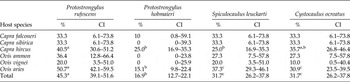
Adult nematodes of four species, namely Protostrongylus rufescens, P. hobmaieri, Spiculocaulus leuckarti, Cystocaulus ocreatus, and one larval nematode of the genus Protostrongylus were identified in the lungs of Caprinae during necropsies. Overall prevalence rates ranged between 16.9% for P. hobmaieri and 45.3% for P. rufescens, and P. rufescens was significantly more prevalent and P. hobmaieri significantly less prevalent than the other parasite species (table 1). Remarkably, Muellerius capillaris was not found in any of the lungs, which is in marked contrast to the results of a previous study on protostrongyles, where 16.8% of the caprine hosts were infected by Muellerius spp. (Kuchboev et al., Reference Kuchboev, Karimova, Ruziev and Azimov2012). This difference might be explained by the fact that caprines for necropsies were obtained from different areas of Uzbekistan.
The prevalence (%) of infection of caprines with four protostrongylid species; CI, 95% confidence intervals; prevalences within the same row marked with the same index letter are not significantly different (P ≥ 0.05), but samples were too few for the analysis of Capra falconeri, Capra sibirica, Ovis ammon and Ovis vignei.

Looking at individual host species, prevalences were only compared statistically for domestic sheep and goats since the number of individuals was too low to allow meaningful results for the other host species. In sheep, again, P. rufescens was significantly the most prevalent parasite and P. hobmaieri the least prevalent parasite. In contrast, the prevalence of P. rufescens in goats was only significantly higher than that of P. hobmaieri and S. leuckarti whereas all other comparisons of prevalences were not significantly different. Comparisons of prevalence for individual parasites between sheep and goats did not reveal any significant differences.
The sex ratio in the population of protostrongylids was studied in detail in naturally infected domestic sheep. Ratios between males and females of 1:3.1 for C. ocreatus, 1:3.2 for S. leuckarti, 1:4.8 for P. rufescens and 1:6.2 for P. hobmaieri were found. All sex ratios were significantly different from 50%, as revealed by bionomial tests, and differences between parasite species were not significantly different (P > 0.05), as determined using a mid-P exact test. Such extreme ratios are likely to improve the egg production of the parasites while limiting the pathogenic consequences by decreasing the total number of living worms. However, the mechanisms that cause this gender ratio are completely unknown.
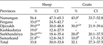
Using the Baermann funnel technique, faecal samples were analysed to determine any geographical differences in prevalence of protostrongyles in domestic goats and sheep (table 2). Prevalences in sheep and goats in the study areas ranged between 10% and 56.4%. Differences in prevalence between various areas were statistically significant. For both sheep and goats, the Namagan province revealed the highest prevalence of protostrongylids, although the difference from Djizzakh province was only significant for sheep. Within a province, no significant differences between sheep and goats were identified.
Prevalence (%) of protostrongylid larvae in faecal samples from sheep and goats as detected in different provinces of Uzbekistan; CI, 95% confidence intervals. Prevalences were compared separately for sheep and goats between the provinces; prevalences for individual provinces within the same column that are marked with the same index letter are not significantly different (P ≥ 0.05); the total prevalence was not included in the statistical analysis.

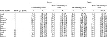
Seasonal and age dynamics of protostrongylid infections in sheep and goats were studied using faecal samples obtained from farms in the Fergana valley of Uzbekistan (table 3). Data show that in lambs below 1 year of age Protostrongylinae and non-Protostrongylinae were both found with prevalences of 3–5% in late spring. By late summer, the prevalence of infection had significantly increased and it peaked in winter, reaching 27% and 26%, although the differences between summer, autumn and winter were not statistically significant. With increasing age, prevalences continued to increase, but all season-related differences within the older age groups were not significant (table 3). In general, the dynamics of the infection in goats and sheep with protostrongylid nematodes was similar. However, the prevalences were always lower in goats, although these differences were not statistically significant (table 3). Larvae from Protostrongylinae include presumably Protostrongylus spp. and S. leuckarti while non-Protostrongylinae likely represent predominantly C. ocreatus but maybe also M capillaris, as identified previously in Uzbekistan (Kuchboev et al., Reference Kuchboev, Karimova, Ruziev and Azimov2012). In a recent publication, internal transcribed spacer (ITS)-2 sequences for protostrongylids identified in the present study have been described (Kuchboev et al., Reference Kuchboev, Krücken, Ruziev and von Samson-Himmelstjerna2015). Since sequences for M. capillaris are also available in GenBank, in the future this will allow determination of prevalences of protostrongylids in faecal samples to the species level.
Seasonal prevalences (%) of larvae from Protostrongylinae and non-Protostongylinae in faecal samples of sheep and goats during 2012/13, relative to host age in years. CI, 95% confidence intervals; prevalences within the same age group and for the same host/parasite combination marked with the same index letter are not significantly different (P ≥ 0.05).

* 2013.
Intermediate hosts of protostrongylids
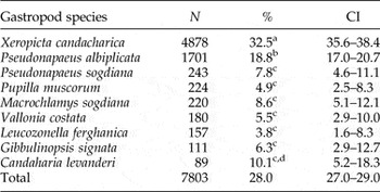
Of 24 gastropod species studied, all belonging to various groups within the Stylommatophora, 9 were found to be infected with protostrongylid larvae (table 4). Gastropod species in which no protostrongylid larvae were found are listed in supplementary table S3. Among those species found to be positive for protostrongyles, prevalence varied between 3.8% and 32.5% with an overall prevalence of 28.0% (table 4). In particular, Xeropicta candacharica is apparently a high-risk factor for transmission of protostrongyles to small ruminants since it was by far the most abundant gastropod species, with 46% of all collected gastropods, and the 32.5% prevalence of protostrongylid larvae was significantly higher than in any other gastropod species.
Prevalence (%) with 95% confidence interval (CI) of protostrongylid larvae in gastropods in Uzbekistan. N, number of gastropods analysed; prevalences marked with the same index letter are not significantly different (P ≥ 0.05).

The second most important intermediate host is apparently Pseudonapaeus albiplicata, which was the second most abundant species, with 16.1% of all samples, and with the second highest prevalence of infection with protostrongylids (18.8%). Prevalence was significantly higher than that of all other gastropods except for X. candacharica and Candaharia levanderi (with a prevalence of 10.1%). The three gastropod species with the highest prevalence, as well as their ranking, were identical to that reported by Kuchboev et al. (Reference Kuchboev, Karimova, Ruziev and Azimov2012).
Obviously, not all species of Stylommatophora are equally susceptible to infection with protostrongylids and consequently their role in the epidemiology of protostrongylidosis in Caprinae differs. It cannot be predicted from the position within the current systematics of Stylommatophora if protostrongylid L3 can be found in the gastropods. On the one hand, some of the gastropod species that were positive were only distantly related, whereas, on the other hand, gastropod species belonging to the same genus were both positive and negative. In comparison to the results presented by Kuchboev et al. (Reference Kuchboev, Karimova, Ruziev and Azimov2012), protostrongylid larvae were not detected in two gastropod species, Bradybaena lantzi and Bradybaena phaezona, which had relatively high prevalences of 12.3% and 6.3% in the previous study. Since the upper limit of the 95% confidence interval for both species is below 1% in the data presented here, this suggests that there are considerable local differences in prevalence. Therefore, the role of intermediate host species might vary between ecosystems and the mere presence of a gastropod species is not always sufficient to predict its role in the transmission cycle.
To obtain a more sustained picture of the role of certain gastropod species in protostrongylid epidemiology, identification of the species combinations of lungworms and gastropods would be required – presumably using the molecular tools for protostrongylid identification mentioned above (Kuchboev et al., Reference Kuchboev, Krücken, Ruziev and von Samson-Himmelstjerna2015). Identification of protostrongylid larvae from gastropods will deepen our understanding of the transmission pathways of protostrongylids. However, a very broad dataset with at least a few hundred sequences from many gastropod species will be required to obtain statistically meaningful data.
Infection of X. candacharica with larvae of protostrongylid nematodes in the region investigated in the present study is rather high. In the period from May to October in 2012 and 2013, the density of gastropod populations on pastures reached 50–60 individuals per m2. The ecological situation in biotopes allows high activity of both gastropods and free L1, promoting contact of parasites and intermediate hosts. It is also noteworthy to mention that the infective protostrongylid L3 were encountered in gastropods throughout the entire collection period. Since Caprinae can be infected during grazing, not only by uptake of entire infected gastropods but also of free L3 that emerged from their intermediate hosts, the risk of infection for definitive hosts is obviously very high.
In order to obtain further insight into the interaction of X. candacharica with protostrongylid larvae, different age groups of gastropods were analysed separately. For this purpose, gastropods were assigned to two age groups, i.e. small juveniles up to 1 year of age and full-sized adults more than 1 year old. Protostrongylid L3 were quantified for 100 randomly chosen individuals from each group. In the juvenile age group, the intensity of infection was significantly lower than in adults, whereas prevalences were very similar in both age groups. The prevalence of L3 was 55% (95% CI = 45.25–64.38%) in juvenile X. candacharica and 57% (95% CI = 47.22–66.27%) in adult X. candacharica, P = 0.778. The median intensity of infection was 1 (range 0–5) in juvenile and 5 (range 0–44) in adult X. candacharica. This difference was statistically significant with P = 0.002 in a Mann–Whitney U test. The intensity of infection in adult X. candacharica might be explained partially by their higher mobility, which increases the possibility of contact with the faeces of small ruminants. Even more importantly, the observed data presumably reflect accumulation of L3 in the intermediate host with increasing age. The high numbers of L3 found in the adults show that these gastropods are much more important for transmission of protostrongylids than the juvenile age group.
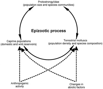
The protostrongylid developmental cycle includes three essential elements for transmission to continue (fig. 1). The interaction between these factors depends on several additional parameters, such as (micro-)climatic conditions that influence gastropod diversity and density, and human activity, including movements of herds between ecosystems or changes in land use that affect population density and community composition of gastropods or domestic and wild Caprinae. In order to understand the prevalence of protostrongylids in Caprinae in depth, the analysis of such interactions in the relevant ecosystems needs to be analysed further. Most importantly, host specificity of the parasites for certain intermediate hosts, and abiotic conditions, particularly temperature, required for development within the intermediate host, should be examined. Many invertebrate species have only limited ranges of favourable environmental factors and small changes might result in dramatic effects for the ecosystem and, in consequence, also for parasite transmission (Kuchboev, Reference Kuchboev2009b). Future studies will need to include the interactions of parasite species with intermediate hosts in more detail, taking advantage of the molecular data published previously (Kuchboev et al., Reference Kuchboev, Krücken, Ruziev and von Samson-Himmelstjerna2015).
A schematic presentation of the factors influencing the transmission and epidemiology of protostrongylids.

Supplementary material
To view supplementary material for this article, please visit http://dx.doi.org/10.1017/S0022149X16000171
Acknowledgements
The authors gratefully acknowledge Professors Georg von Samson-Himmelstjerna and Janina Demeler (Freie Universität Berlin, Germany) and junior scientist Oybek Amirov (Uzbekistan Academy of Sciences) for editing and support. Furthermore, they would like to thank the staff of the Laboratory of Molecular Biology and General Parasitology, Institute of the Gene Pool of Plants and Animals, Uzbekistan Academy of Sciences for helpful advice and for technical assistance.
Financial support
This work has been carried out partially within the frame government research grants of Uzbekistan (FA-A9-T229) and (A7-53) in 2012–2015, and a scholarship grant from the Deutscher Akademischer Austausch Dienst (DAAD, Germany) in 2012.
Conflict of interest
None.
Ethical standards
The authors assert that all procedures contributing to this work comply with the ethical standards of the relevant national and institutional guides regarding the care and use of animals, in particular the law of the Republic of Uzbekistan on ‘Protection and Use of Fauna’.