Introduction
In the vertebrate retina, bipolar cells provide the major pathway through which visual information encoded by photoreceptors is transmitted from the outer retina to the inner retina and ultimately, to the output neurons of the retina, the ganglion cells. While their dendrites reside in the outer plexiform layer, the synaptic terminals of bipolar cells lie in the inner plexiform layer (Fig. 1A), where they are exposed to a variety of circuit interactions that modulate synaptic output. The large and accessible terminals of the rod-dominant Mb1 bipolar cells of the goldfish retina have proven an excellent model system for studying presynaptic mechanisms. Data obtained from these neurons have helped to lay a foundation for our current understanding of presynaptic mechanisms, from calcium entry and the roles of presynaptic calcium, to synaptic vesicle trafficking and neurotransmitter exocytosis, and the role of the synaptic ribbon. Recently, several groups have extended the study of presynaptic mechanisms to the rod bipolar cell of the rodent retina, which has a different connectivity than the Mb1 bipolar cell and experiences different synaptic demands.
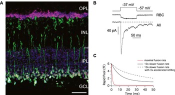
Rod bipolar cells of the mammalian retina. (A) A vertical section of a mouse retina tripled labeled for Protein kinase C-α (a rod bipolar cell marker; green), SV2 (a synaptic vesicle protein; red), and VGluT1 (a vesicular glutamate transporter; blue). A single optical section is shown. VGluT1 and SV2 are selectively found in the outer and inner plexiform layers (OPL and IPL), as expected for proteins located on synaptic vesicles. Photoreceptor terminals, which label for SV2 and VGLuT1 but not PKC-α, are seen as magenta. Rod bipolar cell dendrites, somata and axons, which express PKC-α, are seen as green. Rod bipolar cell terminals, located in the distal IPL, express all three markers and are seen as white. In the IPL, not all SV2 puncta colocalize with VGluT1 in the IPL, consistent with the presence of both glutamatergic and nonglutamatergic synapses. Scale bar: 20 μm. (B) A paired recording from a synaptically coupled rod bipolar and AII amacrine cell in a retinal slice from the mouse retina. A 100-ms depolarization evokes a sustained Ca2+ current in the presynaptic rod bipolar cell. The postsynaptic AII amacrine cell EPSC exhibits both transient and sustained components. Journal of Physiology 587/11 by Snellman et al. (Reference Snellman, Zenisek and Nawy2009) by John Wiley and Sons reproduced with permission from John Wiley and Sons via Copyright Clearance Center. (C) A simple two-step simulation (Innocenti & Heidelberger, Reference Innocenti and Heidelberger2008) depicting the occupancy state of the rapid vesicle pool, which underlies the transient component of the EPSC. With a maximal fusion rate of 1000 s−1, the rapid pool is quickly depleted (red line), consistent with a prominent EPSC transient component. With a fusion rate of 100/s, the depletion of the rapid pool is slower (black line), suggestive of a smaller broader EPSC transient. Increasing the refilling rate, as might occur when increasing the temperature from ambient room temperature to physiological temperature (Dinkelacker et al., Reference Dinkelacker, Voets, Neher and Moser2000; Pyott & Rosenmund, Reference Pyott and Rosenmund2002; Kushmerick et al., Reference Kushmerick, Renden and von Gersdorff2006), reduces the speed with which the rapid pool is depleted. Dashed line shows a simulation with a release rate of 100/s and a 3 times faster refilling rate than that of the solid black line.

Here, we review the current literature on synaptic signaling and neurotransmitter release in the mammalian rod bipolar cell. We compare and contrast results obtained from paired recordings of synaptically connected rod bipolar cells and third-order neurons, which report on transmitter release detected by postsynaptic glutamate receptors, with those obtained using membrane capacitance measurements, which provide a direct presynaptic assay of synaptic vesicle fusion. Where instructive, properties of the mammalian rod bipolar cell will be compared with those of the well-studied rod-dominant Mb1 bipolar cell of the goldfish retina.
Basic physiology and synaptic organization
The mammalian rod bipolar cell is part of a high-sensitivity neural pathway specialized for scotopic vision. In contrast to the Mb1 bipolar cell that receives inputs from both rod and cone photoreceptors and makes chemical synapses directly onto third-order neurons (Stell et al., Reference Stell, Ishida and Lightfoot1977; Ishida et al., Reference Ishida, Stell and Lightfoot1980; Joselevitch & Kamermans, Reference Joselevitch and Kamermans2007), the mammalian rod bipolar cell typically receives inputs exclusively from rod photoreceptors (but see also Pang et al., Reference Pang, Gao, Lem, Bramblett, Paul and Wu2010) and indirectly provides outputs to ganglion cells via a unique mammalian circuit between the rod bipolar cell, AII amacrine cells, and cone bipolar cells. This circuit, which grafts the rod pathway onto the cone pathway at the level of cone bipolar cell axon terminals (Kolb & Famiglietti, Reference Kolb and Famiglietti1974; Kolb, Reference Kolb1979; Bloomfield & Dacheux, Reference Bloomfield and Dacheux2001; Tsukamoto et al., Reference Tsukamoto, Morigiwa, Ueda and Sterling2001; Veruki & Hartveit, Reference Veruki and Hartveit2002), is essential for mammalian scotopic vision. Defects in this pathway are associated with loss of rod-driven on-center responses in ganglion cells (Deans et al., Reference Deans, Volgyi, Goodenough, Bloomfield and Paul2002) and night vision deficits in humans (Berson & Lessell, Reference Berson and Lessell1988; Alexander et al., Reference Alexander, Fishman, Peachey, Marchese and Tso1992; Bech-Hansen et al., Reference Bech-Hansen, Naylor, Maybaum, Pearce, Koop, Fishman, Mets, Musarella and Boycott1998; Strom et al., Reference Strom, Nyakatura, Apfelstedt-Sylla, Hellebrand, Lorenz, Weber, Wutz, Gutwillinger, Ruther, Drescher, Sauer, Zrenner, Meitinger, Rosenthal and Meindl1998; see also Fitzgerald et al., Reference Fitzgerald, Cibis, Giambrone and Harris1994).
The visual response of bipolar cells is predominantly sustained (Nelson, Reference Nelson1982; Saito & Kujiraoka, Reference Saito and Kujiraoka1982; Kolb & Nelson, Reference Kolb and Nelson1983; Dacheux & Raviola, Reference Dacheux and Raviola1986; Bloomfield & Xin, Reference Bloomfield and Xin2000; Euler & Masland, Reference Euler and Masland2000; Pang et al., Reference Pang, Gao and Wu2004; Joselevitch & Kamermans, Reference Joselevitch and Kamermans2007); these neurons encode changes in illumination with a graded change in membrane potential (Dowling & Werblin, Reference Dowling and Werblin1969; Kaneko, Reference Kaneko1973), although under certain conditions, the light response might become regenerative (Burrone & Lagnado, Reference Burrone and Lagnado1997; Zenisek & Matthews, Reference Zenisek and Matthews1998; Protti et al., Reference Protti, Flores-Herr and von Gersdorff2000; Ma & Pan, Reference Ma and Pan2003; Palmer, Reference Palmer2006; Hu et al., Reference Hu, Bi and Pan2009). In contrast to the largely sustained light response of the rod bipolar cell, the light response of the postsynaptic AII amacrine cell exhibits a mixture of transient and sustained components (Nelson, Reference Nelson1982; Dacheux & Raviola, Reference Dacheux and Raviola1986; Bloomfield & Dacheux, Reference Bloomfield and Dacheux2001). While the transient transformation may be sculpted by local circuit interactions such as GABAergic feedback (Tachibana & Kaneko, Reference Tachibana and Kaneko1987; Maguire et al., Reference Maguire, Maple, Lukasiewicz and Werblin1989; Heidelberger & Matthews, Reference Heidelberger and Matthews1991; Maple & Wu, Reference Maple and Wu1998; Vigh & von Gersdorff, Reference Vigh and von Gersdorff2005; Eggers & Lukasiewicz, Reference Eggers and Lukasiewicz2006; Chavez et al., Reference Chavez, Grimes and Diamond2010), the transient component of the AII amacrine cell light response may also arise from intrinsic properties of rod bipolar cell glutamate release.
Rod bipolar cells release the excitatory neurotransmitter glutamate via calcium-dependent exocytosis at ribbon-style active zones (Dowling & Boycott, Reference Dowling and Boycott1966; Dowling, Reference Dowling1987). Conventional synapses are rare, although they have been reported in some species (Miller et al., Reference Miller, Gottesman, Henderson, Sikora and Kolb2001; Midorikawa et al., Reference Midorikawa, Tsukamoto, Berglund, Ishii and Tachibana2007). The synaptic ribbon tethers synaptic vesicles near the sites of fusion. The morphology, molecular architecture, and general function of synaptic ribbons, also found in some primary sensory neurons such as photoreceptors, have been the topics of numerous recent reviews (Heidelberger et al., Reference Heidelberger, Thoreson and Witkovsky2005; Sterling & Matthews, Reference Sterling and Matthews2005; tom Dieck & Brandstatter, Reference tom Dieck and Brandstatter2006; Wu et al., Reference Wu, Ryan and Lagnado2007; Schmitz, Reference Schmitz2009; Zanazzi & Matthews, Reference Zanazzi and Matthews2009). An overall picture emerges in which vesicles that are tethered near the base of a synaptic ribbon and close to the presynaptic plasma membrane, wherein calcium channels and fusion machinery components cluster, contribute to the fastest component of glutamate release. Vesicles located further up the ribbon may contribute to a second, larger and more sustained component of release. Both components of release reflect the fusion of vesicles that have undergone all ATP-dependent priming steps required for fusion (Heidelberger, Reference Heidelberger1998; Heidelberger et al., Reference Heidelberger, Sterling and Matthews2002a). One or both populations may be refilled from those many vesicles that are not associated with synaptic ribbons but are freely diffusible in the synaptic terminal cytosol (Zenisek et al., Reference Zenisek, Davila, Wan and Almers2003; Holt et al., Reference Holt, Cooke, Neef and Lagnado2004; LoGiudice et al., Reference LoGiudice, Sterling and Matthews2008). In addition, some release may occur outside of ribbon-style active zones (Midorikawa et al., Reference Midorikawa, Tsukamoto, Berglund, Ishii and Tachibana2007; Zenisek, Reference Zenisek2008), although the magnitude of this release and its role in synaptic communication is not yet clear.
In the Mb1 bipolar cell, exocytosis is triggered via calcium entry through L-type voltage-gated calcium channels (Tachibana & Okada, Reference Tachibana and Okada1991; Tachibana et al., Reference Tachibana, Okada, Arimura, Kobayashi and Piccolino1993). These channels are preferentially located in the synaptic terminal (Heidelberger & Matthews, Reference Heidelberger and Matthews1992; Tachibana et al., Reference Tachibana, Okada, Arimura, Kobayashi and Piccolino1993) and have been identified as the Cav1.3 (α(1D)) subunit (Logiudice et al., Reference Logiudice, Henry and Matthews2006). This type of L-type calcium channel is less sensitive to dihydropyridine agonists and antagonists, rapidly activates at a relatively negative voltage range, and shows a substantially slower rate of calcium-dependent inactivation than other L-type calcium channels (von Gersdorff & Matthews, Reference von Gersdorff and Matthews1996; Koschak et al., Reference Koschak, Reimer, Huber, Grabner, Glossmann, Engel and Striessnig2001; Xu & Lipscombe, Reference Xu and Lipscombe2001; Logiudice et al., Reference Logiudice, Henry and Matthews2006). The latter properties help to maintain graded exocytosis during a prolonged stimulus (Heidelberger & Matthews, Reference Heidelberger and Matthews1992; von Gersdorff et al., Reference von Gersdorff, Vardi, Matthews and Sterling1996; Sakaba et al., Reference Sakaba, Tachibana, Matsui and Minami1997; Mennerick & Matthews, Reference Mennerick and Matthews1998; Protti & Llano, Reference Protti and Llano1998; Xu & Lipscombe, Reference Xu and Lipscombe2001; Zenisek et al., Reference Zenisek, Davila, Wan and Almers2003; Logiudice et al., Reference Logiudice, Henry and Matthews2006).
Calcium entry through both L- and T-type calcium channels have been proposed to drive exocytosis in mammalian rod bipolar cells (de la Villa et al., Reference de la Villa, Vaquero and Kaneko1998; Hartveit, Reference Hartveit1999; Pan, Reference Pan2000; Pan et al., Reference Pan, Hu, Perring and Andrade2001; Hu et al., Reference Hu, Bi and Pan2009). However, the primary source of calcium that triggers release in rats and mice may be different. In the rat rod bipolar terminal, the T-type calcium currents are prominent (Pan, Reference Pan2000; Pan et al., Reference Pan, Hu, Perring and Andrade2001; Hu et al., Reference Hu, Bi and Pan2009), although the L-type Cav1.2 (α(1C)) and/or Cav1.3 subunit have also been shown to be expressed at low levels (Kamphuis & Hendriksen, 1998; Xu et al., Reference Xu, Zhao and Yang2002). The T-type calcium current in a rat rod bipolar cell terminal is atypical as it exhibits both transient and sustained components (Pan, Reference Pan2000). This channel also has an extremely slow activation and slow time-dependent inactivation kinetics (Pan, Reference Pan2000; Pan et al., Reference Pan, Hu, Perring and Andrade2001; Singer & Diamond, Reference Singer and Diamond2003), possibly arising from a splicing variant, mutation, or other modification of the Cav3.3 (α(1I)) Ca2+ channel subunit (Pan, Reference Pan2000; Hu et al., Reference Hu, Bi and Pan2009). Although the T-type calcium current was originally suggested to drive the transient release of glutamate in the rat rod bipolar cell (Pan et al., Reference Pan, Hu, Perring and Andrade2001), a subsequent study reported transient release in the presence of sustained calcium influx when T-type channels were not activated (Singer & Diamond, Reference Singer and Diamond2003). Activation of T-type calcium channels resulted in larger postsynaptic currents at the rat rod bipolar cell–AII amacrine cell synapse, consistent with a role in boosting glutamatergic synaptic transmission (Singer & Diamond, Reference Singer and Diamond2003).
In mouse rod bipolar cell terminal, the L-type Cav1.2, Cav1.3, and Cav1.4 (α(1F)) channels are expressed (Satoh et al., Reference Satoh, Aoki, Watanabe and Kaneko1998; Berntson et al., Reference Berntson, Taylor and Morgans2003; Baumann et al., Reference Baumann, Gerstner, Zong, Biel and Wahl-Schott2004) . The major subunit, however, is Cav1.4 (Berntson et al., Reference Berntson, Taylor and Morgans2003; Baumann et al., Reference Baumann, Gerstner, Zong, Biel and Wahl-Schott2004), which shows slow voltage-dependent inactivation and lacks calcium-dependent calcium channel inactivation (Koschak et al., Reference Koschak, Reimer, Walter, Hoda, Heinzle, Grabner and Striessnig2003; Baumann et al., Reference Baumann, Gerstner, Zong, Biel and Wahl-Schott2004; McRory et al., Reference McRory, Hamid, Doering, Garcia, Parker, Hamming, Chen, Hildebrand, Beedle, Feldcamp, Zamponi and Snutch2004; Wahl-Schott et al., Reference Wahl-Schott, Baumann, Cuny, Eckert, Griessmeier and Biel2006). The properties of this channel are ideal for supporting continuous calcium signaling during prolonged depolarization in the light. Mutations in Cav1.4 channels, which are also present on rod photoreceptor terminals (Morgans et al., Reference Morgans, Bayley, Oesch, Ren, Akileswaran and Taylor2005), are associated with incomplete X-linked congenital stationary night blindness in humans (Bech-Hansen et al., Reference Bech-Hansen, Naylor, Maybaum, Pearce, Koop, Fishman, Mets, Musarella and Boycott1998; Strom et al., Reference Strom, Nyakatura, Apfelstedt-Sylla, Hellebrand, Lorenz, Weber, Wutz, Gutwillinger, Ruther, Drescher, Sauer, Zrenner, Meitinger, Rosenthal and Meindl1998). Mouse bipolar cells have also been reported to express a transient T-type ICa (Kaneko et al., Reference Kaneko, Pinto and Tachibana1989, Reference Kaneko, Suzuki, Pinto and Tachibana1991), but there is evidence to suggest that these channels are not present in the synaptic endings (Satoh et al., Reference Satoh, Aoki, Watanabe and Kaneko1998). Thus, in contrast to the rat, the T-type calcium channel may not be positioned in the mouse rod bipolar cell to drive exocytosis.
Synaptic vesicle pools
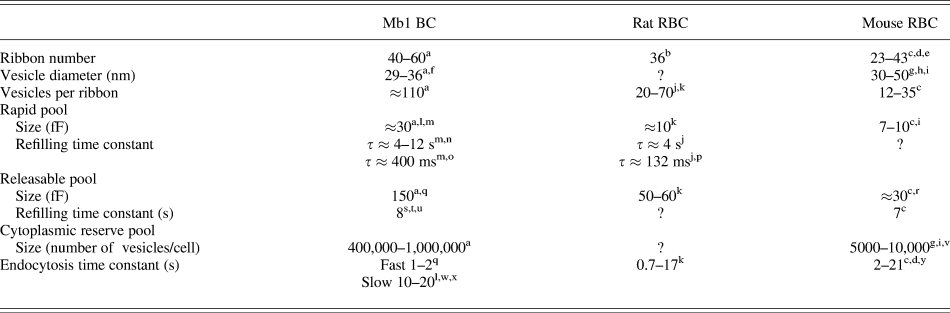
The concept of synaptic vesicle pools, first introduced in the general exocytosis literature, is based upon the physiological ability to identify distinct components of release. These components possess distinct kinetic properties, magnitudes, and proximity to calcium channels. The latter is evidenced by differential sensitivity to the calcium buffers, glycol-bis(2-aminoethylether)-N,N,N′,N′-tetraacetic acid (EGTA) and 1,2-bis(o-aminophenoxy)ethane- N,N,N′,N′-tetraacetic acid (BAPTA), which have similar binding affinities for calcium ions but different on rates (Adler et al., Reference Adler, Augustine, Duffy and Charlton1991; Borst & Sakmann, Reference Borst and Sakmann1996; Mennerick & Matthews, Reference Mennerick and Matthews1996; Sakaba et al., Reference Sakaba, Tachibana, Matsui and Minami1997; Moser & Beutner, Reference Moser and Beutner2000; Burrone et al., 2002). The naming of these pools has been largely descriptive, with the first pool described in a particular neuron under a particular set of conditions being given the name “releasable pool” or “readily releasable pool” and subsequent pools being named relative to the first. Thus, some authors have referred to a particular pool as the releasable pool, while others have called the same pool the reserve pool depending upon the assay. The commonly used abbreviations for the pools such as “RP” and “RRP” are also confusing as the former may be used to denote either the reserve pool or the releasable pool and the latter, the rapidly releasing pool or the readily releasable pool. In the bipolar cell literature, this loose nomenclature has led to some consternation, although in actuality, there is remarkable agreement across laboratories and experimental approaches. Here, we fall back on nomenclature derived from capacitance measurements in other systems and refer to the pool that fuses with properties consistent with vesicles being already docked and primed at the active zone and located closest to calcium hannels, as the rapid or rapidly releasing pool, and the slower releasable pool, which has undergone all ATP-dependent priming steps (Heidelberger, Reference Heidelberger1998; Heidelberger et al., Reference Heidelberger, Sterling and Matthews2002a), is released within several hundred milliseconds, and is similar in size to the ribbon-tethered pool, as the releasable pool. We refer to those vesicles that reside in the cytoplasm, are not attached to synaptic ribbons, and are not recruited to fuse in response to a single 1 s, maximal depolarization as the cytoplasmic reserve or reserve pool. Properties of the different vesicle pools of rod-driven bipolar cells are summarized in Table 1.
Synaptic vesicle pools of rod-driven bipolar cells

BC, bipolar cell, RBC, rod bipolar cell.
Only key references are included in this table.
a von Gersdorff et al. (Reference von Gersdorff, Vardi, Matthews and Sterling1996).
b Singer et al. (Reference Singer, Lassova, Vardi and Diamond2004).
c Wan et al. (Reference Wan, Vila, Zhou and Heidelberger2008).
d LoGiudice et al. (Reference LoGiudice, Sterling and Matthews2008).
e Tsukamoto et al. (Reference Tsukamoto, Morigiwa, Ueda and Sterling2001).
f Lagnado et al. (Reference Lagnado, Gomis and Job1996).
g Logiudice et al. (Reference Logiudice, Sterling and Matthews2009).
h Spiwoks-Becker et al. (Reference Spiwoks-Becker, Vollrath, Seeliger, Jaissle, Eshkind and Leube2001).
j Singer and Diamond (Reference Singer and Diamond2006).
k Oltedal and Hartveit (Reference Oltedal and Hartveit2010).
l Neves and Lagnado (Reference Neves and Lagnado1999).
m Mennerick and Matthews (Reference Mennerick and Matthews1996).
n Palmer et al. (Reference Palmer, Hull, Vigh and von Gersdorff2003).
o Refilling during high Ca2+.
p Refilling during ongoing release.
q von Gersdorff and Matthews (Reference von Gersdorff and Matthews1994a).
r Zhou et al. (Reference Zhou, Wan, Thakur and Heidelberger2006).
s Hull et al. (Reference Hull, Studholme, Yazulla and von Gersdorff2006).
t Heidelberger et al. (Reference Heidelberger, Sterling and Matthews2002a).
u von Gersdorff and Matthews (Reference von Gersdorff and Matthews1997).
v Estimated.
w Hull and von Gersdorff (Reference Hull and von Gersdorff2004).
x Heidelberger et al. (Reference Heidelberger, Zhou and Matthews2002b).
y Llobet et al. (Reference Llobet, Beaumont and Lagnado2003).
The rapidly releasing pool
A depolarizing voltage step applied to a presynaptic rat rod bipolar cell evokes an excitatory postsynaptic synaptic current (EPSC) in a postsynaptic AII amacrine cell (Fig. 1B). This EPSC consists of a prominent fast transient component followed by a small more sustained component (Singer & Diamond, Reference Singer and Diamond2003; Trexler et al., Reference Trexler, Li and Massey2005; Snellman et al., Reference Snellman, Zenisek and Nawy2009). The transient component was observed under exogenous calcium-buffering conditions expected to block all release except that which occurs in the close proximity of an open calcium channel. The activation of this component was limited by the time course of calcium channel activation (Singer & Diamond, Reference Singer and Diamond2003), similar to the rapidly releasing pool of the Mb1 bipolar cell (Mennerick & Matthews, Reference Mennerick and Matthews1996, Reference Mennerick and Matthews1998). The transient component was depleted with a time constant of a few milliseconds (Singer & Diamond, Reference Singer and Diamond2006; Oltedal & Hartveit, Reference Oltedal and Hartveit2010), also consistent with a rapid pool (Mennerick & Matthews, Reference Mennerick and Matthews1996). Thus, the transient component of the EPSC is likely to result from the exocytosis of neurotransmitter from a limited pool of rapidly releasing vesicles. The magnitude of the EPSC transient was estimated to reflect the release of glutamate from about 70 vesicles per terminal, or about 7 vesicles per synaptic ribbon, given 10 ribbon synapses between a rod bipolar cell and AII amacrine cell (Singer & Diamond, Reference Singer and Diamond2006).
Another way to study synaptic vesicle pools is via membrane capacitance measurements. Membrane capacitance measurements permit changes in surface area indicative of membrane addition and retrieval to be monitored as an index of exocytosis and endocytosis (Lindau & Neher, Reference Lindau and Neher1988; Gillis, Reference Gillis, Neher and Sakmann1995). Almost a decade ago, Pan et al. (Reference Pan, Hu, Perring and Andrade2001) used this approach in the isolated synaptic terminals of the rat bipolar cell to show that activation of the T-type calcium channel could trigger exocytosis in this neuron. As with the Mb1 bipolar cell (Mennerick et al., Reference Mennerick, Zenisek and Matthews1997; Palmer et al., Reference Palmer, Hull, Vigh and von Gersdorff2003), the capacitance approach can be applied to rat rod bipolar cells embedded in a retinal slice provided that the methodology is modified to account for the complex morphology of an intact rat rod bipolar cell (Oltedal et al., Reference Oltedal, Morkve, Veruki and Hartveit2007; Oltedal & Hartveit, Reference Oltedal and Hartveit2010). The capacitance approach has also been successfully used to study synaptic vesicle dynamics in isolated intact mouse rod bipolar cells under carefully controlled conditions (Zhou et al., Reference Zhou, Wan, Thakur and Heidelberger2006; Wan et al., Reference Wan, Vila, Zhou and Heidelberger2008, Reference Wan, Zhou, Thakur, Vila, Sherry, Janz and Heidelberger2010). Extra care is warranted when employing capacitance techniques in the mammalian rod bipolar cell when compared with the goldfish Mb1 bipolar cell. This is due to the smaller size of a mammalian rod bipolar cell synaptic terminal, which can hinder direct terminal recordings, particularly in the mouse, and the comparatively long thin axon connecting the soma and the terminal.
Estimates of the magnitude of the rapidly releasing pool made with capacitance measurements are similar between mouse and rat, although the rapid pool of the mouse is a little smaller. The rapid pool is about 10 fF in the rat (Oltedal & Hartveit, Reference Oltedal and Hartveit2010) and approximately 7 fF in the mouse (Wan et al., Reference Wan, Vila, Zhou and Heidelberger2008, Reference Wan, Zhou, Thakur, Vila, Sherry, Janz and Heidelberger2010). As expected for a rapid pool (Mennerick & Matthews, Reference Mennerick and Matthews1996), the rate of exocytosis of this component of release is limited by the activation time course of the presynaptic calcium channels (Oltedal & Hartveit, Reference Oltedal and Hartveit2010).
To relate the magnitude of the rapid pool to the number of vesicles or vesicles released/synaptic ribbon requires knowledge of the vesicle surface area. Synaptic vesicles in mouse rod bipolar cells have a mean diameter of about 30–40 nm in fixed tissue (Spiwoks-Becker et al., Reference Spiwoks-Becker, Vollrath, Seeliger, Jaissle, Eshkind and Leube2001; LoGiudice et al., Reference LoGiudice, Sterling and Matthews2008; Wan et al., Reference Wan, Zhou, Thakur, Vila, Sherry, Janz and Heidelberger2010), somewhat larger than that reported for Mb1 bipolar cells (Lagnado et al., Reference Lagnado, Gomis and Job1996; von Gersdorff et al., Reference von Gersdorff, Vardi, Matthews and Sterling1996). If corrected for systematic underestimation in thin sections (Abercrombie, Reference Abercrombie1946), synaptic vesicle diameter in the mouse rod bipolar cell rises to about 50 nm (Wan et al., Reference Wan, Zhou, Thakur, Vila, Sherry, Janz and Heidelberger2010). If a specific membrane capacitance of 9 fF/μm2 is assumed (Gentet et al., Reference Gentet, Stuart and Clements2000), a 7–10 fF rapid pool would represent about 100–400 vesicles per cell, assuming an average vesicle diameter of 30–50 nm. Given that there are approximately 35 synaptic ribbons per rodent rod bipolar cell (Singer et al., Reference Singer, Lassova, Vardi and Diamond2004; Wan et al., Reference Wan, Vila, Zhou and Heidelberger2008), this corresponds to a rapid pool in the rodent rod bipolar cell of between 3 and 11 vesicles per synaptic ribbon, in general agreement with the estimate of 7 vesicles per synaptic ribbon obtained from analysis of the EPSC transient (Singer & Diamond, Reference Singer and Diamond2006).
It is important to note that there is some play in the numbers, particularly with respect to the total number of rapidly releasing vesicles. Capacitance measurements suggest upwards of 100 vesicles in the rapid pool per cell, while the EPSC transient calculations suggest less than 100. While this discrepancy could reflect the possibility that not all fused vesicles release glutamate or that not all released glutamate is detected by postsynaptic receptors, it may also simply reflect the many assumptions and uncertainties inherent in the calculations. Ultrastructural analysis of the rodent rod bipolar cell synaptic ribbon may provide some answers. Taken as a whole, the data support the hypothesis that the transient component of the EPSC reflects fusion of vesicles that belong to the rapidly releasing pool of vesicles, although, as will be discussed in a subsequent section, the shape of the transient may be influenced by factors that modulate synaptic vesicle dynamics.
The releasable pool
In the Mb1 bipolar cell of the goldfish, a 5:1 relationship between the magnitude of the releasable pool and the rapid pool has been noted (Sterling & Matthews, Reference Sterling and Matthews2005). This relationship led Singer and Diamond (Reference Singer and Diamond2006) to extrapolate from the magnitude of the EPSC transient, representing the rapid pool, a releasable pool of approximately 35 vesicles in the rat rod bipolar cell. More recently, the magnitude of the releasable pool has been directly assayed using capacitance measurements in both mouse and rat rod bipolar cells. In the isolated mouse rod bipolar cell, this pool is approximately 30 fF (Zhou et al., Reference Zhou, Wan, Thakur and Heidelberger2006; Wan et al., Reference Wan, Vila, Zhou and Heidelberger2008), and like the similarly characterized releasable pool of the Mb1 bipolar cell and other ribbon-style synapses (von Gersdorff & Matthews, Reference von Gersdorff and Matthews1994a, Reference von Gersdorff and Matthews1997; Moser & Beutner, Reference Moser and Beutner2000; Heidelberger et al., Reference Heidelberger, Sterling and Matthews2002a; Eisen et al., Reference Eisen, Spassova and Parsons2004; Rabl et al., Reference Rabl, Cadetti and Thoreson2005), this pool in the mouse rod bipolar cell was depleted with a time constant of a few hundred milliseconds (Wan et al., Reference Wan, Vila, Zhou and Heidelberger2008). Also as in the Mb1 bipolar cell (Mennerick & Matthews, Reference Mennerick and Matthews1996; von Gersdorff et al., Reference von Gersdorff, Vardi, Matthews and Sterling1996), this pool is roughly 5 times the ≈7 fF estimate of the rapid pool obtained from a pulse duration plot (Wan et al., Reference Wan, Vila, Zhou and Heidelberger2008). With respect to vesicle number, the results suggest a releasable pool of vesicles between 425 and 1200 vesicles per terminal and between 12 and 35 vesicles per synaptic ribbon, for a 30- to 50-nm range of vesicle diameters (see above). In the rat rod bipolar cell, capacitance measurements indicate a releasable pool of 50–60 fF (Oltedal & Hartveit, Reference Oltedal and Hartveit2010), which is about 5 times their estimate of the magnitude of the rat rapid pool obtained with capacitance measurements. The 50–60 fF corresponds to 700–2400 vesicles, depending upon the assumed diameter and approximately 20–69 vesicles per synaptic ribbon.
It has been suggested that because the upper limit for the estimate of releasable vesicles in the rat rod bipolar cell exceeds 5 times the seven vesicles per ribbon rapid pool estimate of Singer and Diamond (2006), this could indicate that more vesicles are fusing than are tethered to synaptic ribbons (Oltedal & Hartveit, Reference Oltedal and Hartveit2010). Extra-ribbon release has been reported in optical studies of synaptic vesicle fusion in Mb1 bipolar cells (Zenisek et al., Reference Zenisek, Horst, Merrifield, Sterling and Matthews2004; Coggins et al., Reference Coggins, Grabner, Almers and Zenisek2007; Midorikawa et al., Reference Midorikawa, Tsukamoto, Berglund, Ishii and Tachibana2007; Zenisek, Reference Zenisek2008). However, the magnitude of the proposed extra-ribbon release and its significance to synaptic signaling is not well delineated. In addition, there is the added complication that ribbons can detach from the plasma membrane (Zenisek et al., Reference Zenisek, Horst, Merrifield, Sterling and Matthews2004; Midorikawa et al., Reference Midorikawa, Tsukamoto, Berglund, Ishii and Tachibana2007). Given the assumptions inherent in converting capacitance to vesicle number or postsynaptic responses to the number of fused vesicles per ribbon, it would be prudent to await ultrastructural analysis of a ribbon-style active zone in a rodent rod bipolar cell before firmly concluding that there must be extra-ribbon release based solely upon a comparison of results obtained with different approaches. Taken at face value, the numbers do suggest that the synaptic ribbons of the rat rod bipolar cell may tether roughly twice as many vesicles as that of the mouse rod bipolar cell. It will be interesting to see whether this prediction will be confirmed by ultrastructural examination.
The cytoplasmic reserve pool
In addition to the rapid and releasable pools, bipolar cells also contain a large number of cytoplasmic vesicles that are not tethered to synaptic ribbons (von Gersdorff et al., Reference von Gersdorff, Vardi, Matthews and Sterling1996). These cytoplasmic vesicles are much more mobile than those of the conventional synapse and of ribbon-tethered vesicles (Zenisek et al., Reference Zenisek, Davila, Wan and Almers2003; Holt et al., Reference Holt, Cooke, Neef and Lagnado2004; LoGiudice et al., Reference LoGiudice, Sterling and Matthews2008). This enhanced mobility helps support the quick replenishment of releasable pools (Rea et al., Reference Rea, Li, Dharia, Levitan, Sterling and Kramer2004; LoGiudice et al., Reference LoGiudice, Sterling and Matthews2008). The reason underlying enhanced mobility is not clear, but one possibility is that it reflects the ribbon-specific lack of synapsin, a synaptic vesicle phosphoprotein thought to attach vesicles to the actin cytoskeleton (Mandell et al., Reference Mandell, Townes-Anderson, Czernik, Cameron, Greengard and De Camilli1990, Reference Mandell, Czernik, De Camilli, Greengard and Townes-Anderson1992; Von Kriegstein et al., Reference Von Kriegstein, Schmitz, Link and Sudhof1999; but see Gaffield & Betz, Reference Gaffield and Betz2007). In the Mb1 bipolar cell, there are approximately 1000 vesicles in the cytoplasmic reserve for every vesicle on a synaptic ribbon (von Gersdorff et al., Reference von Gersdorff, Vardi, Matthews and Sterling1996). The magnitude of this pool is not yet known for the rod bipolar cell. However, if the density of vesicles per terminal is similar to that of the Mb1 bipolar cell, this would suggest a reserve pool in the mouse rod bipolar cell that is more than 100-fold smaller (Logiudice et al., Reference Logiudice, Sterling and Matthews2009; Wan et al., Reference Wan, Zhou, Thakur, Vila, Sherry, Janz and Heidelberger2010).
Refilling of vesicle pools and the role of calcium
In much of the secretory literature, a paired-pulse paradigm is used to study refilling of a depleted pool. The first pulse is of a magnitude and duration known to deplete a particular pool. The second pulse in the pair is given at a variable time point after the first to assess the state of refilling of the previously depleted pool. Using this type of static assay, the releasable pool in the Mb1 bipolar neuron was found to refill with a time constant of approximately 7–8 s (von Gersdorff & Matthews, Reference von Gersdorff and Matthews1997; Heidelberger et al., Reference Heidelberger, Sterling and Matthews2002a; Hull et al., Reference Hull, Studholme, Yazulla and von Gersdorff2006). Refilling of the rapid pool when assayed with a paired-pulse paradigm also exhibited a time constant of a few seconds (τ ≈ 4 s; Mennerick & Matthews, Reference Mennerick and Matthews1996), reflecting the rate of recovery when calcium channels are closed. However, a small fast component of refilling with a time constant of a few hundred milliseconds has been noted when examining recovery of the releasable pool; this component has been attributed to the refilling of the rapid pool when calcium channels are open (Mennerick & Matthews, Reference Mennerick and Matthews1996). Consistent with a calcium-dependent mechanism, this component is inhibited by intracellular addition of the calcium buffer EGTA (Gomis et al., Reference Gomis, Burrone and Lagnado1999; see also Sakaba et al., Reference Sakaba, Tachibana, Matsui and Minami1997). Together, these observations support that in the Mb1 bipolar cell, as has been described for other neurons and secretory cells, there is calcium-accelerated refilling pool of the rapidly releasing pool (von Ruden & Neher, Reference von Ruden and Neher1993; Dittman & Regehr, Reference Dittman and Regehr1998; Stevens & Wesseling, Reference Stevens and Wesseling1998; Wang & Kaczmarek, Reference Wang and Kaczmarek1998; Gomis et al., Reference Gomis, Burrone and Lagnado1999; Sakaba & Neher, Reference Sakaba and Neher2001).
In the mammalian rod bipolar cell, refilling of vesicle pools has also been assayed using a paired-pulse protocol. In the rat, recovery from paired-pulse depression of the rapidly releasing pool was examined by monitoring the recovery of the transient component of the EPSC. The time constant of recovery is approximately 4 s; refilling was faster following a depleting stimulus of a larger magnitude, suggestive of calcium-accelerated refilling (Singer & Diamond Reference Singer and Diamond2006). In the mouse rod bipolar cell, recovery from paired-pulse depression has been assayed using capacitance measurements. Like the Mb1 bipolar cell, the mouse rod bipolar cell releasable pool refills with a time constant of about 7 s (Fig. 2A). In addition, a small fast component of refilling is present that may reflect calcium-accelerated filling of the rapid pool (Wan et al., Reference Wan, Vila, Zhou and Heidelberger2008). Consistent with this possibility, in response to a brief stimulus train in which each pulse is of a magnitude sufficient to deplete the rapid pool, the evoked cumulative capacitance increase rises more rapidly and to a higher level when the presynaptic calcium concentration is elevated (Fig. 2B; see also Wan et al., Reference Wan, Zhou, Thakur, Vila, Sherry, Janz and Heidelberger2010). Together, these lines of evidence support the hypothesis that, as in the Mb1 bipolar cell and other neurons, refilling of the rapid pool is accelerated by elevated calcium in the mammalian rod bipolar cell.
Recovery from depression and the role of presynaptic calcium. (A) Recovery of the releasable pool from depression was measured in the isolated mouse rod bipolar cell using a paired-pulse paradigm. The second capacitance response as a percentage of the first is plotted against the interpulse interval. The time course of refilling is described by a single exponential function (curve) given by the equation Y = Y 0 + A 1e(−x/τ), where Y 0 = 97.3 ± 5.0, A 1 = 90.2 ± 6.3, τ = 6.8 ± 1.5 s. The nonzero intercept suggests the presence of a fast component of refilling. Reproduced with permission from Wan et al. (Reference Wan, Vila, Zhou and Heidelberger2008), copyright Cambridge University Press. (B) Elevated presynaptic Ca2+ enhances the rate of refilling of the rapid pool in the mouse rod bipolar cell. The cumulative exocytosis, ΔC m, evoked by a brief 4-Hz stimulus train (Wan et al., Reference Wan, Zhou, Thakur, Vila, Sherry, Janz and Heidelberger2010), is plotted as a function of time for each pulse in the train. Each pulse in the train was sufficient to deplete the rapid pool. Filled circles represent data from control cells loaded with standard internal solution (containing 0.5 mM EGTA and no added Ca2+). Open circles represent data from cells loaded with internal solution containing 0.2 mM EGTA and 0.15 mM Ca2+. With the latter solutions, cells had an average resting Ca2+ of ≈115 nM compared with ≈35 nM in controls and higher stimulus-evoked rises in Ca2+ (figure 7A, Wan et al., Reference Wan, Zhou, Thakur, Vila, Sherry, Janz and Heidelberger2010). Data replotted from Wan et al. (Reference Wan, Zhou, Thakur, Vila, Sherry, Janz and Heidelberger2010).

Endocytosis and synaptic vesicle recycling
Following exocytosis, fused vesicles are retrieved from the plasma membrane through a process called endocytosis. In neurons, this process is compensatory, meaning that the endocytosis that follows calcium-evoked exocytosis restores the cell surface area back to baseline. Multiple pathways have been suggested to participate in compensatory endocytosis. These include clathrin-mediated retrieval of single vesicles, bulk retrieval of large cisternae, and kiss-and-run retrieval, which is the rapid reversal of a pore-like connection between the vesicle and plasma membrane. The first two, but not the latter, may also require the activity of dynamin, a GTPase essential for membrane scission (Koenig & Ikeda, Reference Koenig and Ikeda1989; Shupliakov et al., Reference Shupliakov, Low, Grabs, Gad, Chen, David, Takei, De Camilli and Brodin1997; Marks et al., Reference Marks, Stowell, Vallis, Mills, Gibson, Hopkins and McMahon2001; Yamashita et al., Reference Yamashita, Hige and Takahashi2005; Lou et al., Reference Lou, Paradise, Ferguson and De Camilli2008). Significant advances in our general understanding of endocytosis mechanisms have recently been reviewed (Rizzoli & Betz, Reference Rizzoli and Betz2005; LoGiudice & Matthews, Reference LoGiudice and Matthews2007; Wu et al., Reference Wu, Ryan and Lagnado2007; Smith et al., Reference Smith, Renden and von Gersdorff2008; Dittman & Ryan, Reference Dittman and Ryan2009; Royle & Lagnado, Reference Royle and Lagnado2010).
In the Mb1 bipolar cell, capacitance studies have identified two modes of compensatory endocytosis. The fast mode of membrane retrieval has a time constant of a few seconds or less, while the slower phase has a time constant in the 10- to 20-s range (von Gersdorff & Matthews, Reference von Gersdorff and Matthews1994a; Neves & Lagnado, Reference Neves and Lagnado1999; Heidelberger et al., Reference Heidelberger, Zhou and Matthews2002b; Hull & von Gersdorff, Reference Hull and von Gersdorff2004). Both components may be noted following a single stimulus, and the balance may shift from fast to slow in the presence of elevated presynaptic calcium concentration (von Gersdorff & Matthews, Reference von Gersdorff and Matthews1994b; Neves & Lagnado, Reference Neves and Lagnado1999; Heidelberger, Reference Heidelberger2001). With strong stimuli, a calcium-dependent delay in the onset of endocytosis is also observed (von Gersdorff & Matthews, Reference von Gersdorff and Matthews1994bb; Neves & Lagnado, Reference Neves and Lagnado1999; Heidelberger Reference Heidelberger2001).
The fast and slow modes of endocytosis in the Mb1 bipolar cell are thought to reflect distinct mechanistic pathways of membrane retrieval. First, these two pathways are differentially sensitive to blockade by elevations in hydrostatic pressure. Elevated hydrostatic pressure selectively inhibits the slow phase of retrieval, sparing the fast (Heidelberger et al., Reference Heidelberger, Zhou and Matthews2002b). Only the slow phase of endocytosis is inhibited by peptides that interfere with clathrin-dependent membrane retrieval (Jockusch et al., Reference Jockusch, Praefcke, McMahon and Lagnado2005). Thus, the slow mode, but not the fast, has been attributed to the clathrin-dependent retrieval of vesicles following their full collapse. The molecular pathway underlying the fast mode is not yet clear. Optical techniques have failed to detect evidence of kiss-and-run retrieval in the Mb1 bipolar neuron (Zenisek et al., Reference Zenisek, Steyer, Feldman and Almers2002; Llobet et al., Reference Llobet, Beaumont and Lagnado2003), suggesting that the fast mode might represent a mechanism other than kiss and run. The fast mode is selectively inhibited by elevated internal chloride ions (Hull & von Gersdorff, Reference Hull and von Gersdorff2004), but like the slow mode, it requires nucleotide hydrolysis (Heidelberger, Reference Heidelberger2001; Heidelberger et al., Reference Heidelberger, Sterling and Matthews2002a; Jockusch et al., Reference Jockusch, Praefcke, McMahon and Lagnado2005). Thus, the fast mode of endocytosis may represent dynamin-dependent clathrin-independent membrane retrieval. Interestingly, introduction of nonhydrolyzable GTP analogs into the Mb1 terminal was found to inhibit both fast and slow modes of endocytosis following a relatively mild stimulus (Jockusch et al., Reference Jockusch, Praefcke, McMahon and Lagnado2005), but not after a stimulus that depleted the entire releasable pool (Heidelberger, Reference Heidelberger2001). Although other explanations are possible, this differential effect may reflect an activity-dependent transition between dynamin-dependent and dynamin-independent rapid and slow endocytosis, as reported recently in the calyx of Held (Xu et al., Reference Xu, McNeil, Wu, Nees, Bai and Wu2008). Following very strong or prolonged exocytosis, endocytosis in the Mb1 bipolar cell can also occur by a form of bulk retrieval that does not involve clathrin-coated vesicles or pits (Holt et al., Reference Holt, Cooke, Wu and Lagnado2003; Paillart et al., Reference Paillart, Li, Matthews and Sterling2003). Inhibitors of phosphatidylinositol (PI)-3-kinase block bulk retrieval, but not the fast and slow modes (Holt et al., Reference Holt, Cooke, Wu and Lagnado2003), indicating that bulk membrane retrieval may occur by a different biochemical pathway (Clayton et al., Reference Clayton, Sue, Smillie, O’Leary, Bache, Cheung, Cole, Wyllie, Sutherland, Robinson and Cousin2010).
In the mammalian rod bipolar cell, calcium-evoked exocytosis is followed by compensatory endocytosis. Reminiscent of endocytosis in the Mb1 bipolar neuron, in both the mouse and rat rod bipolar cell, the time course of endocytosis is prolonged with stronger stimuli or high presynaptic calcium loads (Wan et al., Reference Wan, Vila, Zhou and Heidelberger2008; Oltedal & Hartveit, Reference Oltedal and Hartveit2010). In addition, the brief delay between the succession of exocytosis and the onset of endocytosis is prolonged with larger presynaptic calcium loads. In response to a single depolarization of a few hundred milliseconds to a second in duration, an average time constant of membrane retrieval of ≈7 s was reported in the mouse rod bipolar cell, with a range of 2–21 s depending upon the depolarization-evoked rise in the presynaptic calcium concentration. In the rat rod bipolar cell, the time constant of retrieval averaged ≈17 s, although values as low as ≈1 s were noted, depending upon the stimulus. A separate fast phase of retrieval was not discernable in either species, although the existence of one cannot be excluded due to resolution issues and the possibility that some amount of exocytosis may occur following calcium channel closure (e.g., Fig. 3, inset). We do note that in both species, the time constant of recovery is faster following milder stimulations (Wan et al., Reference Wan, Vila, Zhou and Heidelberger2008; Oltedal & Hartveit, Reference Oltedal and Hartveit2010). These shorter time constants are similar to those of the fast phase of endocytosis reported in other neurons and could indicate the presence of a fast mode of endocytosis that is limited in capacity. Future experiments will be needed to resolve this issue.
Dynasore inhibits compensatory endocytosis in the mouse rod bipolar cell. (A) Representative example of exocytosis followed by endocytosis evoked by a 500-ms depolarization in a mouse rod bipolar cell. (B) Representative example of the response to a 500-ms depolarization in a mouse rod bipolar cell treated with 160-μM dynasore. Note the slow time course of membrane recovery. Inset shows a small rise in membrane capacitance that occurs after the closure of calcium channels. (C) The extent of membrane retrieval 10 s after calcium channel closure is significantly reduced in rod bipolar cells treated with dynasore (P < 0.05). Ten seconds after calcium channel closure, control cells retrieved ≈76% of membrane added via stimulus-evoked exocytosis. By contrast, the extent of membrane retrieval in this same interval was reduced to ≈40% in cells exposed to dynasore. In 2/14 cells, dynasore fully blocked endocytosis, whereas all control cells exhibited membrane recovery. Error bars represent s.e.m. (Wan & Heidelberger, unpublished data).

Recently, using a combination of electrophysiology, confocal imaging, and electron microscopy, LoGiudice et al. (Reference Logiudice, Sterling and Matthews2009) reported that exocytosis was followed by the retrieval of single clathrin-coated vesicles. Retrieval was dynamin dependent, as evidenced by the fact that terminals treated with a potent inhibitor of dynamin function called dynasore had more vesicles attached to the plasma membrane after stimulation (Logiudice et al., Reference Logiudice, Sterling and Matthews2009). Using membrane capacitance measurements to study membrane retrieval, we have shown directly that compensatory endocytosis is inhibited by dynasore in the mouse rod bipolar cell (Fig. 3). Dynasore had no effect on the magnitude of the stimulus-evoked calcium transient or on the recovery of the presynaptic calcium concentration to baseline (not shown); however, resting calcium was mildly elevated in the presence of dynasore (control: 43 ± 9 nM, n = 16; dynasore: 83 ± 21 nM, n = 14; P = 0.008, Mann–Whitney Rank Sum Test). Whether this elevation is sufficient to impede the recovery of membrane capacitance in the mouse rod bipolar cell is not known, but it is well below the levels that impede endocytosis in the Mb1 bipolar cell (von Gersdorff & Matthews, Reference von Gersdorff and Matthews1994b; Rouze & Schwartz, Reference Rouze and Schwartz1998). Thus, it seems reasonable to provisionally postulate that the dominant mode of compensatory endocytosis under the conditions tested is both dynamin dependent and clathrin dependent in the mammalian rod bipolar cell.
Synaptic vesicle dynamics and the postsynaptic response
It is generally agreed that an understanding of synaptic vesicle dynamics is an essential part of understanding how the pattern of neurotransmitter release is determined and thus, how presynaptic properties dictate the postsynaptic response. For the rod bipolar cell to AII amacrine cell synapse, the evidence presented above suggests that the transient component of the EPSC corresponds to the exocytosis of neurotransmitter from a small rapid pool of vesicles. The structure of the ribbon, combined with recent optical work (Zenisek et al., Reference Zenisek, Steyer and Almers2000, Reference Zenisek, Horst, Merrifield, Sterling and Matthews2004; LoGiudice et al., Reference LoGiudice, Sterling and Matthews2008), provides support for the hypothesis that vesicle replenishment at ribbon-style active zones is important for ongoing release and that these replacement vesicles can come from higher rows of vesicles tethered to the synaptic ribbons.
A related question is the extent to which the experiments used to probe the vesicle pools recapitulate the light-driven responses of the postsynaptic AII amacrine cells. While the light responses of third-order neurons have both transient and sustained components, the transient component is not nearly as prominent under physiological conditions as it is under the types of experimental protocols presented above. A clue to understanding this apparent discrepancy comes from earlier work in the Mb1 bipolar cell in which a depolarization-dependent delay in glutamate exocytosis was reported (von Gersdorff et al., Reference von Gersdorff, Sakaba, Berglund and Tachibana1998). Activation of a glutamate-gated current attributable to fusion of vesicles in the rapid pool was more rapid in onset, peaked faster, and was larger when evoked by a strong depolarizing voltage step. This current was delayed in onset, peaked more slowly, and was smaller when evoked by a voltage step to a less positive potential (von Gersdorff et al., Reference von Gersdorff, Sakaba, Berglund and Tachibana1998). Similarly, even when the voltage response of the Mb1 bipolar cell is regenerative, spike latency decreases with light intensity (Protti et al., Reference Protti, Flores-Herr and von Gersdorff2000). Thus, stimulus strength has the ability to alter the kinetics of membrane depolarization, glutamate release, and the shape of the EPSC, presumably via its effect on calcium channel activity and associated changes in the presynaptic calcium concentration.
In an elegant study, Snellman and colleagues reflected upon the fact that in most studies of mammalian rod bipolar cell of exocytosis, a depolarizing voltage step was used to evoke calcium-triggered glutamate release. The essentially instantaneous depolarization drives a concerted opening of calcium channels, resulting in a relatively rapid rise in the presynaptic calcium concentration. Because the rate of vesicle fusion is steeply calcium dependent (Heidelberger et al., Reference Heidelberger, Heinemann, Neher and Matthews1994), such stimuli will trigger the fast fusion of vesicles in the rapid pool. By contrast, the membrane response of the mammalian rod bipolar cell to light is not instantaneous, but may take 100 ms or more to peak (Euler & Masland, Reference Euler and Masland2000; Berntson et al., Reference Berntson, Smith and Taylor2004; Trexler et al., Reference Trexler, Li and Massey2005). Snellman et al. (Reference Snellman, Zenisek and Nawy2009) varied the properties of the stimuli given to the rod bipolar cell and demonstrated that the shape of the AII amacrine cell EPSC is profoundly influenced by the rate at which the rod bipolar cell is depolarized. Faster membrane depolarizations were associated with a pronounced transient component of the EPSC, while slower rates of membrane depolarization favored more sustained release.
The effect of rate can generally be understood by looking at a simple computer simulation of the occupancy state of the rapid pool (Fig. 1C). In this simulation, we compare the time course of depletion of the rapid pool when the fusion rate is changed 10-fold, keeping all other starting parameters constant. With a fusion rate comparable to that of a nonneuronal secretory cell or asynchronous release in a neuron (Heinemann et al., Reference Heinemann, Chow, Neher and Zucker1994; Sun et al., Reference Sun, Pang, Qin, Fahim, Adachi and Sudhof2007), the rapid pool empties over a period of tens of milliseconds (Fig. 1C; black trace; fusion rate = 100/s). With a fusion rate of 1000 s−1 (Fig. 1C; red trace), which is similar in magnitude to the maximal fusion rates of neurons (Heidelberger et al., Reference Heidelberger, Heinemann, Neher and Matthews1994; Bollmann et al., Reference Bollmann, Sakmann and Borst2000; Schneggenburger & Neher, Reference Schneggenburger and Neher2000; Beutner et al., Reference Beutner, Voets, Neher and Moser2001; Duncan et al., Reference Duncan, Rabl, Gemp, Heidelberger and Thoreson2010), the pool becomes depleted within a few milliseconds, indicative of a fast bolus of glutamate release.
In a similar vein, it is important to note that all of the studies on bipolar cell synaptic vesicle dynamics cited above were performed at room temperature. From a mechanistic viewpoint, this facilitates direct comparisons across cells from different species. However, many aspects of synaptic vesicle dynamics may be temperature sensitive, and thus, caution should be used when extrapolating data obtained at room temperature to physiological temperature. For example, studies in other glutamatergic neurons have indicated that the rate of pool refilling is 2 or 3 times faster at physiological temperature than at room temperature (Dinkelacker et al., Reference Dinkelacker, Voets, Neher and Moser2000; Pyott & Rosenmund, Reference Pyott and Rosenmund2002; Kushmerick et al., Reference Kushmerick, Renden and von Gersdorff2006). It is therefore conceivable that at physiological temperatures, the rates of pool refilling in the mammalian rod bipolar cell will be faster than those reported above and in Table 1, by a factor of 2 or 3. The simulation in Fig. 1C (dashed trace) shows that a threefold increase in the refilling rate delays the depletion of the rapid pool. The predicted response at physiological temperature would therefore be less transient in nature than that observed at room temperature. While this simulation is overly simplistic, it serves to highlight the importance of both synaptic vesicle dynamics and temperature-dependent kinetics in shaping synaptic output.
Concluding remarks
Many of the basic features of synaptic vesicle pools and their dynamics have now been characterized for the mammalian rod bipolar cell. The different vesicle pools, their relationship to each other and the interplay between pools, and the kinetics of vesicle transitions all have the ability to sculpt the pattern of neurotransmitter release. An important goal for the future will be to understand the contributions of synaptic vesicle dynamics to neurotransmitter release under physiological conditions. Furthermore, given that several of the steps in the exocytosis pathway are regulated by calcium, a related topic is the extent to which synaptic vesicle dynamics change with changes in background illumination that may alter steady-state calcium levels. Might the rod bipolar cell secretory machinery adapt to steady illumination and prolonged elevation of the presynaptic calcium concentration, as has been postulated in a recent study (Wan et al., Reference Wan, Zhou, Thakur, Vila, Sherry, Janz and Heidelberger2010)? If so, what advantage does such adaptation provide? Does it preserve the gain of the synapse or change it? Finally, the move into the mammalian system, especially the mouse rod bipolar cell, along with recent explorations into zebrafish bipolar cell physiology (reviewed in Connaughton, Reference Connaughton2011), will open up the field to the identification and study of presynaptic molecules that modulate the exocytotic pathway, and hence synaptic transmission from bipolar cells to second- and third-order neurons.
Acknowledgments
This work was supported by National Eye Institute Award 5R01EY012128. We thank Alejandro Vila for Fig. 1A. We thank Proleta Datta for comments on the manuscript.