Introduction
The most common causes of dementia are characterized by specific profiles of cognitive impairment. Alzheimer's disease (AD) is characterized by prominent episodic memory impairment whereas in other types of dementia, other domains are more primarily affected, such as executive functioning in vascular dementia (VaD) (Karantzoulis & Galvin, Reference Karantzoulis and Galvin2011).
Studies of trajectories of cognitive decline in different types of dementia in a single cohort are scarce. Most studies are not longitudinal, which limits interpretation of the results. Studies with a longitudinal set-up are frequently limited to the Mini-Mental State Examination (MMSE) as a cognitive measure (Hanyu et al. Reference Hanyu, Sato, Hirao, Kanetaka, Sakurai and Iwamoto2009; Tan et al. Reference Tan, Libon, Rascovsky, Grossman and Xie2013). As the MMSE is a global screening test, no domain-specific conclusions can be drawn. Furthermore, the single focus of most studies is AD instead of the various types of dementia, making it difficult to compare these different types. Two previous longitudinal studies investigated decline on several neuropsychological tests in various types of dementia (Rogers et al. Reference Rogers, Ivanoiu, Patterson and Hodges2006; Libon et al. Reference Libon, Xie, Wang, Massimo, Moore, Vesely, Khan, Chatterjee, Coslett, Hurtig, Liang and Grossman2009). One study focused on semantic memory tests and used patients with semantic dementia as the reference group (Rogers et al. Reference Rogers, Ivanoiu, Patterson and Hodges2006). They found that all types of dementia performed worse than controls and AD showed a similar but milder pattern of impairment of semantic memory to semantic dementia. The study by Libon et al. (Reference Libon, Xie, Wang, Massimo, Moore, Vesely, Khan, Chatterjee, Coslett, Hurtig, Liang and Grossman2009) focused on three domains (executive functioning, language and visuoconstruction) in four subtypes of frontotemporal lobar degeneration (FTLD), and concluded that diverse neuropsychological patterns of FTLD subtypes continue to exist.
In the current study we investigated decline in global cognition and five cognitive domains for five types of dementia compared to controls. We aimed to provide a better insight into the trajectories of cognitive decline over time in different types of dementia. We expected that each type of dementia would show decline in those cognitive domains that are associated with the brain areas most prominently involved in each type of dementia.
Method
Subjects
We included 270 patients with a diagnosis of dementia and at least two visits including neuropsychological evaluations (baseline and follow-up) from the Amsterdam Dementia Cohort at the Alzheimer Centre of the VU University Medical Centre (VUmc) Amsterdam, between January 2004 and December 2011 (van der Flier et al. Reference van der Flier, Pijnenburg, Prins, Lemstra, Bouwman, Teunissen, van Berckel, Stam, Barkhof, Visser, van Egmond and Scheltens2014). Patients with available follow-up were younger (65 ± 8 v. 69 ± 9 years), less often female (41% v. 51%), had higher education (5 ± 1 v. 4.7 ± 1) and higher MMSE scores (22 ± 4 v. 19 ± 6) than the 384 persons who presented during the same time period but received no follow-up (all p < 0.05).
All patients underwent a standardized 1-day assessment that included medical history, informant-based history, physical and neurological examination including the Clinical Dementia Rating (CDR) scale, neuropsychological assessment and the Geriatric Depression Scale (GDS), laboratory tests, magnetic resonance imaging of the brain and electroencephalography. The duration of the cognitive complaints as reported by the patient and/or caregiver was recorded to estimate the disease duration at the time of diagnosis. Diagnoses were made in a multidisciplinary consensus meeting using international diagnostic consensus criteria. For a diagnosis of probable AD (n = 199), patients had to fulfil the diagnostic criteria of McKhann et al. (Reference McKhann, Drachman, Folstein, Katzman, Price and Stadlan1984, Reference McKhann, Knopman, Chertkow, Hyman, Jack, Kawas, Klunk, Koroshetz, Manly, Mayeux, Mohs, Morris, Rossor, Scheltens, Carrillo, Thies, Weintraub and Phelps2011), for vascular dementia (VaD; n = 10) the National Institute of Neurological Disorders and Stroke Association (NINDS) Internationale pour la Recherche et l'Enseignement en Neurosciences (AIREN) criteria (Román et al. Reference Román, Tatemichi, Erkinjuntti, Cummings, Masdeu, Garcia, Amaducci, Orgogozo, Brun, Hofman, Moody, O'Brien, Yamaguchi, Grafman, Drayer, Bennett, Fisher, Ogata, Kokmen, Bermejo, Wolf, Gorelick, Bick, Pajeau, Bell, DeCarli, Culebras, Korczyn, Bogousslavsky, Hartmann and Scheinberg1993), for DLB (n = 26) the criteria of McKeith et al. (Reference McKeith, Dickson, Lowe, Emre, O'Brien, Feldman, Cummings, Duda, Lippa, Perry, Aarsland, Arai, Ballard, Boeve, Burn, Costa, Del Ser, Dubois, Galasko, Gauthier, Goetz, Gomez-Tortosa, Halliday, Hansen, Hardy, Iwatsubo, Kalaria, Kaufer, Kenny, Korczyn, Kosaka, Lee, Lees, Litvan, Londos, Lopez, Minoshima, Mizuno, Molina, Mukaetova-Ladinska, Pasquier, Perry, Schulz, Trojanowski and Yamada2005), for behavioural variant frontotemporal dementia (bvFTD; n = 20) the criteria of Neary et al. (Reference Neary, Snowden, Gustafson, Passant, Stuss, Black, Freedman, Kertesz, Robert, Albert, Boone, Miller, Cummings and Benson1998) or Rascovsky et al. (Reference Rascovsky, Hodges, Knopman, Mendez, Kramer, Neuhaus, van Swieten, Seelaar, Dopper, Onyike, Hillis, Josephs, Boeve, Kertesz, Seeley, Rankin, Johnson, Gorno-Tempini, Rosen, Prioleau-Latham, Lee, Kipps, Lillo, Piguet, Rohrer, Rossor, Warren, Fox, Galasko, Salmon, Black, Mesulam, Weintraub, Dickerson, Diehl-Schmid, Pasquier, Deramecourt, Lebert, Pijnenburg, Chow, Manes, Grafman, Cappa, Freedman, Grossman and Miller2011), and for language variant frontotemporal dementia [lvFTD; n = 15, including non-fluent progressive aphasia (n = 7) and semantic dementia (n = 8)], the criteria of Neary et al. (Reference Neary, Snowden, Gustafson, Passant, Stuss, Black, Freedman, Kertesz, Robert, Albert, Boone, Miller, Cummings and Benson1998) or Gorno-Tempini et al. (Reference Gorno-Tempini, Hillis, Weintraub, Kertesz, Mendez, Cappa, Ogar, Rohrer, Black, Boeve, Manes, Dronkers, Vandenberghe, Rascovsky, Patterson, Miller, Knopman, Hodges, Mesulam and Grossman2011). In addition, we included 112 patients with subjective complaints who served as controls. Patients were considered to have subjective cognitive complaints when they had normal laboratory investigations and did not have other known causes of cognitive complaints. Their performances on neuropsychological tests, corrected for age, education or sex, were normal. Furthermore, patients with subjective complaints were only included when they did not progress to mild cognitive impairment (MCI) or dementia within the study period. Pharmacological treatment for dementia was registered: 11 patients with AD used galantamine and 16 patients were treated with rivastigmine (12 AD, three DLB and one bvFTD). Level of education was classified according to the system of Verhage (Reference Verhage1964) ranging from 1 to 7 (low to highly educated). The local medical ethics committee approved the study and all patients gave written informed consent for their clinical data to be used for research purposes.
Neuropsychological assessment
Cognitive functions were assessed with a standardized test battery. We used the MMSE as a measure for global cognitive decline (Folstein et al. Reference Folstein, Folstein and McHugh1975). For memory, we used the Visual Association Test (VAT) and total immediate recall and delayed recall of the Dutch version of the Rey Auditory Verbal Learning Test (RAVLT; Rey, Reference Rey1964; Saan & Deelman, Reference Saan and Deelman1986; Lindeboom et al. Reference Lindeboom, Schmand, Tulner, Walstra and Jonker2002). To examine language, we used the VAT naming, category fluency (animals), the Dutch version of the Controlled Oral Word Association Test (COWAT) (letter fluency), and comparative questions and the naming condition of the Arizona Battery for Communication Disorders (ABCD; Luteijn & Ploeg, Reference Luteijn and van der Ploeg1982; Bayles & Tomoeda, Reference Bayles and Tomoeda1993; Lindeboom et al. Reference Lindeboom, Schmand, Tulner, Walstra and Jonker2002; Schmand et al. Reference Schmand, Groenink and van den Dungen2008). For attention we used the Trail Making Test (TMT) Part A and the forward condition of Digit Span (extended version) (Reitan, Reference Reitan1958; Lindeboom & Matto, Reference Lindeboom and Matto1994). We used TMT Part B, the backwards condition of Digit Span (extended version) and the Frontal Assessment Battery (FAB) to examine executive functioning (Reitan, Reference Reitan1958; Lindeboom & Matto, Reference Lindeboom and Matto1994; Dubois et al. Reference Dubois, Slachevsky, Litvan and Pillon2000). We used three subtests of the Visual Object and Space Perception (VOSP) battery to assess visuospatial functioning, namely (i) incomplete letters, (ii) dot counting and (iii) number location (Warrington & James, Reference Warrington and James1991). TMT A and B scores were log-transformed because they were not normally distributed. TMT A and B scores were inverted by computing −1 × score because higher scores imply a worse performance. There was variability in the number of completed neuropsychological tests. In total, the number of completed tests on baseline and follow-up ranged from 931 (Digit Span forward) to 602 (comparative questions).
Follow-up
All subjects in this cohort (n = 382) underwent follow-up, including physical and neurological examination, and a repeated neuropsychological evaluation. For the total sample, the median number of neuropsychological assessments was 2 (range 2–7) and the mean duration of follow-up was 1.5 ± 0.8 years.
Statistical analysis
PASW Statistics 19.0 for Mac (SPSS Inc., USA) was used. For baseline demographics and raw neuropsychological data, χ2 tests, an independent-samples t test and univariate ANOVAs were performed when appropriate. ANOVAs were conducted with diagnosis as the between-subject factor and neuropsychological test as the dependent variable. Sex, age and education were entered as covariates.
To obtain an unbiased estimation of the cognitive domain scores, we imputed missing neuropsychological test scores by multiple imputation of individual test scores in PASW. The method we used was predictive mean matching because of the non-Gaussian distribution of some of the tests. Predictors for imputation were age (at time of neuropsychological assessment), gender, education, diagnosis, CDR score, GDS score and all available neuropsychological tests. Fifteen imputed data sets were created. Neuropsychological baseline data for controls were standardized into z scores and, based on these z scores, we calculated compound z scores for memory, language, attention, executive functioning and visuospatial functioning for each imputed dataset. Next, we computed z scores relative to the baseline z scores of controls for follow-up of controls and for baseline and follow-up for all dementia groups. We report pooled statistics over 15 imputed data sets.
Linear mixed models (LMMs) with an unstructured covariance pattern were used to assess associations between diagnosis and baseline cognition and cognitive performance over time. The LMM has increased statistical power as it accounts for within-person correlation over time, allows inter-individual differences in number of assessments and differences in time between assessments. The model included terms for diagnosis, time, and the interaction between diagnosis and time. Random effects were subject ID and time. β ± s.e. for diagnosis represents the association between the diagnosis and baseline cognitive performance whereas the interaction between diagnosis and time represents the association between diagnosis and cognitive performance over time. Controls were used as a reference group. Outcome measures were compound z scores and MMSE scores. All analyses were adjusted for age, gender and education. Data are presented as unadjusted β ± s.e. with p values of the adjusted models. We repeated the analyses without the patients who were on pharmacological treatment for dementia. For main effects, the significance level was set at p < 0.05 and for interactions at p < 0.10.
Results
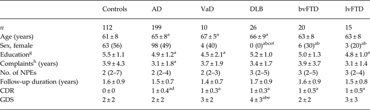
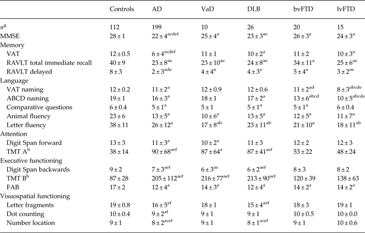
Table 1 summarizes the baseline demographics of the diagnostic groups. In general, patients were older (65 ± 8 v. 61 ± 8 years) and less educated (5.0 ± 1 v. 5.5 ± 1) than controls (both p < 0.001). On the MMSE, patients with dementia scored lower than controls (all p < 0.05). Patients with AD had the lowest MMSE score (22 ± 4). Table 2 lists the raw baseline neuropsychological test performance of controls and patients with different types of dementia.
Baseline demographics of diagnostic groups

NPE, Neuropsychological examination; CDR, Clinical Dementia Rating; GDS, Geriatric Depression Scale; AD, Alzheimer's disease; VaD, vascular dementia; DLB, dementia with Lewy bodies; bvFTD, behavioural variant frontotemporal dementia; lvFTD, language variant frontotemporal dementia.
Values are presented as mean ± standard deviation, number (percentage) or median (range). Significant difference p < 0.05 is compared to acontrols, bAD, cVaD, dDLB, ebvFTD and flvFTD.
gAccording to the Verhage system.
hYears of reported cognitive complaints, an estimation of disease duration.
Baseline raw neuropsychological test performance of diagnostic groups

MMSE, Mini-Mental State Examination; VAT, Visual Association Test; RAVLT, Rey Auditory Verbal Learning Test; ABCD, Arizona Battery for Communication Disorders; TMT, Trail Making Test (Parts A and B); FAB, Frontal Assessment Battery; AD, Alzheimer's disease; VaD, vascular dementia; DLB, dementia with Lewy bodies; bvFTD, behavioural variant frontotemporal dementia; lvTD, language variant frontotemporal dementia.
Values are presented as mean ± standard deviation. Significant difference p < 0.05 is compared to acontrols, bAD, cVaD, dDLB, ebvFTD and flvFTD.
Cognitive profiles are divided into neuropsychological tests. g n differs for every neuropsychological test because raw data are presented. hHigher scores means worse performance.
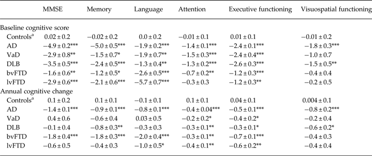
We used LMMs to compare the trajectories of cognitive decline as measured by a comprehensive neuropsychological test battery between patients with different types of dementia and controls. The estimated effects are shown in Table 3. In general, patients with a diagnosis of dementia performed worse than controls at baseline on all cognitive domains except for visuospatial functioning, which was only impaired in AD and DLB.
Estimated effect of diagnosis on baseline and annual change in MMSE score and compound scores for different cognitive domains

MMSE, Mini-Mental State Examination; AD, Alzheimer's disease; VaD, vascular dementia; DLB, dementia with Lewy bodies; bvFTD, behavioural variant frontotemporal dementia; lvFTD, language variant frontotemporal dementia.
a Reference group.
Data are presented as β ± standard error (s.e.). β values represent uncorrected estimated baseline performance or estimated change over time for the diagnoses compared with performance of controls at baseline. p values for differences with controls are given for the models corrected for age, gender and education.
*p < 0.05 for main effect; p < 0.10 for interaction, **p < 0.01, ***p < 0.001.
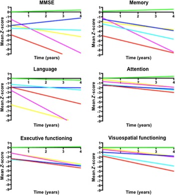
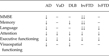
Figure 1 visualizes the trajectories of decline for each of the cognitive domains. Regarding global cognition, we found that, over time, patients with AD and bvFTD declined faster than controls whereas patients with VaD, DLB and lvFTD did not. On memory, patients with AD, DLB and bvFTD declined faster over time than controls whereas patients with VaD and lvFTD did not. For language, patients with AD, bvFTD and lvFTD declined faster over time than controls whereas patients with DLB and VaD did not. We found a significant decline over time for attention in all patients with dementia. Regarding executive functioning, patients with dementia declined over time compared to controls. On visuospatial functioning, only patients with AD and DLB declined over time. When we repeated the analyses without the patients using pharmacological treatment for dementia, the results remained essentially unchanged. Table 4 summarizes the estimated annual cognitive decline for the different dementia groups compared to controls, in a schematic overview.
Estimated cognitive performance over time compared with baseline z scores of controls. Lines represent β values as presented in Table 3. Red lines represent Alzheimer's disease (AD), dark blue lines vascular dementia (VaD), light blue lines dementia with Lewy bodies (DLB), pink lines behavioural variant frontotemporal dementia (bvFTD), yellow lines language variant frontotemporal dementia (lvFTD), and green lines controls. MMSE, Mini-Mental State Examination.

Schematic overview of estimated trajectories of cognitive decline for patients with dementia, compared to controls

MMSE, Mini-Mental State Examination; AD, Alzheimer's disease; VaD, vascular dementia; DLB, dementia with Lewy bodies; bvFTD, behavioural variant frontotemporal dementia; lvFTD, language variant frontotemporal dementia.
↓ Significant annual decline p < 0.10.
↓↓ Significant annual decline p < 0.05 and > 1.5 z score annual decline.
This table is based on the estimated annual cognitive change in Table 3. Patients with AD showed decline on all cognitive domains, and global cognition. Patients with VaD showed a decline in attention and executive functioning. For DLB, we found a decline over time in memory, visuospatial functioning, attention and executive functioning. bvFTD showed a fast decline in global cognition and all cognitive domains with the exception of visuospatial functioning. For lvFTD, we found a decline in language, executive functioning and attention.
Discussion
In this prospective longitudinal study in a large sample of patients with different types of dementia, we found that during an average follow-up time of 1.5 years, patterns of cognitive decline differed between patient groups according to type of dementia. Patients with AD declined in all cognitive domains whereas patients with bvFTD showed the fastest decline over time. Patients with VaD showed decline in attention and executive functioning. Patients with lvFTD showed most decline in attention and executive functioning whereas patients with DLB showed decline over time in several cognitive domains.
bvFTD is neuropsychologically characterized by executive dysfunction with relative sparing of memory and visuospatial functions (Seelaar et al. Reference Seelaar, Rohrer, Pijnenburg, Fox and van Swieten2011). The presence of this cognitive profile, however, is not required according to the revised diagnostic criteria for bvFTD (Rascovsky et al. Reference Rascovsky, Hodges, Knopman, Mendez, Kramer, Neuhaus, van Swieten, Seelaar, Dopper, Onyike, Hillis, Josephs, Boeve, Kertesz, Seeley, Rankin, Johnson, Gorno-Tempini, Rosen, Prioleau-Latham, Lee, Kipps, Lillo, Piguet, Rohrer, Rossor, Warren, Fox, Galasko, Salmon, Black, Mesulam, Weintraub, Dickerson, Diehl-Schmid, Pasquier, Deramecourt, Lebert, Pijnenburg, Chow, Manes, Grafman, Cappa, Freedman, Grossman and Miller2011). Although it has often been reported that memory is relatively spared in bvFTD, prominent amnestic symptoms are present in a subgroup of elderly bvFTD patients (Rascovsky et al. Reference Rascovsky, Hodges, Knopman, Mendez, Kramer, Neuhaus, van Swieten, Seelaar, Dopper, Onyike, Hillis, Josephs, Boeve, Kertesz, Seeley, Rankin, Johnson, Gorno-Tempini, Rosen, Prioleau-Latham, Lee, Kipps, Lillo, Piguet, Rohrer, Rossor, Warren, Fox, Galasko, Salmon, Black, Mesulam, Weintraub, Dickerson, Diehl-Schmid, Pasquier, Deramecourt, Lebert, Pijnenburg, Chow, Manes, Grafman, Cappa, Freedman, Grossman and Miller2011). In our study, executive dysfunction was not the only hallmark of bvFTD; we showed that baseline performance on almost every cognitive domain in bvFTD was worse compared to controls, with language impairment most pronounced. This striking language impairment might be due to fluency tasks that were included in the language domain (Seelaar et al. Reference Seelaar, Rohrer, Pijnenburg, Fox and van Swieten2011). Fluency tasks address both language and executive functioning and therefore how to interpret fluency tasks is open to debate. As expected, lvFTD was, at baseline, most impaired in language (Reilly et al. Reference Reilly, Rodriguez, Lamy and Neils-Strunjas2010; Karantzoulis & Galvin, Reference Karantzoulis and Galvin2011), which declined further over time. In bvFTD and lvFTD, visuospatial functioning was relatively preserved and showed limited decline over time (Reilly et al. Reference Reilly, Rodriguez, Lamy and Neils-Strunjas2010; Karantzoulis & Galvin, Reference Karantzoulis and Galvin2011). This might be due to the sparing of anatomical regions associated with visuospatial abilities, such as the parieto-occipital lobes (McGinnis, Reference McGinnis2012). bvFTD showed the fastest decline of all types of dementia investigated, in all cognitive domains except visuospatial functioning. A previous study showed a faster rate of cognitive decline and shorter survival in FTD compared to AD (Rascovsky et al. Reference Rascovsky, Salmon, Lipton, Leverenz, DeCarli, Jagust, Clark, Mendez, Tang-Wai, Graff-Radford and Galasko2005). In our study, bvFTD patients also showed a faster cognitive decline than AD patients. However, the duration of illness seems to be variable in FTD, ranging from 2 to 20 years (Seelaar et al. Reference Seelaar, Rohrer, Pijnenburg, Fox and van Swieten2011).
DLB is characterized by visuospatial and executive impairment, which are present early in the disease course (Karantzoulis & Galvin, Reference Karantzoulis and Galvin2011). We found that, at baseline, patients with DLB showed most severe impairment on memory and executive functioning. Over time, visuospatial functioning declined fastest after memory. We did not find pronounced impairment in attention in DLB, compared to the other types of dementia. The literature suggests that differences can only be found on more complex attentional tasks (Karantzoulis & Galvin, Reference Karantzoulis and Galvin2011), and it is possible that the tasks we used were insufficiently sensitive. It is often thought that DLB is one of the fastest declining types of dementia but our findings do not support this idea and earlier results seem conflicting (Olichney et al. Reference Olichney, Galasko, Salmon, Hofstetter, Hansen, Katzman and Thal1998; Walker et al. Reference Walker, McKeith, Rodda, Qassem, Tatsch, Booij, Darcourt and O'Brien2012). We observed decline in most cognitive domains but the rate of decline was not much faster than in other types of dementia. An explanation could be that patients who declined too fast were lost to follow-up, and as a result we saw only the milder subgroup.
Memory impairment and executive dysfunction are well-known early features of AD (Karantzoulis & Galvin, Reference Karantzoulis and Galvin2011). We also found more frank visuospatial impairment in AD. This could be due to the relatively young age of our patients with AD. Patients with AD and an earlier age of onset more often have posterior brain damage, including atrophy and functional brain changes (de Waal et al. Reference de Waal, Stam, Blankenstein, Pijnenburg, Scheltens and van der Flier2011; Moller et al. Reference Moller, Vrenken, Jiskoot, Versteeg, Barkhof, Scheltens and van der Flier2013). We found that patients with VaD showed decline in attention and executive functioning. As far as we know, only one study has investigated rate of decline in VaD and AD. However, this study was conducted in the preclinical stage, and persons with preclinical VaD showed a faster rate of cognitive decline than preclinical AD in episodic memory, visuospatial functioning and category fluency (Laukka et al. Reference Laukka, Macdonald, Fratiglioni and Backman2012). This former study did not incorporate tests for executive functioning and attention, the domains in which we found decline in VaD. Another relevant shortcoming of the study by Laukka et al. (Reference Laukka, Macdonald, Fratiglioni and Backman2012) is that no brain imaging was available and about 70% of patients had a diagnosis of VaD, which was primarily a diagnosis of post-stroke dementia. By contrast, the diagnoses of all of our VaD patients were supported by brain imaging, and mostly involved small vessel disease.
The diverse patterns of cognitive decline could not be explained by differences in disease duration at baseline or on the CDR scale, although we found differences on the MMSE. At baseline, patients with AD performed worse on the MMSE and this might indicate that these patients were cognitively more impaired. However, the MMSE is known to rely heavily on memory, the domain on which AD most was impaired (Nieuwenhuis-Mark, Reference Nieuwenhuis-Mark2010). The MMSE also relies on language, and patients with bvFTD declined fastest on language and memory, possibly reflected in their fast decline on MMSE. As a consequence, the MMSE might have some restrictions when it is used to compare global cognition in different types of dementia.
A possible limitation of this study is that, except for AD and controls, sample sizes were relatively small. However, longitudinal studies on non-AD dementias are scarce, and for these more rare types of dementia our group sizes are in fact not so small. We feel that even when reported significance levels are limited by suboptimal power, reporting effect sizes of these groups is very valuable. Our results need to be replicated in larger studies. The sample described in our study was relatively young, which might limit generalizability to dementia in general. Nevertheless, we consider that the relatively young age also has advantages, as non-AD types of dementia often develop at a younger age and hence we were able to include diverse types of dementia. In addition, dementia at a younger age is often thought to be more pure, and less mixed, pathology and hence the patterns of decline may be more specific. Among the strengths of our study is the longitudinal set-up and the comprehensive neuropsychological assessment covering five cognitive domains.
Our findings have considerable impact, as they provide estimates of the natural disease course in different types of dementia. Clinical trials have thus far most frequently used global outcome measures such as the Alzheimer's Disease Assessment Scale – Cognitive subscale (ADAS-Cog; Rosen et al. Reference Rosen, Mohs and Davis1984), the CDR scale, and questionnaires about activities of daily living and behaviour. Shortcomings of the ADAS-Cog include its primary focus on memory and language, while tasks assessing attention, executive or visuospatial functioning are not incorporated in the test (Wesnes & Harrison, Reference Wesnes and Harrison2003). More extensive neuropsychological assessments covering several cognitive domains, such as the Neuropsychological Test Battery, are increasingly being preferred over the ADAS-Cog. These neuropsychological batteries are better at detecting subtle cognitive changes and therefore seem more suitable for cognitive assessments in trials in preclinical/early disease stages. The Food and Drug Administration (FDA) recently proposed revising the criteria for drug approval because persons in very early disease stages have no obvious deterioration in daily functioning. They suggested that, in trials (in very early stages of disease), cognitive tests should be used as end-points (Kozauer & Katz, Reference Kozauer and Katz2013). In addition, clinical trials are currently designed for other types of dementia than AD. Therefore, estimates of cognitive trajectories of different types of dementia are essential for trial design and power calculations.
Acknowledgements
The Alzheimer Centre receives unrestricted funding from various sources through the VUmc Fonds. This study received no specific funding. We thank Dr B. M. Tijms and Dr J. Berkhof for their statistical advice. Research undertaken by the VUmc Alzheimer Centre is part of the neurodegeneration research programme of the Neuroscience Campus Amsterdam. The Alzheimer Centre VUmc is supported by Alzheimer Nederland and Stichting VUmc Fonds. The clinical database structure was developed with funding from Stichting Dioraphte. This research received no specific grant from any funding agency, commercial or not-for-profit sectors.
Declaration of Interest
Dr Prins serves on the advisory board of Boehringer Ingelheim and Envivo. He has been a speaker at symposia organized by Janssen and Novartis. He has a senior fellowship at the Alzheimer Centre VUmc partly supported by Vereniging AEGON and receives research support from the Brain Foundation of The Netherlands (project number H07.03) and Alzheimer Nederland (project number WE.03-2012-02). Dr Scheltens serves/has served on the advisory boards of Genentech, Novartis, Pfizer, Roche, Danone, Nutricia, Jansen AI, Baxter and Lundbeck. He has been a speaker at symposia organized by Lundbeck, Lilly, Merz, Pfizer, Jansen AI, Danone, Novartis, Roche and Genentech. He serves on the editorial board of Alzheimer's Research and Therapy and Alzheimer's Disease and Associated Disorders, is a member of the scientific advisory board of the European Union (EU) Joint Programme on Neurodegenerative Disease Research (JPND) and the French National Plan Alzheimer. Dr van der Flier has received funding and speaker honorarium from Boehringer Ingelheim; all funds were paid to her institution. The other authors have no conflicts of interest to declare.