Implications
The results of this study shows that exposition of rams to high altitude by short or long term induces blood and seminal oxidative stress, and deteriorate some sperm characteristics. These changes may be partly responsible for the low fertility of ovine herds maintained at high altitude. Oral supplementation with vitamins C and E prevents oxidative stress and improves sperm characteristics, especially in rams exposed to high altitude for a short time. This supplementation can be a simple and inexpensive tool to improve the ovine fertility at high altitude.
Introduction
Highland sheep farming is extremely important in the Andean and Qinghai-Tibetan plateaus, where an estimated 25 million people economically depend on sheep breeding (Huddleston et al., Reference Huddleston, Ataman, de Salvo, Zanetti, Bloise, Bel, Franceschini and Fèd´Ostiani2003). However, sheep breeding at high altitude (ha) is hampered by a reduced reproductive efficiency when compared with their low altitude (la) counterparts (Parraguez et al., Reference Parraguez, Urquieta, Pérez, Castellaro, De Los Reyes, Torres-Rovira, Aguado-Martínez, Astiz and González-Bulnes2006).
Most of the studies at ha have been focused in female reproductive efficiency and have shown that low fertility in ewes results from inadequate function of the reproductive axis, as a consequence of hypoxia, oxidative stress or both (Parraguez et al., Reference Parraguez, Atlagich, Araneda, García, Muñoz, De los Reyes and Urquieta2013). In spite of the greater relative importance of males in sheep flocks (one male mates 20 to 25 females), there is a scarcity of similar studies in rams. There appears to be only one report from Peruvian researchers, published in the 1940s, that addressed severe disturbances in the ejaculates and sexual libido of rams taken to ha (Monge et al., Reference Monge, San Martín, Atkins and Castañón1945). Studies in humans (Okumura et al., Reference Okumura, Fuse, Kawauchi and Akashi2003) and rats (Farías et al., Reference Farías, Bustos-Obregón, Tapia, Gutiérrez, Zepeda, Juantok, Cruz, Soto, Benites and Reyes2008) have shown clear evidence of deleterious effects on the male reproductive axis after ha exposure, including deterioration in both testicular and seminal parameters. Interestingly, in human populations living at ha over multiple generations, these negative male reproductive changes are absent, indicating an adaptation to that environmental condition (Gonzales, Reference Gonzales2007).
Previous studies in ewes have demonstrated that most of the deleterious effects of ha on reproductive efficiency, either at short- or long-term exposure, are associated with oxidative stress secondary to hypoxia (Parraguez et al., Reference Parraguez, Urquieta, Pérez, Castellaro, De Los Reyes, Torres-Rovira, Aguado-Martínez, Astiz and González-Bulnes2006, Reference Parraguez, Atlagich, Behn, Bruzzone and Raggi2011 and Reference Parraguez, Atlagich, Araneda, García, Muñoz, De los Reyes and Urquieta2013). Oxidative stress results from unbalanced production of reactive oxygen species (ROS) and the protective effect of the endogenous antioxidant system, where superoxide dismutase (SOD) and glutathione peroxidase (GSH-Px) are protagonist enzymes. Deleterious effects of ROS are mainly related to changes in lipids, protein and DNA, resulting in damage to or cell death. Spermatozoa are highly vulnerable to ROS effects, leading to fertility disorders (Walczak–Jedrzejowska et al., Reference Walczak–Jedrzejowska, Wolski and Slowikowska-Hilczer2013).
There is currently no data regarding redox balance in rams at ha, but recently we have demonstrated that increases in seminal antioxidant status in rams at la can be obtained by supplementation with antioxidant vitamins, leading to increased sperm concentration, motility and viability (Cofré et al., Reference Cofré, Peralta, Urquieta, Raggi, Benavides and Parraguez2016). Likewise, data from human and rodent models point to the main role of oxidative status in the conservation of semen quality (Agarwal and Allamaneni, Reference Agarwal and Allamaneni2011) and have shown the efficacy of antioxidant treatment for improvement semen quality (Agarwal and Sekhon, Reference Agarwal and Sekhon2010).
Thus, having in mind the social and economic importance of sheep production for rural populations living at ha (Huddleston et al., Reference Huddleston, Ataman, de Salvo, Zanetti, Bloise, Bel, Franceschini and Fèd´Ostiani2003), the first aim of the present work was to characterize the effects of short- and long-term exposure to ha hypoxic environment on ram testosterone levels and semen quality. The second objective was to establish whether oral antioxidant vitamin supplementation prevents or attenuates effects associated with ha exposure.
Material and methods
Ethics statement
This study was performed in agreement with the Guide for Care and Use of Laboratory Animals (Eighth Edition, National Research Council, National Institute of Health, Washington, DC, USA). The protocols were approved by the Bioethics Review Committee of the Faculty of Veterinary and Animal Sciences, University of Chile, as well as by the Bioethics Advisory Committee of the Chilean National Commission for Scientific and Technological Research (CONICYT, Chile).
Animal management and sampling
The experiment involved 48 crossbred rams, from the experimental flocks of the University of Chile, with 1 year of age, 45.5±0.9 kg of mean BW and 2.8±0.3 of body condition score in a 1 to 5 scale. In al, 16 native ha rams (group HH; descendants of sheep introduced to the Andean high plateau by Spanish settlers almost 500 years ago) at the beginning of the experiment, were allocated to a single pen at the animal facilities of the International Centre for Andean Studies (INCAS, University of Chile; ~3600 m altitude, barometric pressure ~667 hPa). A total of 32 native la rams (~500 m; barometric pressure ~990 hPa), with similar phenotypes, BWs and age to the males in the HH group were also selected. Four days before starting the experimental protocol, half of the la rams (group LH, n=16) were moved to the INCAS facilities to join the HH rams. The remaining 16 rams were maintained at low altitude (group LL). Animals were fed with alfalfa hay and balanced feed to satisfy requirements (NRC, 2006); water was supplied ad libitum. The experiment began in mid-December, along with the start of the reproductive season in Chile.
Rams in each group were randomly divided into two equal subgroups (n=8 each) and housed in different pens. Half of the animals remained as controls (maintaining the identification as groups HH, LH and LL), whereas the other half of the rams were supplemented daily with 600 mg of vitamin C (ascorbic acid) and 450 IU of vitamin E (alpha-tocopherol) orally, over 60 days (groups HHV, LHV and LLV). Vitamin doses were calculated on the basis of previous experiments on sheep maintained at similar environmental conditions, where the treatment prevented oxidative stress (Parraguez et al., Reference Parraguez, Atlagich, Behn, Bruzzone and Raggi2011).
Assessment of vitamin C and E, oxidative stress biomarkers and testosterone in blood
Blood samples (10 ml) were taken from the left jugular vein at 0, 30 and 60 days after starting vitamin supplementation using heparinized syringes. Blood was centrifuged at 1200×g ×5 min at 4°C; the obtained plasma was stored at −80°C until assayed.
Vitamins C and E were measured by high-performance liquid chromatography as previously described (Parraguez et al., Reference Parraguez, Atlagich, Behn, Bruzzone and Raggi2011). Briefly, vitamin C was measured in plasma samples diluted 10-fold in ultra-pure water, by means of amperometric detection, using a glassy carbon electrode operated at 800 mV and an Ag/AgCl reference electrode. Vitamin E was measured in ethanol/dichloromethane extracted plasma samples by means of spectrofluorimetric detection at 290 and 330 nm wave-length for excitation and emission.
Malondialdehyde (MDA) and protein carbonyls (PC) were measured in blood plasma as oxidative damage biomarkers for lipids and proteins, respectively. Quantification of MDA was made using a colorimetric kit (TBARS, TCA method, Assay Kit; Cayman Chemical Company, Ann Arbor, MI, USA), following the manufacturer instructions. Absorbance was read at 540 nm with a microplate reader (Perlong DNM-9602; Nanjing Perlove Medical Equipment Co. Ltd., Nanjing, China). Quantification of PC was made using the OxiSelectTM Protein Carbonyl ELISA kit (Cell Biolabs Inc., San Diego, CA, USA) according to the protocol provided by the manufacturer. Absorbance was read at 450 nm in the microplate reader. Before the assay, plasma samples were diluted in phosphate buffered saline to achieve a protein concentration of 10 µg/ml. In addition, total antioxidant capacity (TAC) in plasma was assessed using the colorimetric Antioxidant Assay Kit (Cayman Chemical Company), following the instructions of the manufacturer. Absorbance was read at 405 nm in the microplate reader.
Plasma testosterone concentrations were measured by radioimmunoassay (Testo-RIA-CT; DIAsource Immuno Assays S.A., Louvain-la-Neuve, Belgium) in 50 µl of plasma. Measurements were done in duplicate and the minimum concentration detected by the assay was 0.11 ng/ml. The intra- and inter-assay variations coefficients were 4.6% and 6.2%, respectively.
Assessment of semen quality and semen oxidative status
Ejaculates were obtained at 0, 30 and 60 days after starting vitamins supplementation, using an electro-ejaculator standardized for small ruminants (Minitube e320, Tiefenbach, Germany). This technique was preferred to prevent the eventual absence of sexual libido observed by Monge et al. (Reference Monge, San Martín, Atkins and Castañón1945) in rams exposed to HA. Furthermore, as indicated by Matthews et al. (Reference Matthews, Bester and Schwalbach2003), ‘electroejaculation is more practical as it does not require previous training of the rams and can be used for breeding soundness examination’. Before starting the electroejaculation procedure, rams were sedated using xylazine (0.2 mg/kg BW, Xylazine 2%®; Centrovet, Santiago, Chile), then the rectum was emptied and the periprepuceal area was washed with saline solution and carefully dried with paper towel. The probe (diameter: 2.5 cm, length: 16 cm) was lubricated with contact gel, introduced into the rectum with the three longitudinal electrodes ventrally oriented and electric ramps were applied. The electric ramps consisted of consecutive increases of 0.5 V, starting from 0 up to a maximum of 8 V. Each voltage was applied for 3 s followed by a rest period of 3 s. All animals ejaculated between 4 and 8 V. The ejaculates were recovered in clean graduated glass cups, which enabled direct volume measurement. Then, a 100 µl semen sample was taken for pH determination using test strips (MColorpHastTM; Merck Millipore, Billerica, MA, USA). Subsequently, the collection cups were isolated and protected from direct sunlight. Sperm concentration, overall and progressive motility, and viability were evaluated in a CASA system (ISAS-V®, Proiser, Valencia, Spain). For this, semen samples were diluted in Tris/citric acid/fructose medium to obtain a final concentration of about 50×106 sperms/ml (Emmanverdi et al., Reference Emmanverdi, Zhandi, Zareshahneh, Sharafi and Akabar-Sharif2013). Sperm viability was assessed using the Duovital® florescent kit (Proiser, Valencia, Spain). Measurements were done in 4 µl sperm suspension loaded in a 20 µm depth cell counting chamber (Spermtrack®, Proiser) maintained at 37.5°C in a thermal plate, as recommended by the manufacturer.
Afterwards, ejaculates were centrifuged at 1500×g ×10 min at 4°C and seminal fluid was stored at −80°C until assayed for SOD and GSH-Px activities and TAC. Concentrations of SOD were measured in 10 µl seminal plasma diluted 1 : 25 in sample buffer (50 mMTris-HCl, pH 8.0), using the Superoxide Dismutase Assay Kit® (Cayman Chemical Company), following the supplier instructions. Concentrations of GSH-Px were measured in 20 µl seminal plasma, using the Glutathione Peroxidase Assay Kit® (Cayman Chemical Company), following supplier guidelines. Meanwhile, TAC was measured in 10 µl seminal fluid diluted 1 : 10 in assay buffer (5 mM potassium phosphate, pH 7.4, containing 0.9% sodium chloride and 0.1% glucose), using the Antioxidant Assay Kit® (ELISA commercial kit, Cayman Chemical Company), following the supplier instructions. In the SOD and TAC assays, absorbance was measured in a microplate reader (DNM-9602; Perlong Medical Equipment Co. Ltd., Nanjing, China), meanwhile the GSH-Px assay absorbance was read at 340 nm every minute for 5 min with a microplate spectrophotometer (Epoch®, Winooski, VT, USA).
Statistical analysis
Comparisons among groups for each experimental time (0, 30 and 60 days of vitamins supplementation), were done by analysis of variance, using the general linear model procedure (GLM; SAS Institute Inc., Cary, NC, USA). Comparisons were made using two statistical models. The first model was used to test the effect of altitudinal status, including two cross factors: the place of birth or origin of the animals and the place where the samples were obtained and studied. The second model took into consideration the previous two factors in addition to vitamin supplementation. Interactions among factors were also analysed. When significant differences were found, Duncan’s test was used to determine the groups among which the differences were statistically significant. Significant differences were considered when P<0.05. The results are expressed as means±SEM.
Results
Blood plasma concentrations of vitamins C and E, and oxidative stress biomarkers
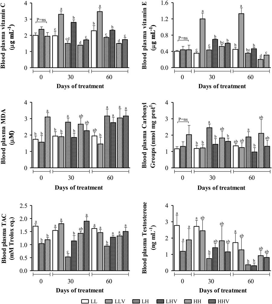
Blood plasma concentrations of vitamins C and E are shown in Figure 1 (upper left and right panels, respectively). There were no differences at day 0; however, vitamin concentrations in the plasma of all supplemented groups increased at subsequent time points (days 30 and 60). However, vitamin E plasma concentrations did not achieve statistical significance between HH and HHV rams at day 30. The same situation was found for groups LH and LHV at day 60 of supplementation. It is noteworthy that the supplemented group LLV reached the highest values for both vitamins.
Blood plasma concentrations of vitamins C and E, oxidative stress biomarkers and testosterone in control and vitamin C/E supplemented rams, exposed to high altitude conditions. Ram groups are as follows: HH, rams native to high altitude, kept at high altitude, without vitamins supplementation; HHV, rams native to high altitude, kept at high altitude, with vitamins supplementation; LH, rams native to low altitude, taken to high altitude, without vitamins supplementation; LHV, rams native to low altitude, taken to high altitude, with vitamins supplementation; LL, rams native to low altitude, kept at low altitude, without vitamins supplementation; LLV, rams native to low altitude, kept at low altitude, with vitamins supplementation. Different letters above the bars indicate significant differences among groups in each experimental time (Duncan test, P<0.05).
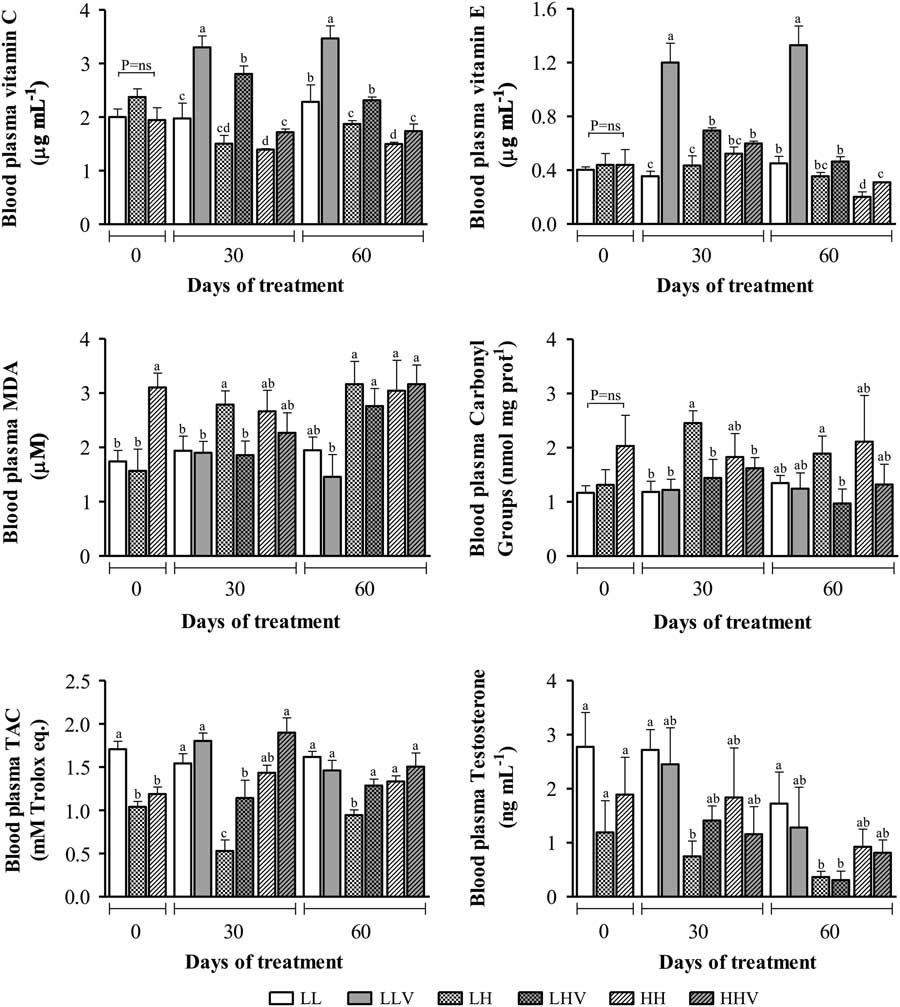
Biomarkers of oxidative damage (MDA, PC) and TAC in plasma are shown in Figure 1 (intermediate left and right, and lower left panels, respectively). In the case of MDA, the values at day 0 were higher in HH animals than in the other groups. At day 30, MDA increased in LH rams, which was prevented in the group with vitamins supplementation (group LHV). At day 60, all the rams exposed to ha showed high levels of MDA, with high dispersion of the values. In the case of PC, there were no differences among groups at day 0. At days 30 and 60, PC had a similar pattern to MDA at day 30, with also high dispersion of the values, especially in ha groups, at day 60. The lowest value for this trait was present in LHV group. Analysis of changes in TAC over time showed, overall, an inverted pattern to PC, being significantly higher in LL rams at day 0. At days 30 and 60, TAC significantly decreased in LH group; an effect not observed in group LHV.
Blood plasma concentrations of testosterone
Plasma testosterone concentrations are shown in Figure 1 (lower right panel). No significant differences in testosterone levels were found at day 0. At day 30 of treatment, LH rams showed the lowest testosterone value; significantly different from LL rams. At day 60 of treatment, the lowest concentrations of testosterone were observed in LH and LHV animals; significantly different to LL group. No effect of vitamin supplementation was observed on the concentration of testosterone at days 30 and 60.
Semen quality parameters
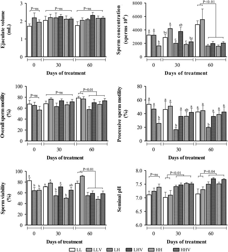
Results on semen characteristics are shown in Figure 2. The mean ejaculate volume was 2.0±0.1 ml, without differences among groups in each experimental time (left upper panel). Assessment of sperm concentration at time 0 showed a lower value in HH rams. At day 30, vitamin supplementation lead to an increased sperm concentration in LLV and LHV rams, but this effect was not present in HHV animals. At day 60, there were no observable effect of vitamin supplementation and only differences according to the altitudinal status were observed; where, animals exposed to ha had lower sperm concentrations (right upper panel).
Characteristics of ejaculates obtained from control and vitamin C/E supplemented rams, exposed to high altitude conditions. Rams groups are as follow: HH, rams native to high altitude, kept at high altitude, without vitamins supplementation; HHV, rams native to high altitude, kept at high altitude, with vitamins supplementation; LH, rams native to low altitude, taken to high altitude, without vitamins supplementation; LHV, rams native to low altitude, taken to high altitude, with vitamins supplementation; LL, rams native to low altitude, kept at low altitude, without vitamins supplementation; LLV, rams native to low altitude, kept at low altitude, with vitamins supplementation. Different letters above the bars indicate significant differences among groups in each experimental time (Duncan test, P<0.05).
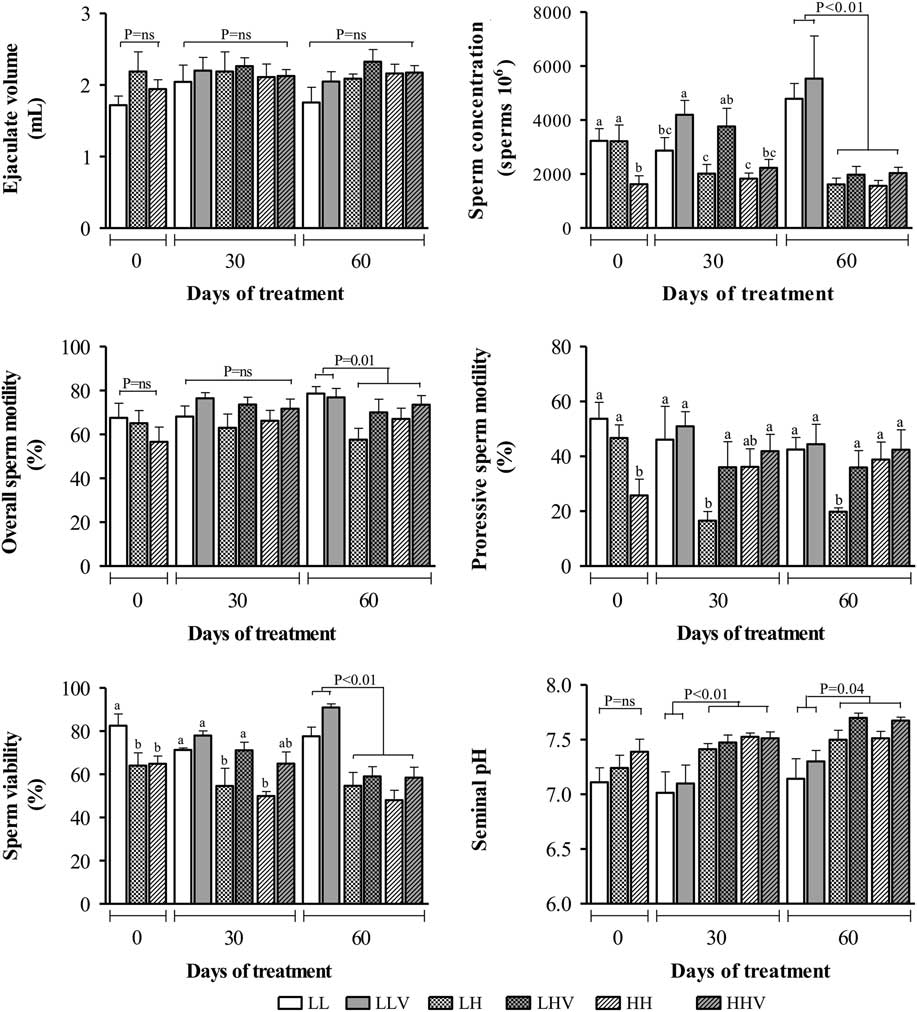
Overall sperm motility (left intermediate panel) showed no differences among groups at days 0 and 30. At day 60, lower overall sperm motility was present in the four groups exposed to ha. Meanwhile, progressive sperm motility (Figure 2, right intermediate panel) was lower in HH rams at day 0. A marked decrease in progressive motility was observed in LH group at days 30 and 60, which was prevented by vitamin supplementation in the LHV group. Sperm viability is presented in the left lower panel. Lower values for this trait were shown by groups exposed to ha, at three experimental time points. Vitamin supplementation was able to prevent the ha effect at day 30 of treatment, but no such effect was observed at day 60. Seminal pH values (right lower panel) ranged from 6.5 to 8.3, without differences among groups at day 0; however, significant increases were detected at 30 and 60 days of exposure to ha. No effect of vitamin supplementation was observed for seminal pH.
Semen oxidative status
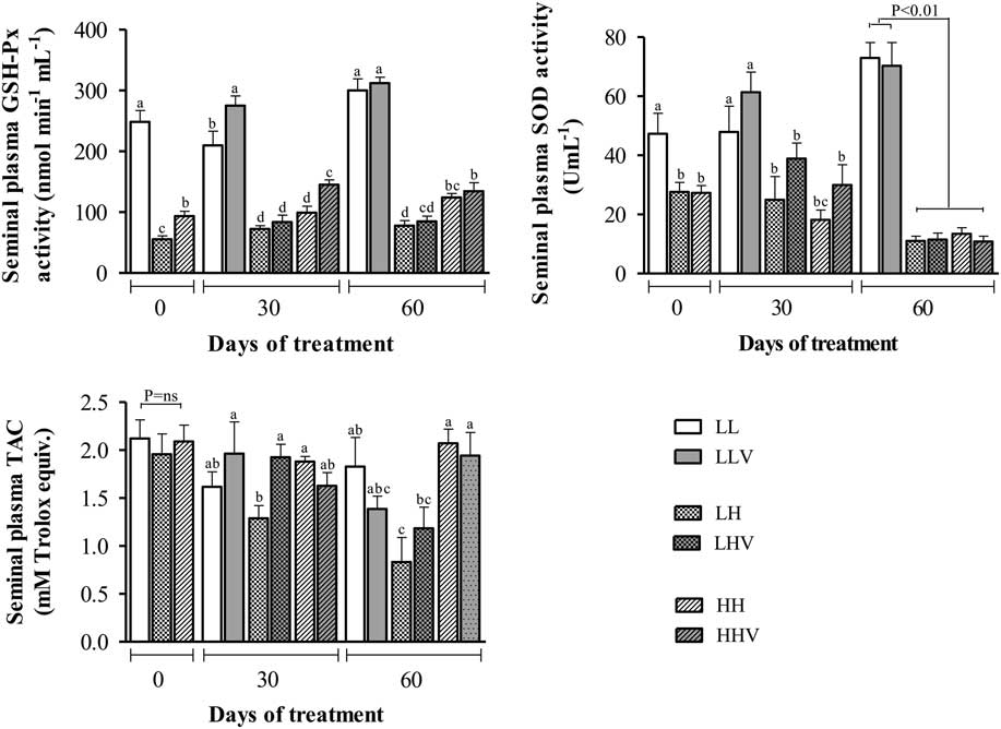
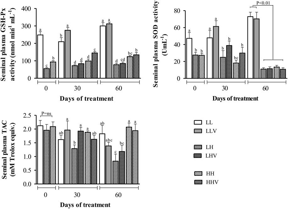
Parameters of oxidative seminal status are shown in Figure 3. Activities of GSH-Px (upper panel) and SOD (intermediate panel) were significantly reduced in ha rams, situation occurring from day 0 onwards. However, for GSH-Px the effect of ha was more accentuated in LH rams. At day 30, vitamin supplementation reduced ha effects in HH rams for GSH-Px activity. At day 60, vitamin supplementation showed no effect on both antioxidant enzymes. In the case of TAC (bottom panel), only an effect of short-term exposure to ha was observed, thus, decreased TAC was present in LH rams at 60 days of treatment. In the same group of rams, vitamin supplementation increased the TAC, but this effect was observed only at day 30.
Antioxidant status of seminal plasma from ejaculates obtained from control and vitamin C/E supplemented rams, exposed to high altitude conditions. Rams groups are as follow: HH, rams native to high altitude, kept at high altitude, without vitamins supplementation; HHV, rams native to high altitude, kept at high altitude, with vitamins supplementation; LH, rams native to low altitude, taken to high altitude, without vitamins supplementation; LHV, rams native to low altitude, taken to high altitude, with vitamins supplementation; LL, rams native to low altitude, kept at low altitude, without vitamins supplementation; LLV, rams native to low altitude, kept at low altitude, with vitamins supplementation. Different letters above the bars indicate significant differences among groups in each experimental time (Duncan test, P<0.05).
Discussion
The results of the present study indicate a state of oxidative stress in rams exposed to ha, with short-term exposed animals being the most affected. Blood concentrations of MDA, CP and TAC are normally used for evaluation of redox status, in the general body or specific tissues (Marrocco et al., Reference Marrocco, Altieri and Peluso2017). In the present work, blood MDA and CP were increased, while TAC was decreased by exposure to ha, evidencing the state of oxidative stress, mainly in the rams exposed to ha during short time. These results are in agreement with those obtained in pregnant ewes (Parraguez et al., Reference Parraguez, Atlagich, Behn, Bruzzone and Raggi2011) and humans (Sinha et al., Reference Sinha, Singh, Saha, Kain and Tyagi2010). Moreover, blood and seminal plasma oxidative stress biomarkers show similar patterns in men with reproductive dysfunctions leading to infertility (Benedetti et al., Reference Benedetti, Tagliamonte, Catalani, Primiterra, Canestrari, De Stefanu, Palini and Bulleti2012). Oral supplementation with vitamin C and E increased plasma levels of these vitamins, confirming previous data in rams (Cofré et al., Reference Cofré, Peralta, Urquieta, Raggi, Benavides and Parraguez2016), and improved the redox status in those recently exposed to ha. These results are consistent with those obtained in ewes maintained under different altitudinal conditions, being also associated with prevention against deleterious effects of ha on reproductive parameters (Parraguez et al., Reference Parraguez, Atlagich, Behn, Bruzzone and Raggi2011), in a similar way to our LHV rams.
Previous studies on the effects of the exposure to ha hypoxic environment in male reproductive function, either for short or long time and either in rams (Monge et al., Reference Monge, San Martín, Atkins and Castañón1945) or men (Donayre et al., Reference Donayre, Guerra-García, Moncloa and Sobrevilla1968), have been based on the evaluation of semen quality and sexual behavior. The current study also evaluated possible effects of the exposure to ha on plasma testosterone concentrations in rams. Our results indicate that ha has a detrimental effect on testosterone concentration, especially in LH rams. Previous studies in rats have shown that blood testosterone decreased after a few days of exposure to simulated ha (Farías et al., Reference Farías, Bustos-Obregón, Tapia, Gutiérrez, Zepeda, Juantok, Cruz, Soto, Benites and Reyes2008). In men, however, results have shown inconsistent effects on testosterone levels, from increases under acute ha exposure (Barnholt et al., Reference Barnholt, Hoffman, Rock, Muza, Fulco, Braun, Holloway, Mazzeo, Cymerman and Friedlander2006), to no changes (Basu et al., Reference Basu, Pal, Prasad, Malhotra, Rao and Sawhney1997) or decreases (Benso et al., Reference Benso, Broglio, Aimaretti, Lucatello, Lanfranco, Ghigo and Grottoli2007) after long-term exposure. Such differences may be related to different stages of adaptation to the altitude (Gonzales, Reference Gonzales2013), a hypothesis consistent with the results obtained in the present study, where the most affected males were those recently exposed to ha.
The present study showed no effects of supplementation with vitamin C or E on testosterone levels in ha rams, which is in agreement with previous data on the use of oral supplementation with only vitamin E (Rekkas et al., Reference Rekkas, Kokolis, Belibasaki, Tsantarliotou and Smokovitis2000).
Sperm characteristics were highly sensitive to ha. Sperm viability in LH rams decreased, becoming similar to HH animals, after only 4 days of ha exposure. Progressive and overall sperm motility in LH rams decreased after 30 and 60 days of exposure to ha, respectively. These results are consistent with previous data in men after 7 to 14 days of ha exposure (Donayre et al., Reference Donayre, Guerra-García, Moncloa and Sobrevilla1968). When considering possible causes for these changes in sperm characteristics, it needs is important to know that the overall process of spermatogenesis lasts around 47 days in the ram (Sharpe, Reference Sharpe1994) and more than 70 in men (Hess and de Franca, Reference Hess and de Franca2008). Hence, these quick effects of ha exposure would be related to deleterious effects of the exposure to hypoxic environment on mature semen. In agreement with this observation, previous studies have found impaired sperm motility and increased apoptosis in mature sperm exposed to small increases in the amount of ROS in testis and epididymis (Aitken and Koppers, Reference Aitken and Koppers2011; Bansal and Bilaspuri, Reference Bansal and Bilaspuri2011). The data obtained in the current study would support this hypothesis. Information on seminal oxidative/antioxidant status in individuals exposed to ha is scarcely available in the literature. Studies in humans and rats exposed to natural or artificial ha conditions showed decreased levels/activities of GSH-Px and SOD in different tissues (Dosek et al., Reference Dosek, Ohno, Acs, Taylor and Radak2007), consistent with our findings in seminal fluid. On the other hand, data from studies at la showed a positive correlation among TAC and activity of GSH-Px and SOD in seminal fluid and semen quality in both men (Khosrowbeygi et al., Reference Khosrowbeygi, Nosratollah and Deldar2004) and rams (Cofré et al., Reference Cofré, Peralta, Urquieta, Raggi, Benavides and Parraguez2016), with SOD and GSH-Px having a crucial protective role against ROS in ram-lamb’s semen (Kasimanickam et al., Reference Kasimanickam, Pelzer, Kasimanickam, Swecker and Tatcher2006).
Supplementation with vitamin C and E showed short-term beneficial effects on sperm viability, concentration and progressive motility, especially in LH rams, in agreement with previously reported data in rams maintained at la (Cofré et al., Reference Cofré, Peralta, Urquieta, Raggi, Benavides and Parraguez2016). Previous studies in rams supplemented with vitamin C doses equivalent to the current study, resulted in significantly increased semen volume and sperm concentration, motility and viability (Jafaroghli et al., Reference Jafaroghli, Abdi-Benemar, Zamiri, Khalili, Farshad and Shadparvar2014), whilst supplementation with vitamin E increased ejaculate volume, sperm concentration and sperm motility (Yue et al., Reference Yue, Yan, Luo, Xu and Jin2010).
Moreover, results from the current study also suggest short-term beneficial effects of oral vitamin C and E supplementation on the oxidative/antioxidant status of the rams, which is in agreement with previous studies that report a significant increase in GSH-Px and SOD activities in seminal plasma and a significant improvement in semen quality parameters after treatment of bucks with vitamin E (Majid et al., Reference Majid, Qureshi and Khan2014).
In agreement with the present results obtained in rams, as well as those previously obtained in sheep (Parraguez et al., Reference Parraguez, Atlagich, Araneda, García, Muñoz, De los Reyes and Urquieta2013 and Reference Parraguez, Díaz, Cofre, Urquieta, De Los Reyes, Astiz and González-Bulnes2014), it is feasible to think that the observed seminal modifications at ha are partially responsible for the low fertility rate of ovine species raised under this condition. This is also supported by similar changes in both the semen and sperm characteristics present in human anomalies leading to subfertility or infertility, which are frequently associated to the presence of oxidative stress (Agarwal and Allamaneni, Reference Agarwal and Allamaneni2011; Benedetti et al., Reference Benedetti, Tagliamonte, Catalani, Primiterra, Canestrari, De Stefanu, Palini and Bulleti2012).
Conclusions
Rams under both short- and long-term exposure to ha conditions have impairments of oxidative/antioxidant status in plasma and seminal fluid, effects which concurrently decrease semen quality. These effects may contribute to the previously reported decreased fertility of ovine species at ha. Oral supplementation with vitamins C and E had beneficial effects on blood and semen oxidative status, as well as some of the male semen characteristics. Thus, oral supplementation with vitamins C and E constitute a simple and cheap alternative to improve semen quality of rams at ha, primarily in short-term exposed animals. Future works should assess whether changes in seminal quality affect the rate of fertilization of rams moved to high altitudes.
Acknowledgements
The authors thank Veterinary students Ignacio Ceppi, Carolina Rojas, Catalina Chappuzeau and Mabel Pinto for their assistance during animal sampling and Mr Gabino Llusco for his assistance with animal care. The authors also thank Dr Quentin L. Sciascia (Leibniz Institute for Farm Animal Biology, Institute for Nutritional Physiology, Germany) for expert language reviewing of the text. This work was supported by grant FONDECYT 1130181 from CONICYT, Chile.