Introduction
Schizophrenia is an incredibly complex syndrome, with a group of signs and symptoms of unknown etiology mainly defined by observed signs of psychosis; in its most common form, it presents itself with paranoid delusions and auditory hallucinations late in adolescence or early adulthood.Reference Insel1 Currently available pharmacological treatments have limited efficacy, and we have seen little substantial improvement since the advent of the first antipsychotics in the 1950s.Reference Hallak, de Paula, Chaves and Bressan2
Unconjugated bilirubin (UCB) is the water-insoluble fraction of total bilirubin in the serum. After binding to albumin and being transported to the liver, it is processed by the hepatic 1A1 isoform of uridinediphosphate-glucuronosyltransferase (UGT1A1) and becomes conjugated bilirubin (direct bilirubin), which can, in turn, be excreted into the bile.Reference Hirschfield and Alexander3 In practice, unconjugated bilirubin is calculated by finding the difference between directly measurable levels of serum total bilirubin (TB) and conjugated bilirubin (CB); for this reason, UCB is also sometimes called indirect bilirubin. Some studies have found a possible correlation between UCB levels and an increased incidence of schizophrenia.Reference Miyaoka, Seno, Itoga, Iijima, Inagaki and Horiguchi4–Reference Miyaoka, Wake, Hayashida and Horiguchi7 UCB levels may have both direct and indirect toxic effects on the central nervous system (CNS),Reference Brites8 with animal models (Gunn rat) presenting schizophrenia-like behavior.Reference Hayashida, Miyaoka and Tsuchie9 Therefore, measurement of serum concentrations of UCB has been proposed as a possible biomarker for the management of schizophrenia.Reference Gama-Marques, Tinoco, Pedro, Leote, Silva and Ouakinin10
While UCB concentrations below 100 nM are able to protect the CNS against oxidative damage,Reference Doré, Takahashi, Ferris, Hester, Guastella and Snyder11 moderate and high levels preferentially cause early necrosis.Reference Hankø, Hansen, Almaas, Lindstad and Rootwelt12 Neurons are more susceptible to UCB-induced damage than astrocytes,Reference Brito, Rosa and Falcão13 as neurons commonly experience higher levels of reactive oxygen species (ROS), protein oxidation, and lipid peroxidation upon UCB exposure than astrocytes.Reference Conforti, Adalbert and Coleman14 Interaction with UCB causes increased polarity and fluidity,Reference Rodrigues, Solá, Castro, Laires, Brites and Moura15 disrupting the neuronal membrane structure. This is accompanied by reduced activity of Mg2+-ATPase aminophospholipid translocase (flippase), Na+, and K+-ATPase, along with enhanced intracellular levels of Ca2+, effects which can lead to cell destruction.Reference Khan and Poduval16 Developing neurons are particularly susceptible to UCB interaction, with neurogenesis impairment, neuritic atrophy, and cell death by both necrosis and apoptosis.Reference Brito, Silva and Brites17, Reference Falcão, Silva, Pancadas, Fernandes, Brito and Brites18 The contact results in a sudden and lasting decrease in neuronal arborization,Reference Fernandes, Falcão and Abranches19 and cells remain vulnerable to a secondary toxic stimulus.Reference Falcão, Silva, Pancadas, Fernandes, Brito and Brites18 UCB also causes synaptotoxicity, which has been associated with the potentiation of inhibitory synaptic transmissionReference Shi, Kakazu, Shibata, Matsumoto, Nakagawa and Komune20 and impairment of the induction of long-term potentiation.Reference Chang, Lee, Huang and Hsu21 An N-methyl-d-aspartate (NMDA) receptor-mediated excitotoxic mechanism is another pathway leading to UCB-induced neuronal injury.Reference Bellefontaine, Hanchate and Parkash22 UCB has been demonstrated to raise the extracellular concentration of glutamate, by enhancing its release, mainly from immature neurons and overstimulating NMDA receptors.Reference Falcão, Fernandes, Brito, Silva and Brites23 UCB also causes mitochondrial swelling,Reference Solá, Diógenes, Brites and Rodrigues24 Bax translocation, and caspase-3 activation.Reference Rodrigues, Solá and Brites25
The survival and proper functioning of neurons are ensured by the large number of glial cells,Reference Streit26 which include oligodendrocytes (OLGs), astrocytes, and microglial cells. Therefore, UCB-induced glial damage must be considered an important event in CNS injury. Astrocytes are the most abundant type of gliaReference Nair, Frederick and Miller27 and have multiple homeostatic properties.Reference Orellana, Sáez and Shoji28 UCB causes a rapid increase in the extracellular glutamate content,Reference Fernandes, Silva, Falcão, Brito and Brites29 mainly in immature astrocytes,Reference Falcão, Fernandes, Brito, Silva and Brites30 that appears to result from the inhibition of its uptake by UCBReference Silva, Mata, Gulbenkian, Brito, Tiribelli and Brites31 and also from enhanced reactive astrocyte secretionReference Falcão, Fernandes, Brito, Silva and Brites23; excessive exposure to glutamate causes neuronal injury, and this “excitotoxicity” can be harmful to neurons and OLGs.Reference Brites8 Elevated concentrations of UCB can cause astrocytes to secrete TNF-α and IL-1βReference Fernandes, Silva, Falcão, Brito and Brites29; it can also cause inhibition of cell endocytosisReference Silva, Mata, Gulbenkian and Brites32 and upregulation of both TNF-α receptor (TNFR)1 and IL-1β receptor (IL-1R)1.Reference Fernandes, Barateiro and Falcão33 UCB also has deleterious effects on OLGs. Moderate perinatal systemic inflammation is related to deficient maturation of OLG precursors, with diminished density of myelinating, therefore, causing long-lasting effects.Reference Favrais, van de Looij and Fleiss34 Under these conditions, vulnerable microglia may modify their functional capabilities and become more susceptible to injury or aging.Reference Harry and Kraft35 UCB also compromises the mechanism of myelinogenesis.Reference Brites8 Microglia, which account for around 12% of the total glial population,Reference Ladeby, Wirenfeldt and Garcia-Ovejero36 were shown to be involved in neurogenesis, postlesional tissue repair, and “synaptic stripping.”Reference Graeber37 They play a major role in synaptic pruning during postnatal development.Reference Paolicelli, Bolasco and Pagani38 The first reaction of microglia against UCB insult is an increase in phagocytosis.Reference Brites8 Microglial cells can be activated or overactivated, depending on the stimuli, and, consequently, become neurotoxicReference Carson, Bilousova, Puntambekar, Melchior, Doose and Ethell39; the cause of increased microglial activation is still unclear, but oxidative stress appears to have a central role.Reference Floyd and Hensley40 The most marked effect of UCB on microglia was shown to be elevated levels of IL-1β that was related to the loss of neuronal integrityReference Depino, Ferrari, Pott Godoy, Tarelli and Pitossi41; these cells also release the cytotoxic molecule, glutamate, when exposed to UCB.Reference Gordo, Falcão, Fernandes, Brito, Silva and Brites42 Additionally, microglia contribute to the generation of nitric oxide (NO), an important component in microglial-induced neuronal death.Reference Graber, Snyder-Keller, Lawrence and Turner43 Finally, these cells undergo cytoplasmic fragmentation and nuclear condensation when exposed to UCB for longer periods,Reference Silva, Vaz and Barateiro44 thus thwarting any beneficial effect they might have in the protection against neuronal damage. Neuroinflammation plays a role in UCB-induced neuronal damage as well. UCB inhibits the function of polymorphonuclear leukocytes and lymphocytesReference Brites8 and stimulates the release of the proinflammatory cytokines, ROS and NO, which can disrupt nerve terminals and impair synaptic activity.Reference Haustein, Read, Steinert, Pilati, Dinsdale and Forsythe45 Neurons previously exposed to UCB have demonstrated an increased vulnerability to subsequent inflammatory injury with lipopolysaccharide or TNF-α,Reference Falcão, Silva, Pancadas, Fernandes, Brito and Brites18 implying that the priming of neurons by UCB may cause long-term neurological dysfunction. A recent study showed that UCB can induce pyroptosis, a highly inflammatory form of cell death, in cultured rat cortical astrocytes.Reference Feng, Li, Wei, Li, Song and Hua46 It reported that UCB caused in vitro activation of caspase-1, known as an important mediator of pyroptosis,Reference Bergsbaken, Fink and Cookson47 which was, in turn, involved in plasma membrane rupture and cell viability loss.
In this paper, we will review the relation between UCB and schizophrenia.
Methods
We used the Preferred Reporting Items for Systematic Reviews and Meta-Analyses (PRISMA) available at http://www.prisma-statement.org/. As the available literature on the subject of the relationship between UCB and schizophrenia is not vast, we considered any relevant studies published in English, regardless of date of publication, sample size, outcomes, comparators, or length of follow-up as potentially eligible for this article. As the potential number of available articles was small, we have also included single case reports. The studies which considered the role of UCB in CNS inflammation in vivo and in vitro and/or the correlation between UCB levels or its oxidative metabolite biopyrrin and schizophrenia were included. The studies which only screened its samples for TB were not included to keep this article focused. PubMed and Cochrane Library databases were used for the search, as well as ClinicalTrials.gov. The search on the platforms was last done on February 26, 2018. The terms included were the truncated terms (bilirubin*[All fields] AND schizo*[All fields]). The research yielded 75 results from PubMed and 15 from Cochrane Library; no results came up in ClinicalTrials.gov. After the inclusion of 1 extra paper (found in the bibliography of the abovementioned articles) and exclusion of case reports, duplicates, and off-topic articles, 13 articles remained for inclusion in the review. Figure 1 shows the article selection process for review.
Articles selection process for PRISMA systematic review.

Results
We found 3 studies that reported high levels of UCB in 26 Gunn rats as in vivo models of schizophrenia and 10 studies reporting high levels of UCB in 473 patients with schizophrenia.
High levels of UCB in in vivo models of schizophrenia
In 1944, Gunn reportedReference Gunn48 a new type of mutant rat that had unconjugated hyperbilirubinemia and jaundice. Gunn rats have since been used in several previous studies as the classic animal model of UCB encephalopathy.Reference Rice and Shapiro49 Homozygotic recessive–jaundiced (jj) Gunn rats have a congenital deficiency of the bilirubin liver conjugating enzyme, UGT1A1, and consistently develop acute CNS dysfunction and kernicterus when administered sulfadimethoxineReference Rose and Wisniewski50 or phenylhydrazine.Reference Rice and Shapiro49
Hayashida et al.Reference Hayashida, Miyaoka and Tsuchie9 analyzed the behavioral changes induced by high serum UCB in Gunn rats and considered the possibility of using it in new in vivo models for schizophrenia.Reference Carpenter and Koenig51 The animals used in the study were male hyperbillirubinemic j/j Gunn rats (with severely high levels of UCB), nonjaundiced +/j male Gunn rats (with non-severely high levels of UCB), and male Wistar rats. Several behavioral studies have been conducted to assess certain specific actions (such as stretching, grooming, sniffing, and defecation, among others) and social interactions that were counted during a specific amount of time in controlled environments (open field, home cage, and new cage). To simulate environmental stress, the rats were exposed to a 20-ms burst of white noise at 120 dB (pulse test) and 20-ms burst of white noise at 70 dB (70 prepulse + pulse test), followed by a 20-ms white noise burst at 120 dB or 20-ms white noise burst at 80 dB (80 prepulse + pulse test) and 20-ms white noise burst at 120 dB. The start response was measured using a startle reflex measurement system. Both j/j and +/j Gunn rats exhibited hyperlocomotion, as well as a higher frequency of sniffing and lower frequency of defecation. These stereotypical behaviors (hyperlocomotion, continuous sniffing, licking, and gnawing) are thought to be animal equivalents of the positive symptoms of schizophrenia.Reference Nabeshima, Yamada, Yamaguchi, Hiramatsu, Furukawa and Kameyama52 In the social interaction tests, frequently observed behaviors of j/j and +/j Gunn rats were attacking. Additionally, j/j rats showed impaired prepulse inhibition; hypersensitivity to psychostimulants is suspected to be one of the features in schizophrenia, and patients with schizophrenia often perform poorly in prepulse inhibition tests.Reference Braff and Geyer53
In a follow-up study,Reference Liaury, Miyaoka and Tsumori54 Liaury et al. set out to examine the effects of UCB in the morphology of the microglial cells of Gunn rats. In it, the hippocampal dentate gyrus (DG) of 10 Gunn rats was compared with those of 10 Winstar rats. Microglial cells were marked with antibodies in an immunohistochemistry analysis; rabbit anti-ionized calcium-binding adaptor molecule 1 (Iba1) and mouse anti-CD11b were used. With light and confocal microscopy, Iba1-marked cells were found to have elongated cell somata with fewer branches but thicker processes in the hilus, subgranular zone (SGZ), granular layer (GL), and molecular layer (ML) of the hippocampal DG. This type of morphology is known as an activated morphology, a term that describes proliferating cells that demonstrate changes in their immunophenotype but have not undergone transformation into brain macrophages. Such a transformation can be stimulated by neuronal death, but not by sublethal neuronal injury.Reference Streit, Graeber and Kreutzberg55 The ML cells showed small rod-shaped somata with numerous thin and highly ramified processes. This kind of morphology is known as a ramified/resting morphology and is similar to the morphology found in Wistar rats.
After counting the Iba1-labeled cell numbers, no significant difference between Gunn and Wistar rats in the estimated numbers of Iba1-labeled cells in any area of the DG was found. Using an electron microscopy technique, activated Iba1-labeled cells were shown to have an elongated cell body and enlarged cytoplasm. An organelle-rich cytoplasm that includes the Golgi apparatus, lysosomes, rough endoplasmic reticulum, and some small vesicles containing low-density material was also observed. Next, the expression of a marker that increases during microglia activation, CD11b (integrin aM),Reference Sierra, Encinas and Deudero56 was examined. Microglial cells in Gunn rats were reported to express high levels of CD11b immunoreactivity, and microglial cells in Wistar rats did not show significant CD11b expression. A statistical analysis showed that areas of CD11b expression were significantly greater in Gunn rats than Wistar rats in the hilus (P < 0.005), SGZ (P < 0.001), and GL (P < 0.005). There was no significant difference in CD11b expression in the ML between Gunn rats and Wistar rats.
We have previously discussed the activation of microglial cells and their role in CNS toxicity. This mechanism is a probable indicator of neurotoxicity and oxidative stress in the hippocampus. Indeed, a significant reduction in hippocampal volume has been a frequent finding in both postmortem and in vivo brain imaging studiesReference Shenton, Dickey, Frumin and McCarley57, Reference Velakoulis, Wood and Wong58; it has also been suggested that schizophrenia pathology could begin with excitotoxic damage to the hippocampus.Reference Schobel, Chaudhury and Khan59
Tsuchie et al.Reference Tsuchie, Miyaoka and Furuya60 published a study that focused on the potential effects of antipsychotic therapy on the behavioral abnormalities of Gunn rats, as previously mentioned. In this study, Gunn and Winstar rats (serving as the control group) were administered 0.1 mg/kg of risperidone, 0.4 mg/kg of aripiprazole, 0.2 mg/kg of haloperidol, or saline as the control; the drugs were orally administered 1 day before the open-field and prepulse inhibition tests and were continued for 14 days for the social interaction test. Six of each rat type (12 in total) were used in each medication group. Rats from each medication group were tested for their horizontal (ambulation) and vertical (rearing) locomotor activity in a novel environment (open-field test), their level and quality of social interaction when paired together in a single cage, and their startle response prepulse inhibition (with no stimulus; 120-dB pulse of a 20-ms duration; 120-dB pulse preceded 100 ms by 70-dB or 80-dB pulses of 20 ms in duration).
In the open-field test, there was a significant difference between the Gunn rat control group (GC) (10,595.4 ± 964.3) and Wistar control group (WC) (7988.0 ± 1164.8) in the time spent ambulating (P < 0.05). The period of ambulation for the Gunn rat risperidone group (GR) (7341.8 ± 1930.6) was significantly lower than that of the GC (P < 0.05), and although there was no significant difference among the Gunn rat haloperidol group (GH) (8740.7 ± 1493.5), the Gunn rat aripiprazole group (GA) (9371.3 ± 1869.5), and GC, there was a tendency for the activity to be reduced in the GH and GA groups. The activity of the Wistar rat medication groups was slightly decreased, but there was no significant difference between the WC and Wistar medication groups (WR, 6812.5 ± 1812.8; WH, 6563.2 ± 900.4; and WA, 6487.3 ± 4056.4); there was also no substantial difference between the WC and Gunn rat medication groups. As for rearing frequency, there was a significant difference between the GC (217.5 ± 27.3) and WC (109.2 ± 34.4) groups (P < 0.01). The frequency of rearing for the Gunn rat medication groups (GR, 159.5 ± 45.5; GH, 141.8 ± 28.4; and GA, 149.3 ± 46.2) was significantly lower than that of the GC (P < 0.01). There was no significant difference between the WC and Wistar rat medication groups (WR, 97.8 ± 35.2; WH, 70.2 ± 30.6; and WA, 100.7 ± 55.3).
The purpose of the open-field test was to measure behavioral changes when the animals were placed in new environments. Exploratory activities increase when rats feel anxiety or fear or have problems adapting to new environments. Patients with schizophrenia have a lower capability of adapting to their surroundings when placed in a foreign environment, and a subset of patients exhibits psychomotor agitation; thus, increased locomotor activity might mimic the psychomotor agitation seen in humans with schizophrenia.Reference Powell and Miyakawa61 Indeed, the activity of the GC group was increased compared with that of the WC group. Additionally, when the Gunn rats were given antipsychotics, their rearing activity was significantly decreased, implying that the drugs tested in Gunn rats had a therapeutic effect on their hyperactivity and ability to adapt.
In the social interaction test, Gunn rats (49.0 ± 15.9) exhibited significantly fewer social behaviors than the Wistar rats (236.2 ± 37.8) (P < 0.01) and also exhibited more antisocial behaviors (227.8 ± 60.3) than the Wistar rats (8.8 ± 8.9) (P < 0.01). The social behaviors of the Gunn rat medication groups were increased (GR, 94.5 ± 38.2; GH, 74.8 ± 30.5; and GA, 93.2 ± 38.0), and although the improvement in the social behavior of the Gunn rat after treatment with antipsychotics was not significant, the medication still tended to have a positive effect. The antisocial behaviors of the Gunn rat medication groups were improved (GR, 187.8 ± 60.3; GH, 180.7 ± 37.9; and GA, 157.0 ± 39.1), and there was a significant difference between the GC group and the medicated groups (P < 0.01). The purpose of this test was to measure any decrease in motivation or social behaviors in the rats, mimicking the behavior frequently seen in patients with schizophrenia. Antipsychotic drugs significantly improved the antisocial behaviors of the Gunn rats.
In the prepulse inhibition (PPI) test, there was a significant difference in the 70-dB prepulse test between the GC (13.2 ± 21.2) and WC (40.3 ± 28.9) (P < 0.01); although there was no significant difference between the GC and the Gunn rat medication groups (GR, 20.3 ± 27.0; GH, 25.0 ± 9.2; and GA, 24.2 ± 22.2), the Gunn rat PPI deficit tended to be ameliorated by the administered antipsychotics. During the 80-dB prepulse, there was a significant difference between the GC (43.7 ± 13.9) and the WC (77.1 ± 8.2) (P < 0.01), and there was a tendency for the PPI deficit in the Gunn rat medication groups to be inhibited (GR, 54.5 ± 21.6; GH, 51.7 ± 19.7; and GA, 47.2 ± 8.9).
This study showed that antipsychotic medications affected the behaviors of Gunn rats in a similar way as demonstrated in schizophrenia. The findings of this study and the previously mentioned studies suggest that the Gunn rat represents a promising new model for schizophrenia and that this model might help further the investigation of schizophrenia and the role of UCB in its pathogenesis.
High levels of UCB in humans with schizophrenia
A considerable number of published studies have demonstrated an increase in serum TB in patients with schizophrenia, but few have specified the correlation between the unconjugated form of the protein and schizophrenia. In 1991, Muller et al. demonstrated a significantly higher incidence of hyperbilirubinemia in patients with schizophrenia than patients who had other disorders,Reference Müller, Schiller and Ackenheil62 being one, if not the first, study to propose a relationship between the molecule and illness.
Considering the specific correlation between UCB and schizophrenia, there were few, but interesting, studies published. In 2000, Miyaoka et al.Reference Miyaoka, Seno, Itoga, Iijima, Inagaki and Horiguchi4 published a study tackling the association between Gilbert’s syndrome (GS), a disease which has a course with high plasma UCB and schizophrenia. In that study, the plasma bilirubin concentration was collected from a sample of 290 patients admitted to the Department of Psychiatry at the Shimane Medical University, and the TB and CB levels were subsequently determined using the bilirubin oxidase method. UCB was calculated based on the difference between both parameters. The patients were subdivided into 3 groups based on their diagnosis (ICD-10): schizophrenia (97 patients), affective psychosis (145 patients), and neurosis/personality disorders (48 patients). A total of 9% of all patients and 20.6% of patients with schizophrenia were found to have elevated plasma bilirubin levels; the normal prevalence of chronic mild hyperbilirubinemia (Gilbert’s syndrome) in the general population is 2.4–7.0%. All patients with schizophrenia and hyperbilirubinemia had increased UCB, but CB was within the normal range. Statistically significant differences were found between patients with schizophrenia and affective psychosis (χ2 = 18.8, P < 0.0001), between patients with schizophrenia and mania (χ2 = 3.991, P < 0.0457), between patients with schizophrenia and psychotic depression (χ2 = 15.66, P < 0.0001), and between patients with schizophrenia and neurosis/personality disorders (χ2 = 5.543, P < 0.0186). The mean ± standard deviation (SD) for the plasma bilirubin level in patients with schizophrenia and hyperbilirubinemia was 2.05 ± 0.15 mg/dL. During hospitalization, hyperbilirubinemia decreased in 80% (n = 16), fluctuated in 20% (n = 4), and increased in 0% (n = 0) of the 20 patients administered antipsychotic treatment. Of the patients with schizophrenia and hyperbilirubinemia, 45% (n = 9) had been drug-free for at least 6 to 8 weeks prior to their admission. Statistically, the patients with schizophrenia and hyperbilirubinemia experienced a significant decrease in plasma bilirubin concentration during the course of neuroleptic treatment.
Next, a subgroup of patients with schizophrenia and hyperbilirubinemia had their psychiatric symptoms evaluated using the Positive and Negative Syndrome Scale (PANSS). Positive PANSS scores differed significantly between patients with schizophrenia with and without hyperbilirubinemia (t = 8.057, P < 0.0001). Similarly, a significant difference was found in the general PANSS scores between the patients with schizophrenia with and without hyperbilirubinemia. However, no difference was found between subjects with schizophrenia with and without hyperbilirubinemia on the negative subscale. It is important to note that this study did not have a healthy or non-psychiatric control group, which increases the risk of bias.
In 2008, Miyaoka et al.Reference Miyaoka, Wake, Hayashida and Horiguchi7 published a more in-depth study. In it, the team expanded their research on the same sample group using several imaging techniques and testing for clinical features using different scales. It again showed an increase in positive and general PANSS scores in patients with schizophrenia and an idiopathic increase in plasma UCB (Gilbert’s syndrome [GS]) during the acute phase of the psychotic episodes, as compared with those without GS. It also did not demonstrate a significant difference in negative scores between patients with and without GS during the acute phase. In the stable phases, however, it showed a relevant increase in all PANSS subscales scores in patients with schizophrenia and GS, as compared with those without GS.
Studying the brain morphology of all patients with schizophrenia using computed tomography (CT) scans, the same article reported diffuse enlargement in almost all internal and external components of cerebrospinal fluid (CSF) spaces in patients with schizophrenia and GS relative to patients with schizophrenia without elevated UCB and controls. The study also included a fluid-attenuated inversion-recovery magnetic resonance (FLAIR MR) analysis: axial 5-mm-thick FLAIR MR images from patients with schizophrenia and GS (n = 18) and those with schizophrenia without GS (n = 18) who were all diagnosed according to DSM-IV criteria and were compared with age- and sex-matched non-psychiatric controls (n = 18); signal intensities in the hippocampus, caudate, thalamus, anterior cingulated gyrus, and insula were graded relative to cortical signal intensity in the frontal lobe. Compared with both patients with schizophrenia without GS and normal controls, the patients with schizophrenia and GS had significantly increased signal intensities in almost all regions studied, suggesting specific changes in the frontotemporal cortex, limbic system, and basal ganglia. Lastly, to confirm this metabolic alteration, levels of N-acetylaspartate (NAA), choline (Cho), and myoinositol (ml) in the hippocampus, basal ganglia, anterior cingulate gyrus (ACC), and insula of the cerebellum in patients with schizophrenia and GS were analyzed using proton magnetic resonance spectroscopy (1H-MRS). According to the article, the major finding was that patients with schizophrenia and GS, as compared with both patients with schizophrenia without GS and normal controls, had significant decreases in NAA/Cr and ml/Cr in the hippocampus. In the basal ganglia, patients with schizophrenia and GS had significant decreases in NAA/Cr and ml/Cr, as compared with normal controls, and demonstrated a significant decrease in ml/Cr, as compared with patients with schizophrenia without GS. In the ACC, patients with schizophrenia and GS experienced significant decreases in NAA/Cr, Cho/Cr, and ml/Cr, as compared with healthy subjects and patients with schizophrenia without GS. In the insular cortex, patients with schizophrenia and GS showed a significant decrease in NAA/Cr, Cho/Cr, and ml/Cr, as compared with healthy subjects and patients with schizophrenia without GS. Patients without GS also showed significant decreases in NAA/Cr, Cho/Cr, and ml/Cr, as compared with healthy subjects. Assuming that NAA is a neuron number, viability marker, or both, a decrease in NAA/Cr in patients with schizophrenia and GS might suggest the effect of UCB on the structure, function, or both of the hippocampus, basal ganglia, ACC, and insular cortex. Further, a decrease in ml/Cr, as a glial marker, viability marker, or both, in patients with schizophrenia and GS might suggest the effect of UCB on the glial structure, function, or both of the hippocampus.
Semnani et al.Reference Semnani, Nazemi, Azariyam and Ardakani5 published a study in which 162 patients with schizophrenia, 155 patients with bipolar disorder (BD) type I, and 95 patients with cardiac disease had their serum levels of TB and CB measured both at admission and at discharge and after treatment. The initial mean TB level of patients with schizophrenia was in the normal range but significantly higher than that of patients with BD, and the same parameter was higher in patients with BD than patients with cardiac disease. All patients with schizophrenia were administered antipsychotic medications including haloperidol (100 patients), risperidone (53 patients), and clozapine (9 patients). Fifty-seven patients with BD also underwent antipsychotic therapy: haloperidol (42 patients), perphenazine (1 patient), and risperidone (14 patients). The mean UCB level was decreased in all 3 groups at discharge (P < 0.15), so the mean UCB level at discharge was not significantly different among the 3 groups, but the mean rate of decrease during the course of treatment was higher in patients with schizophrenia than those with BD (post hoc Bonferroni, P < 0.0001) and in patients with BD than patients with cardiac disease (post hoc Bonferroni, P < 0.0001). Twenty-three of the patients with schizophrenia (5.6%) and 5 of the patients with BD (1.2%) had Gilbert’s disease; no patient with cardiac disease was diagnosed with GS. The frequency of GS was significantly higher in patients with schizophrenia than in either patients with cardiac disease (Fisher’s exact test, P < 0.0001) or patients with BD (χ2 = 11.8, df = 1, P < 0.001). The frequency of Gilbert’s disease was not significantly higher in patients with BD than those with cardiac disease (Fisher’s exact test, P < 0.16). The mean scores for the positive symptoms and general psychopathology subscales were not significantly different between patients with schizophrenia and those with BD (t = 0.89, df = 315, P < 0.37). The decreased levels of bilirubin demonstrated during treatment in both group S and group B patients was primarily correlated by the improvement in the general psychophysiology scores of PANSS (type III sum of squares = 1.12, F = 23.8, P < 0.0001) rather than in the positive symptom scores (type III sum of squares = 0.01, F = 0.27, P < 0.6). GS was associated with higher general psychopathology subscale scores in both the schizophrenia and BD groups, and the mean score of positive symptoms was not significantly different among GS and non-GS associated patients in both the schizophrenic and BD groups.
A study by Radhakrishnan et al.Reference Radhakrishnan, Kanigere, Menon, Calvin, Janish and Srinivasan6 examined the association between TB, CB, UCB, and schizophrenia, as compared with patients with BD. A total of 140 patients, 69 with BD and 71 with schizophrenia (65 of whom were diagnosed with paranoid schizophrenia and 6 with non-paranoid schizophrenia), were analyzed. The study showed significantly higher levels of TB and UCB (in mg/dL) in the schizophrenic group, as compared with the BD group (mean TB = 0.62 ± 0.44 vs 0.47 ± 0.26, respectively; t(138) = 2.37; P = 0.02; mean UCB 0.46 ± 0.44 vs 0.32 ± 0.24, respectively; t(138) = 2.26; P = 0.03). There was no significant difference between the schizophrenic and BD groups in CB levels (mean CB = 0.16 ± 0.07 vs 0.15 ± 0.07, respectively; t(138) = 0.642; P = 0.52). Using regression models (ANCOVA), the authors found that gender had a significant effect on TB (β = 0.13, t(137) = 2.1, P = 0.05, R2 = 0.06, F(2,137) = 5.1, P < 0.05) and UCB (β = 0.19, t(137) = 2.24, P < 0.05, R2 = 0.07, F(2,137) = 5.2, P < 0.05); the effects of medication status and dose of antipsychotics were not significant on TB (β = 0.05, t(137) = 0.619, P > 0.05, R2 = 0.042, F(2,137) = 2.9, P = 0.05; β = 0.004, t(137) = 0.042, P > 0.05, R2 = 0.039, F(2,137) = 2.79, P > 0.05, respectively) and UCB levels (β = 0.06, t(137) = 0.680, P > 0.05, R2 = 0.039, F(2,137) = 2.78, P > 0.05; β = 0.01, t(137) = 0.116, P > 0.05, R2 = 0.036, F(2,137) = 2.55, P > 0.05, respectively). While comparing the paranoid schizophrenic and non-paranoid schizophrenic groups, the mean serum TB and UCB (in mg/dl) were significantly higher in the paranoid schizophrenic (mean TB = 0.60 ± 0.43, t(69) = 2.08, P = 0.03; mean UCB = 0.44 ± 0.43, t(69) = 1.95, P = 0.05, respectively) and non-paranoid schizophrenic groups (mean TB = 0.80 ± 0.49, t(69) = 2.76, P = 0.03; mean UCB = 0.65 ± 0.43, t(69) = 2.99, P = 0.004), as compared with the BD group (mean TB = 0.47 ± 0.07, mean UCB = 0.32 ± 0.23), but there was no significant difference between the paranoid and non-paranoid schizophrenic groups (mean TB = 0.60 ± 0.43 vs 0.8 ± 0.48, respectively; t(69) = 1.07; P = 0.29; mean UCB = 0.44 ± 0.43 vs 0.65 ± 0.43, respectively; t(69) = 1.11, P = 0.26). As in the aforementioned research by Miyaoka et al., there was no healthy or non-psychiatric control group in this study.
A more recent study by Gama-Marques et al.Reference Gama-Marques, Tinoco, Pedro, Leote, Silva and Ouakinin10 set out to evaluate whether high, yet within normal range, UCB levels could be considered a potential biomarker in the differentiation among acute psychotic patientsReference Gama Marques64 with schizophrenia, schizoaffective disorder, and bipolar disease. Two hundred four acute patients (50 with schizophrenia, 69 with schizoaffective disorder, and 85 with BD) and 55 healthy controls were included. Patients with schizophrenia had higher mean UCB values (0.39 mg/dL, SD 0.16 mg/dL), patients with schizoaffective disorder had intermediate mean UCB values (0.36 mg/dL, SD 0.13 mg/dL), and patients with BD had lower mean UCB values (0.29 mg/dL, SD 0.13 mg/dL). A Kolmogorov-Smirnov test was applied to confirm a normal distribution for age and UCB in all groups, and a one-way analysis of variance (ANOVA) test was used to demonstrate a significant difference (P ≤ 0.0001) between mean UCB values. In the post hoc Bonferroni multiple comparison tests, there was a significant difference between patients with schizophrenia and BD (P ≤ 0.0001), as well as between patients with schizoaffective disorder and patients with BD (P ≤ 0.01). Both patients with schizophrenia (P ≤ 0.0001) and patients with schizoaffective disorder (P ≤ 0.01) patients demonstrated a significant difference, as compared with controls. A univariate corrected ANOVA model using age and gender as covariates was employed to test for possible bias; no influence was found.
The reaction of bilirubin with ROS produces many kinds of biopyrrins, also known as bilirubin oxidative metabolites (BOMs). In 1994, Yamaguchi et al. determined the chemical structures of 2 biopyrrins (biotripyrin-a and -b) purified from the urine of healthy volunteers.Reference Yamaguchi, Shioji, Sugimoto, Komoda and Nakajima67 Yamaguchi et al.Reference Yamaguchi, Shioji, Sugimoto and Yamaoka68 reported in a later study that emotional stimuli are associated with an increase in the BOMs in human urine and thus could be useful markers of psychological stress. Following this logic, some studies have analyzed the urinary secretion of BOMs in patients with schizophrenia to examine the antioxidative activity of bilirubin in this group.
Miyaoka et al.Reference Miyaoka, Yasukawa and Yasuda69 studied 15 patients with acute schizophrenia and 10 patients with depression (both groups were diagnosed according to DSM-IV) who were compared with controls including 48 men and 48 women and determined that the mean levels of urinary BOMs in patients with schizophrenia and depression were significantly higher (11.3 ± 7.1 and 4.74 ± 1.1 μmol/g creatinine, respectively) than those of healthy controls (2.5 ± 1.3 μmol/g creatinine). Moreover, the mean level of urinary BOMs in patients with schizophrenia was significant higher than that of patients with depression, and the level of urinary BOMs showed correlations with Brief Psychiatric Rating Scale (BPRS) scores in patients with schizophrenia (r = 0.671, P < 0.0001).
Yasukawa et al.Reference Yasukawa, Miyaoka, Yasuda, Hayashida, Inagaki and Horiguch70 published a similar study, in which 15 patients with schizophrenia (diagnosed using DSM-IV criteria) and 100 healthy controls (50 men and 50 women) had their urinary BOM levels analyzed. They reported significantly higher urinary BOM concentrations in patients with a diagnosis of schizophrenia on admission (15.9 ± 8.2 μmol/g creatinine), as compared with the controls (2.2 ± 1.3 μmol/g creatinine; P = 0.0164). Treatment response was associated with significant decreases in the BOM concentration.
Miyaoka et al.Reference Miyaoka, Ieda and Hashioka71 published an article in which the urinary BOMs of 29 patients with schizophrenia (DSM-IV-TR) were compared with those of 30 healthy controls. The mean levels of urinary BOMs in patients with schizophrenia and normal controls were 1.51 ± 0.74 and 1.11 ± 0.54 μmol/g creatinine, respectively; the mean levels of urinary BOMs in patients with schizophrenia were significantly higher than those of the control subjects (P = 0.019). The level of urinary BOMs showed a correlation with the duration of illness (r = −0.419, P = 0.024) and BPRS scores (r = 0.583, P = 0.001).
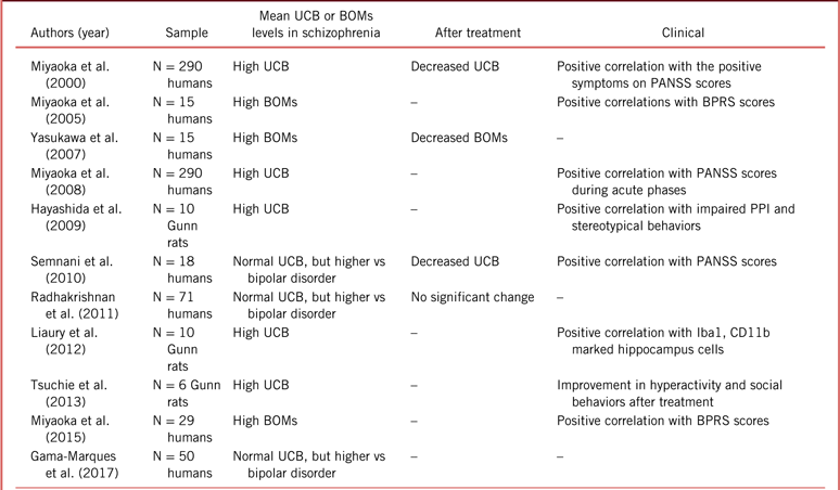
The main findings of our systematic review are chronologically gathered in Table 1.
The main positive findings of the systematic review suggesting a positive correlation between UCB levels and schizophrenia.

BOMs, bilirubin oxidative metabolites (urine); BPRS, Brief Psychiatric Rating Scale; PANSS, Positive and Negative Syndrome Scale; PPI, prepulse inhibition test; UCB, unconjugated bilirubin (plasma).
Discussion
The topic of the correlation between UCB and the incidence of schizophrenia cannot be said to be a completely new one; many studies have been conducted to elucidate this specific aspect of a particularly complex disorder. The interest of this research lies mainly in the possibility of finding an objective, measurable, and potentially influenceable parameter which could help complement the still somewhat subjective approach to the diagnosis, management, and even prevention of schizophrenia. If this possibility turned out to be true, it would imbue medical professionals with a powerful weapon against schizophrenia.
Many studies have found that a statistical relationship exists between UCB and schizophrenia. However, this link remains vague, as it is so far unclear if there is a solid causal connection and, if present, which is the precedent event. It is also uncertain whether there is direct or inverse proportionality between the serum UCB levels and the incidence of schizophrenia. Some studies have reported an increased incidence of schizophrenia with higher levels of UCB,Reference Miyaoka, Seno, Itoga, Iijima, Inagaki and Horiguchi4–Reference Miyaoka, Wake, Hayashida and Horiguchi7, Reference Gama-Marques, Tinoco, Pedro, Leote, Silva and Ouakinin10 and others have reported the opposite: an increase in schizophrenia among individuals with lower UCB levels.Reference Yao, Reddy and van Kammen65, Reference Vítek, Novotná and Lenícek66 Some studies demonstrated a reduction in plasma TB levels with treatment,Reference Miyaoka, Seno, Itoga, Iijima, Inagaki and Horiguchi4, Reference Semnani, Nazemi, Azariyam and Ardakani5 and some reported a correlation between symptomatic scales and plasma UCB concentrations.Reference Miyaoka, Seno, Itoga, Iijima, Inagaki and Horiguchi4, Reference Semnani, Nazemi, Azariyam and Ardakani5, Reference Miyaoka, Wake, Hayashida and Horiguchi7 However, some studies reported the absence of a significant effect from antipsychotic therapy on TB levels.Reference Radhakrishnan, Kanigere, Menon, Calvin, Janish and Srinivasan6, Reference Bach, Kindler and Strik63 This could thus point to a nonlinear, complex link between UCB and the pathophysiology of schizophrenia, a hypothesis which certainly does not appear farfetched if one considers the complicated role of UCB in antioxidant and inflammatory responses, as well as the multifactorial nature of schizophrenia.
As shown by various articles cited in this review, UCB has been linked to in vitro and in vivo neurotoxicity. It appears to have direct and indirect nefarious effects toward most (if not all) cell types in the CNS. The threshold above which UCB begins to lose its beneficial antioxidant effects on CNS cells appears to be rather small,Reference Doré, Takahashi, Ferris, Hester, Guastella and Snyder11 so even small imbalances in the regulation of this molecule could potentially change its positive properties to deleterious ones. In both study “groups,” the studies which report a direct correlation between increased UCB levels and the incidence of schizophrenia and those which report an inverse correlation, the crux of the matter appears to be a dysfunction in the inflammatory mechanisms in the brain. If the concentration of plasma UCB were too high, it would appear to directly cause neuroinflammation, ROS production, and cell apoptosis; if it were too low, however, it could impair the antioxidative defenses of the body and also result in increased inflammation and ROS levels. Therefore, UCB could be both the cause and the effect of the alterations seen in patients with schizophrenia.
Currently, there are 2 main models used to explain the possible causes of schizophrenia. The neurodevelopmental model posits that schizophrenia is the consequence of an anomaly in the developmental stages of the brain which leads to problems in acquiring cognitive skills,Reference Bora and Murray72 a process that starts long before the onset of clinical symptoms.Reference Rapoport, Giedd and Gogtay73 The neurodegenerative model hypothesizes that the dysfunctional development is progressive and persists after the onset of the illness, resulting in structural changes in the brain; it involves a cytopathological process with the loss of neurons and synapses and subsequent gliosis.Reference Rund74 Additionally, some studies support the theory that the psychotic episode is, in itself, neurotoxic and contributes to the structural changes seen after symptom onset.Reference Rund75 It appears plausible that the pathophysiology of schizophrenia could be explained by a combination of these models; it could be seen as an early neurodevelopmental aberration exacerbated by a limited ongoing neurodegenerative process,Reference Lieberman76 recurring neurotoxic psychotic episodes,Reference Buoli, Serati, Caldiroli, Cremaschi and Altamura77 or both.
UCB could potentially play different roles in these hypotheses. As previously stated, neurons previously exposed to elevated concentrations of UCB become more susceptible to further inflammatory damage and death by apoptosisReference Falcão, Silva, Pancadas, Fernandes, Brito and Brites18 that could point to early priming, leading to faults later in life. A subsequent decrease in the cortical cytoarchitecture of mice previously exposed to high levels of UCB was also reported.Reference Falcão, Silva, Pancadas, Fernandes, Brito and Brites18 As neuritic breakdown is believed to underlie the clinical and cognitive symptoms of schizophreniaReference Gilmore, Fredrik Jarskog, Vadlamudi and Lauder78 and a decrease in cortical areas of the brains of patients with schizophrenia has been previously reported,Reference Haukvik, Hartberg and Agartz79 this could signify that the progressive impairment of neuronal development by UCB might explain the proposed link between schizophrenia and hyperbilirubinemia. As shown above, UCB can also directly cause an inflammatory reaction and could be one of the main culprits in the neurodegeneration model. The lower UCB levels, reported by some studies, could indicate a chronic proinflammatory state caused by increased ROS and the depletion of the antioxidant potential, thus leading to the progressive loss of neurons. However, some studies have shown a decrease in UCB levels in patients with previously high plasma UCB with the initiation of antipsychotic treatment.Reference Miyaoka, Seno, Itoga, Iijima, Inagaki and Horiguchi4, Reference Semnani, Nazemi, Azariyam and Ardakani5 The fluctuation of UCB concentrations, be it an increase or a decrease, could explain some of the mechanisms involved in the neurotoxicity of psychotic episodes.
A study by Radhakrishnan et al.Reference Radhakrishnan, Kanigere, Menon, Calvin, Janish and Srinivasan6 has the particularity of making a distinction among different types of patients with schizophrenia, comparing the UCB levels of patients with paranoid schizophrenia with those of patients with non-paranoid schizophrenia; however, it did not find any statistically relevant differences between the 2 groups.
Studies focusing on fluctuations in the biopyrrin concentration are also potentially elucidating. As we have stated, biopyrrins are the product of the oxidation of UCB by ROS. We have also discussed how an elevation in UCB levels can independently cause the production of ROS. This could mean the existence of a buffer system of sorts between UCB and ROS: a surplus of UCB causes inflammation and ROS production, which, in turn, oxidizes UCB, decreasing its plasma concentration (Figure 2).
UCB physiologic (left) and pathologic (right) metabolic pathways.

Biopyrrins were found to be correlated with rating scoresReference Miyaoka, Yasukawa and Yasuda69, Reference Miyaoka, Ieda and Hashioka71 and response to treatment,Reference Yasukawa, Miyaoka, Yasuda, Hayashida, Inagaki and Horiguch70 as well as the duration of the disorder.Reference Miyaoka, Ieda and Hashioka71 The variation in the oxidative metabolism of bilirubin logically indicates the differences in the antioxidant activity of the molecule. However, this could mean the increased recruitment of healthy antioxidant defenses or be the result of compensation by the UCB-ROS buffer system, as previously discussed. To clarify this point, future studies should consider comparing mean plasma UCB levels with mean urinary excretion of biopyrrins in acute and chronic episodes.
Also noteworthy are the comparisons between the bilirubin levels of patients with schizophrenia and those of patients with BD and schizoaffective disorder. Several studies showed higher UCB levels in patients with schizophrenia, as compared with patients with BD,Reference Semnani, Nazemi, Azariyam and Ardakani5, Reference Radhakrishnan, Kanigere, Menon, Calvin, Janish and Srinivasan6, Reference Gama-Marques, Tinoco, Pedro, Leote, Silva and Ouakinin10 and higher UCB levels among patients with BD, as compared with controls.Reference Semnani, Nazemi, Azariyam and Ardakani5 A study by Gama-Marques et al. also showed higher UCB levels in patients with schizoaffective disorder, as compared with patients with BD.Reference Gama-Marques, Tinoco, Pedro, Leote, Silva and Ouakinin10 This could just be the translation of greater subjective stress among patients with schizophrenia. It could, nonetheless, be evidence of a similar pathophysiological background between the diseases. The DSM-5 criteria already consider schizoaffective disorder as part of the schizophrenic spectrum. Additionally, some studies have reported a significant overlap in phenomenological, biological, and genetic characteristics between schizophrenia and BD,Reference Potash80, Reference Potash and Bienvenu81 suggesting that they could also represent different points along the same continuous pathological spectrum.Reference Keshavan, Morris and Sweeney82
Conclusion
As we have seen, evidence supporting the existence of a correlation between UCB and schizophrenia is considerable. The vast majority of studies included in this systematic review have managed to report a statistically relevant link between the 2 variables, thus making it possible for us to affirm that it seems likely that UCB is in some way implicated in the pathology of schizophrenia. However, there existed a discrepancy in this relation, with some studies claiming that schizophrenia was correlated with higher levels of UCB and some claiming that schizophrenia was linked with lower levels of the molecule. We did not see clear reasons why one hypothesis might be more probable than the other, be it biases, statistical errors, bad sample selection, or other reasons. Considering the complexity of schizophrenia, we have conjectured that this association might be multifactorial and nonlinear, with UCB and the pathophysiology of schizophrenia being mutually influenced by each other. In this more holistic hypothesis, schizophrenia is viewed simultaneously as the cause and effect of fluctuations in UCB levels and vice versa, creating a vicious cycle that would perpetuate the symptoms of schizophrenia. However, no studies have yet pointed unequivocally toward this answer. Perhaps, as research on the mechanisms and causes underlying schizophrenia advances, we will better understand the role of UCB in this disease.
Despite the present lack of clarity, we believe there is great potential in the research of UCB as a biological marker for the management of schizophrenia. UCB is likely implicated in many different stages of the progression of the illness. It could be responsible for making one’s nervous system more susceptible to neurodevelopmental faults, consequently qualifying its fluctuations as a promising risk factor. It seems to be linked with the acute phases of the disease, thus making it eligible as a possible tool for early diagnosis and subsequent follow-up. Finally, it appears to be intimately related to the subjective stress and intensity of symptoms, therefore, being a propitious guide for treatment selection or even a possible target for neuroprotective, antioxidant, and anti-inflammatory adjunctive therapies for bilirubin-induced neurological dysfunctionReference Kapitulnik and Maines83 in schizophrenia and spectrum-related disorders. We truly believe there will be, in a near future, interesting opportunities for research on this fascinating topic.Reference Gama Marques and Arantes-Gonçalves84
Disclosures
Erik Pommerening Dornelles, João Gama Marques, and Sílvia Ouakinin have nothing to disclose.