Introduction
The larval stage of the dog tapeworm Echinococcus granulosus causes the life-threatening zoonotic disease called cystic echinococcosis/hydatidosis. This disease is still a major public health problem and economically important in many countries, especially in the Middle East, central Asia, and northern and eastern Africa (Eckert & Deplazes, Reference Eckert and Deplazes2004; Dakkak, Reference Dakkak2010). The liver, lungs and other organ systems of livestock and humans, as its intermediate hosts, are affected by the parasitic cysts (Thompson, Reference Thompson2008). Sheep and camels (with 88% and 70% of fertilized cysts, respectively) are the most important intermediate hosts, with cattle (with 19% of fertilized cysts) being considered as less likely intermediate hosts of E. granulosus (Moks et al., Reference Moks, Jogisalu, Valdmann and Saarma2008; Rokni, Reference Rokni2009). Hydatidosis is endemic in Iran and is responsible for about 1% of admissions to surgical wards (Hadighi et al., Reference Hadighi, Mirhadi and Rokni2003; Samadian et al., Reference Samadian, Alavi, Sharifi, Rezaee and Faramarzi2010; Bagheri et al., Reference Bagheri, Haghi, Amini, Fattahi and Noorshafiee2011).
There are different strains of E. granulosus in the world that can affect the epidemiology, pathology, control and prevention of cystic hydatid disease (Lymbery & Thompson, 1988). To date, 10 genotypes (G1–G10) of E. granulosus have been recognized using molecular tools (Nakao et al., Reference Nakao, McManus, Schantz, Craig and Ito2007; Thompson, Reference Thompson2008). Based on mitochondrial data, the taxonomic position of E. granulosus has been revised: E. granulosus sensu stricto (G1–G3); E. equinus (G4); E. ortleppi (G5); E. canadensis (G6–G10); and E. felidis (‘lion strain’) (Nakao et al., Reference Nakao, McManus, Schantz, Craig and Ito2007; Hüttner et al., Reference Hüttner, Wassermann, Siefert, Boomker, Dinkel, Sako, Mackenstedt, Romig and Ito2008; Moks et al., Reference Moks, Jogisalu, Valdmann and Saarma2008). The large number of E. granulosus-infected livestock animals can act as reservoirs for human infection. Studies in several parts of the world indicate that all genotypes, except E. equinus (G4), have been isolated from human populations and this confirms the importance of characterizing strains of E. granulosus in distinct areas so that relevant control programmes can be performed (Nakao et al., Reference Nakao, McManus, Schantz, Craig and Ito2007; Thompson, Reference Thompson2008; Himsworth et al., Reference Himsworth, Jenkins, Hill, Nsungu, Ndao, Andrew Thompson, Covacin, Ash, Wagner, McConnell, Leighton and Skinner2010).
The south-west of Iran (Khuzestan Province), with an area of about 640,000 km2, an estimated population of 4,275,000 and neighbouring Iraq to the west, is one of the most important endemic areas of hydatidosis in Iran (Rafiei et al., Reference Rafiei, Hemadi, Maraghi, Kaikhaei and Craig2007). Strain characterization of E. granulosus in different parts of Iran indicates that hydatid cysts are commonly found in sheep, camel, cattle and goat populations (Ahmadi & Dalimi, Reference Ahmadi and Dalimi2006; Rostami Nejad et al., Reference Rostami Nejad, Nazemalhosseini Mojarad, Nochi, Fasihi Harandi, Cheraghipour, Mowlavi and Zali2008; Kia et al., Reference Kia, Rahimi, Sharbatkhori, Talebi, Fasihi Harandi and Mirhendi2010; Sharbatkhori et al., Reference Sharbatkhori, Mirhendi, Harandi, Rezaeian, Mohebali, Eshraghian, Rahimi and Kia2010, Reference Sharbatkhori, Fasihi Harandi, Mirhendi, Hajialilo and Kia2011; Pour et al., Reference Pour, Hosseini and Shayan2011; Shahnazi et al., Reference Shahnazi, Hejazi, Salehi and Andalib2011; Sharifiyazdi et al., Reference Sharifiyazdi, Oryan, Ahmadnia and Valinezhad2011). However, there are no genetic data for E. granulosus strains in the south-west of Iran (Khuzestan Province). The aim of the present study was to determine the molecular characterization of E. granulosus isolated from livestock and human subjects in this part of Iran.
Materials and methods
Collection and examination of cysts
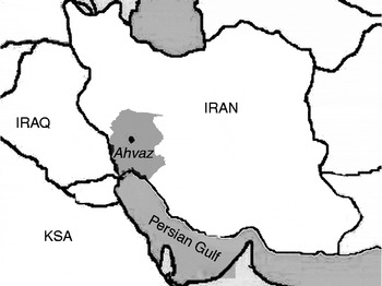
In this study, isolates of 329 hydatid cysts (211 hepatic and 118 pulmonary samples) were collected from the central slaughterhouse of Khuzestan (located in Ahvaz city), including 141 sheep, 104 cattle and 84 goat cases (fig. 1). To determine the fertility status of cysts, hydatid fluid was examined under an optical microscope. In addition, human samples were collected from five patients who had had operations in hospitals of the Ahvaz Jundishapur University of Medical Sciences, Iran. All samples were processed separately and fixed in 80% (v/v) ethanol for final evaluation.
Map of Iran showing study locations in the Khuzestan province. KSA, Saudi Arabia.

DNA extraction and nested polymerase chain reaction
Cyst layers and protoscoleces from each isolate were washed three times in phosphate-buffered saline (pH 7.2). Total genomic DNA extraction from individual isolates was conducted using a standard extraction procedure by QIA DNeasy blood and tissue kit (QIAGEN, Hilden, Germany) according to the manufacturer's instructions. DNA concentration was measured using spectrophotometric determination at A260 according to Karcher (Reference Karcher1995). An internal transcribed spacer 1 (ITS1) fragment was amplified from each sample by nested polymerase chain reaction (PCR). The first-step PCR amplification was performed using primer pair EGF 1 (5′-CCA AAC TTG ATC ATT TAG AGG AAG-3′) and EGR 2 (5′-TAT GGG CCA AAT TCA CTC ATT ACC-3′) as outer primers. The thermocycler (MyCycler; Bio-Rad, Hercules, California, USA) was set to give 6 min at 95°C, followed by 30 cycles, each of 45 s at 94°C, 1 min and 30 s at 55°C and 2 min at 72°C, and then a final extension for 5 min at 72°C. For the second PCR, the internal primers EgF (5′-GTC GTA ACA AGG TTT CCG TAGG-3′) and EgR (5′-TAG ATG CGT TCG AAG TGT CG-3′) were used; these had been described previously for amplification of a 1000 bp fragment (Rahimi et al., Reference Rahimi, Kia, Mirhendi, Talebi, Fasihi Harandi, Jalali-zand and Rokni2007). The PCR product of the first step was diluted 1/10 with ultrapure water and then 1 ml of this dilution was used for the second round of PCR. The thermal profile in this step was as follows: one cycle of 95°C for 2 min, 94°C for 30 s, 55°C for 45 s and 72°C for 1 min, all steps repeating for 30 cycles. For the final extension 72°C for 5 min was used (Rahimi et al., Reference Rahimi, Kia, Mirhendi, Talebi, Fasihi Harandi, Jalali-zand and Rokni2007). The concentration of the materials for both steps of PCR were similar, containing 5 μl template DNA, 0.1 mm deoxynucleoside triphosphates (dNTP) (Roche, Penzberg, Germany), 2 mm MgCl2, 0.5 units Taq polymerase, 50 mm Tris–HCl (pH 7.6) and 25 pm each of forward and reverse primers. Electrophoresis was performed by adding 7 μl sample of the second PCR to 1.5% (w/w) agarose gel, stained with ethidium bromide for 45 min at 100 V. Bands were observed by ultraviolet transillumination.
Restriction fragment length polymorphism polymerase chain reaction and nucleotide sequences
All PCR products were digested for 6–8 h with the restriction endonucleases AluI, MspI and RsaI using buffers recommended by the manufacturer (Fermentas, Vilnius, Lithuania) in a final volume of 20 μl including 5 μl of PCR product, 5 units of the restriction enzymes, 2 μl of the supplied buffer and 8 μl of molecular-grade water. Restriction fragments of amplicons were electrophoresed using a 2% (w/v) agarose gel at 100 V for 180 min. DNA derived from individual hydatid cysts was subjected to sequencing by MWG (Germany; http://www.eurofinsdna.com/home.html) by the primers employed in the nested PCR. All data were analysed using Chromas software (http://www.technelysium.com.au/Chromas.html) and compared with the NCBI nucleotide gene bank (National Center for Biotechnology Information; www.ncbi.nlm.nih.gov/BLAST/).
Results
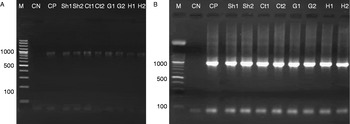
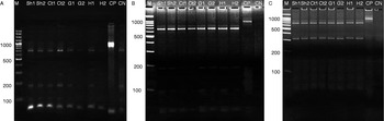
In this study, all isolates of hydatid cysts belonging to human subjects, sheep, goats and cattle were collected from the south-west of Iran and processed for nested PCR. The first-step PCR, using outer primers, showed one heavy fragment (about 1800 bp), which was not sharp enough. The second step of nested PCR was performed and the amplified genomic DNA showed sharp and visible bands. However, no amplification was observed in the negative controls (fig. 2). Restriction fragment length polymorphism (RFLP)-PCR was carried out on the amplified ITS1 region of all isolates by using AluI, MspI and RsaI restriction enzymes. The RFLP patterns used in this study could detect E. granulosus sensu stricto, E. equinus, E. ortleppi and G6/G7 genotypes. One distinguishable pattern was observed with AluI, MspI and RsaI restriction enzymes. In this study, all of the 334 isolates examined were identified as corresponding to the common sheep strain (G1) of E. granulosus (fig. 3) and no other genotypes were detected.
Internal transcribed spacer 1 (ITS1) nested polymerase chain reaction (PCR) results of extracted DNA of Echinococcus granulosus isolates. (A) First-step PCR using outer primer; (B) nested PCR using inner primers. Lanes: M, DNA marker (100 bp); CN, negative control; CP, positive control; and isolates from sheep (Sh), cattle (Ct), goat (G) and human (H).

Restriction fragment length polymorphism (RFLP) polymerase chain reaction (PCR) digests with: (A) AluI, (B) MspI and (C) RsaI enzymes. Lanes: M, DNA size marker (100 bp); isolates from sheep (Sh), cattle (Ct), goat (G) and human (H); CP, internal transcribed spacer 1 (ITS1)-PCR products without restriction endonuclease; CN, Master mix containing restriction endonuclease without PCR products.

The ITS1 gene fragments of three sheep, three cattle, three goat and three human isolates (randomly selected) were sequenced. GenBank was searched for similar sequences published for the G1 genotype (Bowles et al., Reference Bowles, Blair and McManus1995; Van Herwerden et al., Reference Van Herwerden, Gasser and Blair2000; Zhang et al., Reference Zhang, Hu, Jones, Allsopp, Beveridge, Schindler and Gasser2007) with the BLAST program and a significant homology was detected with E. granulosus sequences. The sequences of all 12 examined isolates were highly homologous with a small number of differences, corresponding to punctual base substitutions (data not shown).
Discussion
Many studies indicate that hydatid cysts are frequently found in sheep, camel, cattle and goat populations in Iran (Ahmadi, Reference Ahmadi2005; Rokni, Reference Rokni2009; Pour et al., Reference Pour, Hosseini and Shayan2011; Shahnazi et al., Reference Shahnazi, Hejazi, Salehi and Andalib2011; Sharbatkhori et al., Reference Sharbatkhori, Fasihi Harandi, Mirhendi, Hajialilo and Kia2011). The rate of infection with E. granulosus in sheep, goats, cattle, camels and humans shows a prevalence of 5.1–74.4%, 1.7–20%, 3.5–38.3%, 11.4–70% and 1.2–21.4%, respectively, in different parts of Iran (Ahmadi, Reference Ahmadi2005; Rokni, Reference Rokni2009).
The characterization of E. granolosus strains is important for implementation of control programmes and chemotherapy, and understanding life-cycle patterns. Morphological studies can be unreliable and are not efficient for the determination of genetic diversity (Lymbery & Thompson, Reference Lymbery and Thompson1988; McManus & Bowles, Reference McManus and Bowles1996). Molecular-based methods provide more accurate and reliable information about the E. granulosus isolate taxonomy at the genus, species and subspecies levels, but the application of molecular methods often needs the use of suitable primers and access to sufficient amounts of DNA (McManus & Bowles, Reference McManus and Bowles1996).
In the current research, we performed RFLP-PCR for the identification of E. granolosus strains based on a certain ITS1 primer pair that was able to thoroughly amplify DNA (Rahimi et al., Reference Rahimi, Kia, Mirhendi, Talebi, Fasihi Harandi, Jalali-zand and Rokni2007). The ribosomal DNA (rDNA) repeat unit has different regions and non-specific primers, like those derived from Schistosoma mansoni which have been used widely to study variation and phylogeny in E. granulosus (Bowles et al., Reference Bowles, Blair and McManus1995; Zhang et al., Reference Zhang, Eslami, Hosseini and McManus1998; Kedra et al., Reference Kedra, Swiderski, Tkach, Dubinsky, Pawlowski, Stefaniak and Pawlowski1999; Utuk et al., Reference Utuk, Simsek, Koroglu and McManus2008). The ITS1-RFLP patterns in this study showed that the dominant genotype prevailing in livestock and humans is the G1 genotype. We also demonstrated that the sheep strain (G1) was well adapted to the intermediate hosts in the region; this agrees with other findings concerning hydatidosis infection in central Iran (Shahnazi et al., Reference Shahnazi, Hejazi, Salehi and Andalib2011).
In previous studies, sheep (G1) and camel (G6) strains were identified in central and western areas of Iran (Harandi et al., Reference Harandi, Hobbs, Adams, Mobedi, Morgan-Ryan and Thompson2002; Ahmadi & Dalimi, Reference Ahmadi and Dalimi2006, Parsa et al., Reference Parsa, Haghpanah, Pestechian and Salehi2011; Pour et al., Reference Pour, Hosseini and Shayan2011; Sharbatkhori et al., Reference Sharbatkhori, Fasihi Harandi, Mirhendi, Hajialilo and Kia2011). Dalimi et al. (Reference Dalimi, Motamedi, Hosseini, Mohammadian, Malaki, Ghamari and Ghaffarifar2002) showed that in the south-west and western parts of Iran the contamination rate of cattle compared with sheep is higher; however, the small number of samples decreased the reliability of the study. In the present study, all cattle isolates also showed G1 strain patterns. Although cattle can be contaminated with other strains of E. granulosus, several studies in different countries have demonstrated that the G1 strain is dominant in these animals (Romig et al., Reference Romig, Dinkel and Mackenstedt2006; Thompson, Reference Thompson2008; Parsa et al., Reference Parsa, Haghpanah, Pestechian and Salehi2011). Our results are in accordance with previous studies in other parts of Iran (Sharbatkhori et al., Reference Sharbatkhori, Mirhendi, Jex, Pangasa, Campbell, Kia, Eshraghian, Harandi and Gasser2009, Reference Sharbatkhori, Fasihi Harandi, Mirhendi, Hajialilo and Kia2011; Parsa et al., Reference Parsa, Haghpanah, Pestechian and Salehi2011; Shahnazi et al., Reference Shahnazi, Hejazi, Salehi and Andalib2011), suggesting that sheep strain transmission occurs actively in the south-west of Iran.
According to the results of RFLP patterns, the camel strain (G6) was not detected in the region. Due to the rare occurrence of camels being slaughtered and the low consumption of camel meat, we could not obtain these samples. The absence of the G6 strain in all isolates concurs with the study performed by Parsa et al. (Reference Parsa, Haghpanah, Pestechian and Salehi2011) which only found the sheep strain in the west of Iran (Lorestan region), near the south-west. Migration of livestock takes place from this region to the cooler parts of Iran and, consequently, contact is made with livestock in other provinces of the country. In the current study, the G1 genotype was also found among all five human cases as well as all other livestock animals.
In a similar study, performed by Utuk et al. (Reference Utuk, Simsek, Koroglu and McManus2008) using CfoI, MspI, RsaI and AluI restriction enzymes, it was found that the predominant genotype involved in E. granulosus transmission in Turkey is the common sheep strain (G1 genotype), infecting human, cattle, sheep, goat, and camel populations, as well as the dog definitive host.
As was performed previously (Bowles et al., Reference Bowles, Blair and McManus1995; Van Herwerden et al., Reference Van Herwerden, Gasser and Blair2000), the ITS1 amino acid sequence data showed that different isolates of Echinococcus could possess similar sequence variants. The ITS1 sequence data obtained in this study confirmed RFLP patterns and were identified as corresponding to the G1 strain of E. granulosus. The sheep strain, confirmed as G1 at the DNA sequence level, showed remarkable uniformity with isolates from sheep and a number of other hosts from geographically diverse regions.
In conclusion, this first comprehensive genotypic analysis of E. granulosus shows that the common sheep strain (G1) is predominant in the south-west of Iran, infecting human, sheep, cattle and goat populations. The infected livestock can act as potential reservoirs of human infection and the present results will have an important impact on control programmes against echinococcosis, understanding life-cycle patterns and improving public health.
Acknowledgements
This study was conducted and financially supported by grants from Ahvaz Jundishapur Medical Sciences. Grateful thanks are extended to Mr Nezam Salehi, Dr Kambiz Masoumi and Dr Iran Rashidi for their collaboration in sample collection. The authors declare no conflict of interests.