1. Introduction
Somewhere in the region of 3 billion years ago an enzyme emerged which would dramatically change the chemical composition of our planet and set in motion an unprecedented explosion in biological activity. This enzyme used solar energy to power the thermodynamically and chemically demanding reaction of water splitting. In so doing this enzyme provided biology with an unlimited supply of reducing equivalents (high-energy electrons) needed to convert carbon dioxide (CO2) into the organic molecules of life. Prior to this, biological organisms had been dependent on hydrogen/electron donors such as H2S, NH3, organic acids and Fe2+, which were in limited supply compared with the ‘oceans’ of water with which planet Earth is blessed. The by-product of the water splitting reaction is molecular oxygen. The release of this gas also had dramatic consequences for the development of life, since it created an oxygenic atmosphere and at the same time allowed the ozone layer to form. With oxygen available, the efficiency of cellular metabolism increased dramatically, since for a given amount of substrate, aerobic respiration provides in the region of 20 times more energy than anaerobic respiration. It was probably this improved efficiency of cellular bioenergetics, which paved the way for the subsequent evolution of eukaryotic cells and multicellular organisms. The establishment of the ozone layer provided a shield against harmful UV radiation allowing organisms to exploit the terrestrial as well as the aquatic environments. Consequently life has been able to prosper and diversify on an enormous scale as testified by the fossil records and by the extent and variety of living organisms on our planet today. The enzyme that gave rise to this ‘big bang of evolution’ is known as Photosystem II (PSII) and is truly the ‘engine of life’ (Barber, Reference Barber2003).
2. Photosystem II
That plants could produce oxygen stems back to experiments first conducted by Priestley (Reference Priestley1772) and elaborated on by van Ingen-Housz (Reference van Ingen-Housz1779) who showed that the phenomenon was driven by sunlight falling on leaves and other green plant tissue. Senebier (Reference Senebier1788), de Saussure (Reference De Saussure1804) and later, Bossingault (Reference Bossingault1854) concluded from their experiments that the production of oxygen by plants was matched by the uptake of CO2 and water. The question then arose as to the origin of the oxygen; was it derived from CO2 or H2O? Because it is possible to biochemically separate the H2O and CO2 consuming reactions of photosynthesis, it was clearly shown by Hill (Reference Hill1937) that the O2 was derived from the light-driven splitting of water, a conclusion also reached by Ruben et al. (Reference Ruben, Randle, Kaman and Hyde1941) using isotopic oxygen analyses.
The naming of the oxygen-evolving enzyme as PSII is credited to L.M.N Duysens (see Duysens et al. Reference Duysens, Amesz and Kamp1961) soon after the realisation that there were two photosystems (PSII and Photosystem I (PSI)) working together in series to bridge the redox gap between water oxidation and CO2 reduction. The arrangement is usually called the Z-scheme and was conceived by Hill & Bendall in 1960. It explained the so called ‘red drop’ where the quantum yield for O2 evolution was low at long wavelengths (<700 nm) although chlorophyll was still absorbing light. This light was preferentially absorbed by PS1 but when shorter wavelength light was superimposed, being preferentially absorbed by PSII, the rate of O2 produced was greater than the sum of the two rates for the two light conditions separately. This is known as the ‘enhancement effect’. Both phenomena were discovered by Emerson & Arnold (Reference Emerson and Arnold1932) and Emerson & Lewis (Reference Emerson and Lewis1943) respectively. The Z-scheme also explained the existence of redox active cytochromes with redox potentials intermediate to that of PSII and PSI (Hill & Bendall, Reference Hill and Bendall1960) and that PSII light brought about their reduction, while superimposition of PSI light caused their oxidation (Duysens et al. Reference Duysens, Amesz and Kamp1961). Another important mile stone was the finding by Boardman & Anderson (Reference Boardman and Anderson1964) that PSI and PSII could be physically separated which was the starting point for detailed biochemical, molecular biological and functional studies of both photosystems stretching over the following 30 years. The realisation that PSII was a discrete biochemical entity established that manganese, long known to be required for photosynthetic activity (Pirson et al. Reference Pirson, Tichy and Wilhelmi1951), was intimately involved in the water splitting/oxygen evolving reaction. Similarly it became established that calcium and chloride ions were also cofactors for oxygen evolution (see Debus, Reference Debus1992; Yocum, Reference Yocum2008). The role of Mn as a multivalent cation in water oxidation started to emerge from the ground breaking work of Joliot & Kok, where it became clear that oxidised intermediates had to accumulate prior to dioxygen formation (Joliot et al. Reference Joliot, Barbieri and Chabaud1969; Kok et al. Reference Kok, Forbush and McGLOIN1970).
They demonstrated that it took four flashes of light to obtain the maximum yield of molecular oxygen which was interpreted as a catalytic cycle consisting of five states (S0–S4), whereby oxidising equivalents of about the same potential were sequentially accumulated at the catalytic site with each flash (Fig. 1b ). Thus the S4-state stores the four oxidising equivalents needed to oxidise two water molecules and generate dioxygen and in so doing reverts back to the S0-state. The obvious candidates for storing these oxidising equivalents were the Mn ions shown to be bound within PSII of which there seemed to be four. The role of Mn in the catalytic cycle was consistent with the findings of Dismukes & Siderer (Reference Dismukes and Siderer1980) who discovered a multiline electron spin resonance (EPR) signal in the S2-state typical of a Mn species. The discovery of the primary electron donor to the PSII reactor centre and therefore the primary oxidant driving the S-state cycle was made by Horst Witt. He showed that this was a special chlorophyll species he called P680 (Doring et al. Reference Doring, Stiehl and Witt1967).
(a) An energy-level diagram for light-induced electron transfer in PSII from water oxidation to plastoquinone (PQB) reduction. (b) The S-state cycle showing how the absorption of four photons of light (hν) drives the splitting of two water molecules and formation of O2 through a consecutive series of five intermediates (S0, S1, S2, S3 and S4). Protons (H+) are released into the lumen during this cycle except for the S1 to S2 transition and electrons are donated to P680•+ via the redox active TyrZ (Yz in Figs. 1a & 2).

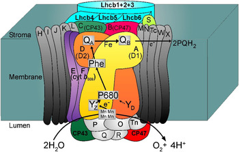
These major contributions, along with other extensive studies, provided by the mid-1990s a detailed understanding of the biophysical, molecular biology and biochemistry of PSII. By this time it was well established that this enzyme is a discrete pigment–protein complex in the thylakoid membranes of all types of plants, algae and cyanobacteria and was composed of many different subunits (Fig. 2). At the heart of the PSII complex there is a reaction centre (RC) where light energy is converted to electrochemical potential energy and where the water splitting reaction occurs in the oxygen-evolving complex (OEC).
Cartoon of the structure and subunit composition of PSII of higher plants and green algae. The intrinsic and extrinsic proteins are labelled according to the gene nomenclature as in Table 1 (e.g., psbA = A) with common nomenclature given for the major subunits (e.g., A = D1 protein). The outer light-harvesting Chla/Chlb-binding proteins are coloured light green (intrinsic Lhcb/Cab proteins). The electron transfer pathway from water oxidation to plastoquinone reduction (see text) is indicated by arrows.

Based on the determination of the 1988 Nobel Prize winning crystal structure of the isolated RC of the anaerobic purple photosynthetic bacterium, Rhodopseudomonas viridis (Diesenhofer et al. Reference Diesenhofer, Epp, Miki, Huber and Michel1985) and comparison of gene sequences, it became clear that there were significant structural and functional homologies between the bacterial RC and that of PSII (Barber, Reference Barber1987; Michel & Deisenhofer, Reference Michel and Deisenhofer1988). This homology did not only relate to similar structural features of the RC, but also suggested cofactor organisation and implied details of the charge separation process on the reducing side of PSII. The difference, of course, was on the oxidation side where there was no oxygen-evolving centre (OEC) in R. viridis and it did not have the many protein subunits found in the hugh PSII–RC core complex (see Fig. 2). Indeed, the PSII–RC core, has in addition to the D1 and D2 RC proteins, two further closely related chlorophyll (Chl)-binding proteins (CP), called CP43 and CP47 which each have six (I–VI) transmembrane helices (Hankamer et al. Reference Hankamer, Morris, Nield, Carne and Barber2001a, Reference Hankamer, Morris, Nield, Gerle and Barberb; Rhee et al. Reference Rhee, Morris, Zheleva, Hankamer, Kuhbrandt and Barber1997, Reference Rhee, Morris, Barber and Keubrandt1998). There are also a number of other intrinsic low-molecular weight subunits, which are rather featureless except for two that bind the high potential haem of cytochrome b559 (Cyt b559). Finally the PSII–RC core complex has several extrinsic proteins attached to its lumenal surface, which form a protein shield over the catalytic site of the OEC. Although one of these proteins, PsbO, is ubiquitous to all oxygenic photosynthetic organisms, the others vary between different types of organisms. Plants and green algae bind the PsbP and PsbQ proteins, while the PsbU and PsbV proteins are only found in red algae and cynanobacteria (De Las Rivas et al. Reference De Las Rivas, Balsera and Barber2004). Other extrinsic regulatory proteins are transiently bound during the life cycle of PSII. This core complex is serviced by an outer light-harvesting system which, in the case of plants and green algae, is composed of intrinsic Lhcb proteins which bind Chl a and Chl b as shown in Fig. 2. In red algae and cyanobacteria, this outer antenna is replaced by phycobiliproteins located in phycobilisomes bound extrinsically to the stromal surface of the PSII core.
The way PSII uses light energy to generate an electrochemical potential in the form of a charge transfer state across the RC is essentially the same as in anaerobic purple photosynthetic bacteria (Barber, Reference Barber1987; Michel & Deisenhofer, Reference Michel and Deisenhofer1988). Chl molecules and other pigments (e.g. β-carotene) absorb visible light and transfer the excitations to the RC where charge separation occurs. The striking difference is that in the case of PSII, the oxidising ‘hole’ left by charge separation is highly oxidative and is sufficient to drive water splitting. All the redox active cofactors involved in the energy conversion process are bound to the D1 and D2 proteins and the following sequence of reactions occurs (see Fig. 1a ). The special form of Chla (P680) acts as an exciton trap and is converted to a strong reducing agent after excitation to P680*. P680* reduces a pheophytin a molecule (Pheo) in a few picoseconds to form the radical pair state P680•+Pheo•−. Within a few hundred picoseconds, Pheo•− reduces a firmly bound plastoquinone molecule QA (PQA) to produce P680•+PheoQA −. P680•+, with its very high redox potential (>1 V), oxidises a tyrosine residue (YZ) to form YZ +P680PheoQA − on a nanosecond timescale. YZ + is deprotonated to form a neutral radical YZ •. In the millisecond time domain QA − reduces a second plastoquinone QB (PQB) to form YZ + •P680PheoQAQB − and at about the same time YZ • extracts an electron from the cluster of four manganese atoms that bind the two substrate water molecules. A second photochemical turnover reduces QB − to QB 2− which is then protonated to plastoquinol (PQH2) and released from the QB-binding site of PSII into the lipid bilayer where it is subsequently oxidised by PSI via the cytochrome b6f complex. Two further photochemical turnovers provide the cluster of four manganese ions and a calcium ion (Mn4Ca2+) with a total of four oxidising equivalents, which are used to oxidise two water molecules to form dioxygen (Eqs. (1) and (2))
 $$2{\rm H}_{\rm 2} {\rm O}\ \mathop{\longrightarrow}\limits^{4{\rm h\nu}} \ \ {\rm O}_2 + \,4{\rm e}\, + \,4{\rm H}^ +. $$
 $$2{\rm H}_{\rm 2} {\rm O}\, + \,{\rm PQ}\ \mathop{\longrightarrow}\limits^{4{\rm h\nu}} \ \ {\rm O}_2 \, + \,2{\rm PQH}_2. $$
As mentioned above, each oxidation state generated within the OEC is represented as an intermediate of the S-state cycle (see Fig. 1b ). In addition to these reactions, side reactions can occur under some conditions, including the oxidation of Cyt b559, a β-carotene molecule and two Chl a molecule (ChlZ and ChlD.) (see Fig. 3 b). These side reactions occur on the tens of millisecond timescale and therefore do not normally compete with the electron transfer pathway leading to water oxidation. Indeed, they probably only occur when the rate of water oxidation becomes limited and thus provide a protective mechanism against the detrimental reactions resulting from the very high redox potential of the long lived P680 radical cation (P680•+) (see Stewart & Brudvig, Reference Stewart and Brudvig1998).
As with the studies of other enzymes, the reactions of PSII could not be fully understood in molecular terms without the detailed knowledge of its structure. There are many techniques, which give structural information at different degrees of complexity but none match the overall level of detail gained from X-ray diffraction analyses of a wide range molecules and complexes. PSII is a membrane protein complex making it more difficult to crystallise. Nevertheless progress during the past dozen years has been substantial and is the focus of this paper. The structural information obtained has improved our understanding of the many reactions of PSII and most importantly, of the water splitting reaction itself, knowledge which not only has far reaching implications for understanding the molecular basis for the energy cycle of our planet, but also for the development of new non-polluting technologies for production of fuel.
3. Crystal Structure Of PSII
3.1 Proteins
The first X-ray derived structure of PSII used a preparation isolated from the cyanobacterium Thermosynechococcus elongatus and was only partially solved by Zouni et al. (Reference Zouni, Witt, Kern, Fromme, Krauss, Saenger and Orth2001). This structural study was initiated by Horst Witt after solving the crystal structure of PSI (Jordon et al. Reference Jordan, Fromme, Witt, Klukas, Saenger and Krauß2001) and was limited to a resolution of 3.8 Å and the C-alpha backbones of many of the subunits were not fully traced. Although no side chain positioning was deduced, their model confirmed the dimeric organisation of the isolated complex (see Hankamer et al. Reference Hankamer, Barber and Boekema1997) and the relative positioning of the major subunits and their transmembrane helices within each monomer derived earlier from electron crystallography (Hankamer et al. Reference Hankamer, Morris, Nield, Carne and Barber2001a, Reference Hankamer, Morris, Nield, Gerle and Barberb; Rhee et al. Reference Rhee, Morris, Zheleva, Hankamer, Kuhbrandt and Barber1997, Reference Rhee, Morris, Barber and Keubrandt1998). It also provided information on the positioning of cofactors involved in excitation transfer and charge separation. Most importantly the analysis of the diffraction data gave the first direct structural hints of the Mn-cluster of the OEC. Two years later Kamiya & Shen (Reference Kamiya and Shen2003) reported a 3.7 Å crystal structure of PSII isolated from Thermosynechococcus vulcanus, a cyanobacterium closely related to T. elongatus and provided additional information to that revealed by Zouni et al. (Reference Zouni, Witt, Kern, Fromme, Krauss, Saenger and Orth2001). The tracing of the C-alpha backbones was more complete and there was some effort made to assign a few amino acids, particularly those of the D1- and D2-proteins, as well as to some regions of the chlorophyll-binding proteins CP43 and CP47. However, it was not until the work of Ferreira et al. (Reference Ferreira, Iverson, Maghlaoui, Barber and Iwata2004) that all the protein subunits of PSII were assigned and their structures revealed. The PSII complex crystallised was isolated from T. elongatus and its X-ray diffraction derived structure was refined to a resolution of 3.5 Å. Over 5000 amino acids (close to 100% of this huge complex) were mapped within the dimeric complex thus revealing for the first time the protein environments of all the major cofactors as well as assigning the location of 18 of the 19 subunits, which made up each monomer. As a consequence, a host of outstanding questions were answered; not least was direct evidence that the phenolic group of YZ (D1Y161) is H-bonded to nearby D1His190, a necessary requirement for this redox active tyrosine to function in electron transfer between P680 and the Mn4Ca2+-cluster. Also the finding that glutamate 354 of CP43 (CP43Glu354) was a ligand to the Mn4Ca2+-cluster highlighted another striking discovery among many others.
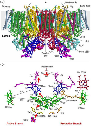
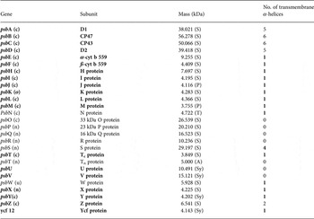
The side view of the PSII RC dimeric core complex of T. elongatus shown in Fig. 3a is taken from the crystal structure of Ferreira et al. (Reference Ferreira, Iverson, Maghlaoui, Barber and Iwata2004). The PSII dimer has dimensions of 105 Å depth (45 Å in spanning the membrane), 205 Å length and 110 Å width and has a calculated total molecular mass of about 700 KDa. There were in total 35 transmembrane helices per monomer depicted as cylinders in Fig. 3a . Details of the 19 different subunits are listed in Table 1 with one, Ycf12 (originally very tentatively suggested to be possibly PsbN but unassigned in the data base), being included, based on more recent biochemical analysis of PSII (Kashino et al. Reference Kashino, Takahashi, Inoue-Kashino, Ban, Yohei- Ikeda, Satoh and Sugiura2007) and confirmed later by the 1.9 Å crystal structure (Umena et al. Reference Umena, Kawakami, Shen and Kamiya2011). Note that the other, non-highlighted proteins in Table 1 are exclusive to the higher plant PSII–RC core complex.
(a) Side view of the structure of Photosystem II, the water splitting enzyme of photosynthesis. This structure was determined by X-ray crystallography (Ferreira et al. Reference Ferreira, Iverson, Maghlaoui, Barber and Iwata2004). The complex is embedded in the thylakoid membrane spanning between its lumenal and stromal surfaces. It is composed of two monomers related to each other by a two-fold axis. Each monomer contains 19 different protein subunits with 16 being located within the membrane matrix and having α-helices (depicted by cylinders). In total, there are 35 transmembrane helices per monomer. The D1- and D2-proteins that compose the RC are shown in yellow and orange respectively. The Mn4Ca-catalytic centre of the OEC is located on the lumenal side of the complex and is stabilised and shielded by three extrinsic proteins, PsbO, PsbU and PsbV. (b) Organisation of the electron transfer cofactors that make up the RC of Photosystem II as revealed by X-ray crystallography (Ferreira et al. Reference Ferreira, Iverson, Maghlaoui, Barber and Iwata2004). Excitation of the RC via the chlorophylls (Chl) shown in green leads to the reduction of pheophytin (Pheo) resulting in the formation of the radical pair P680•+Pheo•−. The radical cation of P680 is localised on PD1, while the radical anion is located on PheoD1. The electron on PheoD1 •− is rapidly donated to a firmly bound plastoquinone QA (shown in purple) and then transferred to a second plastoquinone QB (also shown in purple). This electron transfer is aided by the presence of a non-haem iron located mid-way between them. When the QB plastoquinone has been doubly reduced and protonated the resultant plastoquinol (PQH2) diffuses from the QB-binding site into the lipid matrix of the membrane, P680•+ is reduced by a redox active tyrosine (TyrZ) which then extracts electrons from the Mn4Ca-cluster that constitutes the OEC. These electron transfer processes occur mainly on the D1-side of the RC as shown by the red arrow. Some of the symmetrically related cofactors located on the D2-side (PheoD2) are non-functional. Other cofactors on the D2-side (haem of cytochrome b559 shown in red,the β-carotene molecule shown in brown and ChlZD2), however, are redox active and seem to play a role in protecting PSII against photoinduced damage.

PSII proteins are products of the psbA to psbZ plus ycf 12 genes that occur in oxygenic organisms.

In eukaryotic organisms the psb genes are located in either the chloroplast (c) or nuclear (n) genes. The molecular masses of the mature PsbA–PsbZ/Ycf12 proteins are calculated from the protein sequences reported in the SwissProt database using the MacBioSpec program (Sciex Corp., Thornhill, ON, Canada) for spinach (S), pea (P), tobacco (T), Arabidopsis (A) and Synechocystis PCC6803 (Sy). The proteins assigned in X-ray structure of cyanobacterial PSII are shown in bold.
The three extrinsic proteins, PsbO, Psb U and PsbV, form a cap over the catalytic site where oxygen evolution occurs preventing access by reductants other than water. Some of the low molecular weight subunits are located on the peripheral side of the CP43/D1/D2/CP47 cluster where they probably help to stabilise the binding of Chl and β-carotene molecules contained within the complex. The exception to this are the PsbE and PsbF proteins, which provide histidine ligands for the haem of Cyt b559, and the PsbL, Psb M and Psb T proteins located at the monomer–monomer interface where they possibly play a role in stabilising the dimeric nature of the PSII complex. All the small subunits have a single transmembrane helix except for the PsbZ, which has two (see Table 1). Subsequent X-ray models of cyanobacterial PSII by Loll et al. (Reference Loll, Kern, Saenger, Zouni and Biesiadka2005), Guskov et al. (Reference Guskov, Kern, Gabdulkhakov, Broser, Zouni and Saenger2009) and Umena et al. (Reference Umena, Kawakami, Shen and Kamiya2011) agreed with the assignments of the small subunits made by Ferreira et al. with the 1.9 Å confirming the location of Ycf12 adjacent to PsbK and PsbZ. The 2.9 Å crystal structure of Guskov et al. (Reference Guskov, Kern, Gabdulkhakov, Broser, Zouni and Saenger2009) contained an additional transmembrane helix assigned to PsbY but this peripheral subunit was absent in the structures of Ferreira et al. (Reference Ferreira, Iverson, Maghlaoui, Barber and Iwata2004) and Umena et al. (Reference Umena, Kawakami, Shen and Kamiya2011).
All the crystal structures of PSII confirmed that the transmembrane helices of the D1 and D2 proteins are arranged in an almost identical way to those of the L and M subunits of the purple bacterial RC as anticipated by homology (Barber, Reference Barber1987; Michel & Deisenhofer, Reference Michel and Deisenhofer1988). The six transmembrane helices of CP43 and CP47 are arranged in three pairs around a pseudo-threefold axis as first shown by electron crystallography (Hankamer et al. Reference Hankamer, Morris and Barber1999; Rhee et al. Reference Rhee, Morris, Zheleva, Hankamer, Kuhbrandt and Barber1997, Reference Rhee, Morris, Barber and Keubrandt1998).
These two Chl-binding proteins, CP43 and CP47, are located on each side of the D1 and D2 subunits in such a way that their transmembrane helices are related by the same pseudo-twofold axis that relates the five transmembrane helices of the D1/D2 heterodimer, an organisation very similar to that of the RC core of PSI (Murray et al. Reference Murray, Duncan and Barber2006; Schubert et al. Reference Schubert, Klukas, Saenger, Witt, Fromme and Krauss1998). They are both characterised by having a very large extrinsic loop joining the lumenal ends of helices V and VI and indeed, the OEC ligand CP43Glu354 is located in a 310 - helix of the large loop of CP43.
3.2 Cofactors
3.2.1 Chlorophylls and carotenoids
The Ferreira et al. structure (Reference Ferreira, Iverson, Maghlaoui, Barber and Iwata2004) identified per monomer, 36 Chls and tentatively assigned 7 carotenoids, assumed to be all trans β-carotene (β-Car). It was concluded that CP43 and CP47 bound 14 and 16 Chl a molecules respectively and in the majority of cases these light-harvesting Chls were ligated by either α- or β-linkages to conserved histidine side chains and related by the same pseudo-twofold symmetry axis that relate their transmembrane helices. The Chls were arranged in layers towards the lumenal and stromal surfaces with one Chl in each case located mid-way between the layers. Remarkably the majority of the Chls bound to CP43 and CP47 have counterparts bound in the N-terminal domains of the PsaA and PsaB RC proteins of PSI (Murray et al. Reference Murray, Duncan and Barber2006). The more recent crystal structures of cyanobacterial PSII at 2.9 and 1.9 Å resolution have extended and corrected Ferreira et al.'s assignment of 7 β-Car molecules to 11 all trans β-carotenes and concluded that CP43 binds 13 Chls rather than 14. The higher resolution crystal structures also confirmed that the D1/D2 heterodimer bound six symmetrically related Chls as detailed earlier (Ferreira et al. Reference Ferreira, Iverson, Maghlaoui, Barber and Iwata2004; Kamiya & Shen, Reference Kamiya and Shen2003; Zouni et al. Reference Zouni, Witt, Kern, Fromme, Krauss, Saenger and Orth2001) and extended the assignment of β-Car by Ferreira et al. from one located within the D2 protein to a second non-symmetry related β-Car on the D1 side of then D1/D2 heterodimer.
An important contribution of the 1.9 Å PSII crystal structure (Umena et al. Reference Umena, Kawakami, Shen and Kamiya2011) is that it was at sufficient resolution to identify the location of 2795 water molecules in the dimeric complex. Most of these water molecules form two layers at the stromal and lumenal surfaces. However, a few molecules are located in the membrane spanning region of the complex with seven of them acting as ligands to the Mg ions of chlorophylls, which are not ligated by amino acids (histidines in all cases except one where CP43Asn39 is a ligand). Water ligands were found for the two accessory Chls (ChlD1 and ChlD2) of the D1/D2 heterodimer as originally suggested by Ferreira et al. (Reference Ferreira, Iverson, Maghlaoui, Barber and Iwata2004) while the others are for three Chls of CP47 and two Chls of CP43. Umena et al. (Reference Umena, Kawakami, Shen and Kamiya2011) noted that most of the vinyl groups of the CP43 and CP47 Chls are positioned in, or near, the same plane of the tetrapyrrole ring which would be expected to facilitate energy migration between adjacent chlorophylls. While the role of the Chls and β-Car bound to CP43 and CP47 is to capture photons and transfer excitation to the RC, β-Car has an additional function, namely to quench harmful Chl triplet states and any singlet oxygen that might be generated. For Chl triplet quenching, they must be located close to the porphyrin head group of Chl and this is borne out in the crystal structures. The only exceptions are the two β-Car molecules bound to the D1/D2 heterodimer where their role seems to be to act as electron transfer agents and possibly as singlet oxygen quenchers (Telfer, Reference Telfer2005).
3.2.2 Redox active cofactors
The D1 and D2 proteins bind the cofactors that bring about charge separation leading to the oxidation of water and reduction of the terminal electron/proton plastoquinone acceptor, QB. Together they bind six Chla, two Pheo, two PQ two β-Car and a non-haem Fe. With the exception of β-Car, the Ferreira et al. crystal structure (Reference Ferreira, Iverson, Maghlaoui, Barber and Iwata2004), and also those reported by others (Kamiya & Shen, Reference Kamiya and Shen2003; Zouni et al. Reference Zouni, Witt, Kern, Fromme, Krauss, Saenger and Orth2001), clearly showed that these cofactors are arranged around the same pseudo-two-fold axis which relates the transmembrane helices of D1 and D2, CP43 and CP47 (see Fig. 3b ). The axis passes through the non-haem Fe and through the middle of a cluster of four Chl a molecules called PD1, PD2, ChlD1 and ChlD2 where the suffix denotes binding to the D1 and D2 proteins. Similarly the two pheophytins are referred to as PheoD1 and PheoD2. The QA and QB are positioned on each side of the non-haem iron and are bound to sites located within the D2 and D1 proteins respectively. The remaining two Chls (ChlzD1 and ChlzD2) are also symmetrically related, being ligated to histidines located in the B-transmembrane helices of the D1 and D2 proteins.
Photons are captured by the light harvesting pigments and transferred, as excitation energy, via the Chls of CP43 and CP47 to the PD1/PD2/ChlD1/ChlD2-cluster. Although the PD1 and PD2 are located adjacent to each other, excitonic interaction between their tetrapyrolle head groups is weak and they do not form a ‘special pair’ as found in other types of RCs. Moreover the high resolution structure (Umena et al. Reference Umena, Kawakami, Shen and Kamiya2011) has shown that PD1 and PD2 are not perfectly symmetry related, since the vinyl group of PD1 is approximately in the plane of the membrane with its terminal C-atom close to the Mg of PD2 while the PD2 vinyl group is out of the chlorin plane and is not located close to the Mn of PD1. The monomeric character of PD1 and PD2 makes them approximately isoenergetic with each other and also with the two accessory Chls, ChlD1 and ChlD2. Indeed, according to the 1.9 Å structure (Umena et al. Reference Umena, Kawakami, Shen and Kamiya2011) the π–π and CH-π stacking ranges from 3.3 to 3.5 Å with the shortest distance being between PD1 and ChlD1 Therefore it seems that excitation energy arriving at this cluster is delocalised over all four Chls to form the P* state (Durrant et al. Reference Durrant, Kwa, Van Grondelle, Porter and Klug1995). The initial electron transfer probably occurs from ChlD1 since it is the closest to PheoD1 to form the charge transfer state ChlD1 •+ Pheo•− D1 (Holzwarth et al. Reference Holzwarth, Muller, Rees, Nowaczyk, Sander and Rogner2006). This radical pair state is short lived with migration of the ‘hole’ to PD1 to form PD1 •+Pheo•− where PD1 •+ corresponds to the long lived P680 radical cation (Barber & Archer, Reference Barber and Archer2001).
The electron on PheoD1 then proceeds down the thermodynamic gradient to the terminal PQ electron acceptor bound to the QB site within the D1 protein. As mentioned earlier in Section 2, this electron transfer occurs in a few milliseconds and is aided by a fast intermediate electron transfer from Pheo•− to the PQ bound in the QA site of the D2 protein and by the non-haem iron midway between QA and QB. On receiving two electrons, the PQ is protonated to plastoquinol PQH2 and leaves the PSII complex by detachment from the QB site. A quinone/quinol diffusion channel leading from the QB site has been identified (Loll et al. Reference Loll, Kern, Saenger, Zouni and Biesiadka2005; Murray & Barber, Reference Murray and Barber2007). This channel exits between the subunits of Cyt b559 and PsbJ and is coated with the phytol chains of several Chls and acyl chains of lipids as well as the close proximity of two carotenoids thus imposing a high degree of lipophylicity. A second potential quinone channel has been postulated based on the assignment of a third PQ molecule in the 2.9 Å PSII structure refined by Guskov et al. (Reference Guskov, Kern, Gabdulkhakov, Broser, Zouni and Saenger2009). However, this additional PQ molecule was not identified in the 1.9 Å structure of Umena et al. (Reference Umena, Kawakami, Shen and Kamiya2011) or earlier in the Ferreira et al. (Reference Ferreira, Iverson, Maghlaoui, Barber and Iwata2004) structure.
The reduction of PD1 •+ is brought about by removing an electron from the OEC thus driving a manganese ion of the Mn4Ca2+-cluster into a higher oxidation state. This electron transfer process is aided by a redox active TryZ (YZ) (D1Tyr161). This tyrosine has a symmetrically related counterpart within the D2 protein, TyrD (YD) (see Figs 2 and 3b ) which can also be oxidised by PD1 •+ but is not directly involved in the water splitting reaction. It may, however, because of its long lifetime, help to direct primary charge separation to the D1 side of the RC by electrostatic biasing (Faller et al. Reference Faller, Debus, Brettel, Sugiura, Rutherford and Boussac2001). This symmetrical relationship ends here since the Mn4Ca-cluster is only located on the D1 side while the haem of Cyt b559 is located on the D2 side. Therefore the D1-side of the RC functions directly in energy conversion and water splitting, while the D2-side is, in part, involved in protection against photoinduced damage.
Even with this protective mechanism and also others, the high oxidative chemistry of PSII makes it vulnerable to frequent photoinduced damage, especially at high light intensities when the overall photosynthetic reactions are saturated. Consequently an elaborate and rapid repair mechanism operates to maintain maximum energy conversion efficiency (see Barber & Andersson, Reference Barber and Andersson1992).
Despite the symmetric arrangement of cofactors on the reducing side, electron transport from PD1 to QB involves only PheoD1, while the other possible route via PheoD2 does not normally occur; a situation which is also found for electron transfer in the RC of purple photosynthetic bacteria (Rutherford, Reference Rutherford1989). In fact, the arrangement of the cofactors on the reducing side of the PSII–RC is essentially identical to their bacterial counterparts. The only clear exceptions being that one of the ligands for the non-haem Fe of PSII is a bicarbonate ion and not a glutamate as in the purple bacteria RC and that the QB site is a little larger and in closer contact with the stromal surface than in the bacterial RC. That bicarbonate provides a bidentate ligand for the non-haem iron has been known for some time (Hienerwadal & Berthomieu, Reference Hienerwadal and Berthomieu1995) and the assignment by Ferreira et al. (Reference Ferreira, Iverson, Maghlaoui, Barber and Iwata2004) in their PSII structure has been confirmed in the 1.9 Å crystal structure (Umena et al. Reference Umena, Kawakami, Shen and Kamiya2011). The reason for the difference between PSII and its bacterial counterpart is unknown, but the bicarbonate may function as a feedback mechanism from CO2 fixation to regulate PSII activity since its removal from PSII slows the kinetics of QA to QB electron transfer (Govindjee & Van Rensen, Reference Govindjee, Van Rensen, Deisenhofer and Norris1993).
Cytb559 has a high mid-point potential of +0.4 V and therefore is ideally poised to act as electron donor to PD1 •+ (E~1.2 V) and as an electron acceptor from the reduced PQ in the QB site (E~ 0 V) (Stewart & Brudvig, Reference Stewart and Brudvig1998). This cyclic electron transfer does not normally compete with the water oxidation reactions, having kinetics in the msec timescale, and only becomes relevant when the oxidising PD1 •+ state is long lived and therefore potentially dangerous. Because of the large spatial separation of Cytb559 and PD1 •+, electron flow from the haem to primary donor is made possible by the presence of a redox active β-Car identified by Ferreira et al. (Reference Ferreira, Iverson, Maghlaoui, Barber and Iwata2004) which is positioned strategically between them (see Fig. 3b ). Additionally the two peripheral ChlzD1 and ChlzD2 (see Fig. 3b ) show light-induced redox activity and seem also to provide electrons for neutralising long lived harmful PD1 •+ states. Again the β-Car on the D1 identified by Loll et al. (Reference Loll, Kern, Saenger, Zouni and Biesiadka2005) is probably also required to facilitate the long range electron transfer from ChlzD1 (Telfer, Reference Telfer2005).
3.2.3 Lipids and detergents
The first attempt to assign lipid and detergent molecules in the crystal structure of PSII was made by Loll et al. (Reference Loll, Kern, Saenger, Zouni and Biesiadka2005). They assigned 14 lipids within the monomer: four digalactosyl-diacylglycerol (DGDG), six monogalactosyl-diacylglycerol (MGDG), three sulphoquinovosyl-diacylglycerol (SQDG), one phosphatidyl-glycerol (PG) and three detergent molecules (β-dodecyl maltoside used for isolating the complex).
Using the same data set, assignment was further refined by Guskov et al. (Reference Guskov, Kern, Gabdulkhakov, Broser, Zouni and Saenger2009) raising the total to 25 (seven DGDG, eleven MGDG, five SQDG, two PG) and eight β-dodecyl maltoside molecules. The high lipid content is very unusual for a membrane protein complex and possibly relates to the fact that the D1 protein turns over regularly due to light-induced damage (Barber & Andersson, Reference Barber and Andersson1992). Moreover, it reflects the same relative levels of these lipids in the thylakoid membrane. It was noted that the charged lipids (SQDG and PG) are exclusively located at the outer stromal surface while the neutral DGDG and MGDG were located at both surfaces. Assigning specific lipids and detergents using 3.0–2.9 Å data is difficult and it is not surprising that the 1.9 Å crystal structure significantly modified the original assignments to five DGDG, six MGDG, four SQDG, five PG and more than 15 β-dodecyl maltoside molecules with further refinement required to assign additional densities, which are either lipid or detergent.
3.2.4. Mn4–Ca cluster
The 3.8 Å X-ray diffraction data generated from crystals of PSII isolated from T. elongatus and analysed by Zouni et al. (Reference Zouni, Witt, Kern, Fromme, Krauss, Saenger and Orth2001) provided hints of the position of the Mn4-cluster within the PSII complex. The electron density assigned to the metal cluster was consistent with four Mn ions based on anomalous diffraction data collected at 1.894 Å and was found to be towards the lumenal surface of PSII on the D1-side of the pseudo-twofold axis of the RC. The electron density had a ‘pear-shape’ having dimensions of 6.8 × 4.9 × 3.3 Å, with its longest axis tilted at about 23° to the membrane plane. Because of the low resolution and the absence of side chain positioning, the assignment of the four Mn ions within the density was rather arbitrary. A Mn ion was positioned in the three bulges of the ‘pear-shaped’ density to form an isosceles triangle with a fourth Mn ion placed in the centre of the triangle. The 3 + 1 organisation of the four Mn ions gave support to the arrangement previously suggested by Peloquin et al. (Reference Peloquin, Campbell, Randall, Evanchik, Pecoraro, Armstrong and Britt2000) and earlier by Hasegawa et al. (Reference Hasegawa, Ono, Inoue and Kusunoki1999), and was also a feature of the crystallographic model of the Mn-cluster derived by Kamiya & Shen (Reference Kamiya and Shen2003). However, Kamiya and Shen modelled the four Mn ions so that they were approximately in the same plane, whereas Zouni et al. (Reference Zouni, Witt, Kern, Fromme, Krauss, Saenger and Orth2001) had placed the central Mn ion protruding towards the lumenal surface. Importantly, Kamiya and Shen's map contained electron density connecting to that of the Mn-cluster that was tentatively assigned to side chains of the D1-protein, including Ala344, Asp170, Glu333 (or His332). They also suggested that, based on weak electron density, D1His337, D1Asp189 (or D1His190) and D1Tyr73 may also be coordinated to Mn ions of the cluster. Mutagenesis studies already supported the idea that several of these residues could be ligands for the Mn ions (Debus, Reference Debus2001; Diner, Reference Diner2001) including those located in C-terminal domain of the D1-protein (Diner et al. Reference Diner, Nixon and Farchaus1991). As in the case of the earlier crystal structure (Zouni et al. Reference Zouni, Witt, Kern, Fromme, Krauss, Saenger and Orth2001), the model of Kamiya and Shen did not include a Ca2+ bound close to the Mn-cluster.
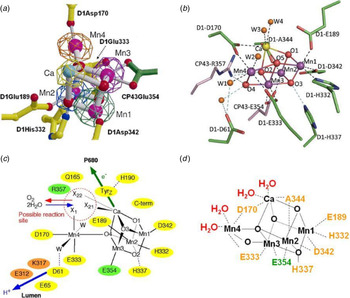
This was rectified in the crystal structure reported by Ferreira et al. (Reference Ferreira, Iverson, Maghlaoui, Barber and Iwata2004). Anomalous diffraction data was collected at a wavelength of 1.89 Å to obtain an electron density profile for the Mn ions and at 2.25 Å to locate Ca2+ (at 2.25 Å Ca has an anomalous difference almost four times stronger than Mn). Again the electron density attributed to the four Mn ions was ‘pear-shaped’ indicative of the 3 + 1 organisation and again one Mn was assigned to the small domain and three in the large globular domain, whereas the 2.25 Å wavelength map covers one metal ion in the large domain of the native density. From this data the three Mn ions (Mn1, Mn2 and Mn3) and the Ca2+ in the large domain were modelled with a cubane organisation with bridging oxygens, an organisation which was compatible with the native electron density. The fourth Mn ion (Mn4) located in the small domain was modelled so that it was linked to the cubane by one of its bridging oxygens (see Fig. 4a ). Based on values determined by EXAFS measurements, the three Mn–di-μ-oxy–Mn bonds of the cubane were spaced at 2.7 Å while the three Mn–di-μ-oxy–Ca2+ bonds at 3.4 Å. The dangler Mn was positioned 3.3 Å from the closest Mn ion of the cubane and about 4 Å from the Ca2+. Although the distances chosen were compatible with distances derived from EXAFS (Yachandra, Reference Yachandra2002) the numbers Mn–di-μ-oxy–Mn bonds was greater than predicted by EXAFS. Nevertheless, density function theory (DFT) and QM/MM calculations (Batista et al. Reference Batista, Sprovier, Gascon, McEvoy and Brudvig2008; Lunberg & Siegbahn Reference Lunberg and Siegbahn2004; Sproviero et al. Reference Sproviero, Gascon, McEvoy, Brudvig and Batista2006, Reference Sproviero, Gascon, McEvoy, Brudvig and Batista2007) and chemical synthesis of mixed Mn/Ca complexes (Misra et al. Reference Misra, Wernsdorfer, Abboud and Christou2005) and more recently by Kanady et al. (Reference Kanady, Tsui, Day and Agapie2011) and Mukherjee et al. (Reference Mukherjee, Stull, Yano, Stamatatos, Pringouri, Stich, Abboud, Britt, Yachandra and Christou2012) suggested that the Mn3CaO4 cubane was chemically feasible in the absence of a protein scaffold despite there being no known similar structure in biology or chemistry at the time of conception. Importantly, the complete Mn4CaO4 cluster with essentially identical geometry to that suggested by Ferreira et al. (Reference Ferreira, Iverson, Maghlaoui, Barber and Iwata2004) has been very recently synthesised by Zhang et al. (Reference Zhang, Chen, Dong, Shen, Dau and Zhao2015) (see Fig. 6a ). (see the discussion of this in Section 3.2.4).
(a) Cubane structural model of the Mn4Ca cluster of the OEC and identification of amino acids within the first coordination sphere deduced from X-ray diffraction analyses at 3.5 Å by Ferreira et al. (Reference Ferreira, Iverson, Maghlaoui, Barber and Iwata2004). (b) Structure of the Mn4CaO5 cluster and its ligand environment as determined at a resolution of 1.9 Å by Umena et al. (Reference Umena, Kawakami, Shen and Kamiya2011). (c) Schematic view of the OEC published in Ferreira et al. (Reference Ferreira, Iverson, Maghlaoui, Barber and Iwata2004). Residues in D1, D2 and CP43 subunits are shown in yellow, orange and green, respectively. X11, X21 and X22 were proposed to be substrate water binding positions to Mn4 (X11) and to Ca (X21 and X22), identified from the position of the non-protein ligand and coordination pattern of Mn and Ca ions. Possible water molecules, which were not visible at the 3.5 Å resolution were suggested and indicated as W. Possible hydrogen bonds were also proposed and shown as light-blue dotted lines. (d) Diagrammatic representation of the Mn4CaO5 cluster and coordinating ligands determined by Umena et al. (Reference Umena, Kawakami, Shen and Kamiya2011).

The cubane model of Ferreira et al. (Reference Ferreira, Iverson, Maghlaoui, Barber and Iwata2004) provided an important basis for its further refinement by quantum mechanical calculations and for developing detailed mechanisms for the water splitting reaction leading to dioxygen formation (Brudvig, Reference Brudvig2008; Dau et al. Reference Dau, Grundmeier, Loja and Haumann2008; McEvoy & Brudvig et al. Reference McEvoy and Brudvig2004, Reference McEvoy and Brudvig2006; Siegbahn, Reference Siegbahn2006, Reference Siegbahn2008, Reference Siegbahn2009, Reference Siegbahn2012).
Based on this organisation of the metal ions, the Mn3CaO4 cubane had four protein side chains as potential ligands: D1Asp342, D1Glu189, D1His332 and CP43Glu354. Identification of the glutamate of CP43 as a Mn-ligand was a surprise and is a residue of a conserved motif Gly–Gly–Glu–Thr–Met–Arg–Phe–Trp–Asp which forms a 310 helix in the large extrinsic loop joining the lumenal ends of transmembrane helices V and V1 of this protein. In addition to these four apparent protein ligands it was noted that the C-terminal residue of the D1 protein, D1Ala344, is located close to Ca2+ and that D1His337 could be hydrogen bonded to one of the bridging oxo-bonds of the cubane. Two side chain densities were available as ligands for the ‘dangler’ Mn4 outside the cubane cluster, D1Asp170 and D1Glu333 and it was suggested that D1Asp61 might also function as a second sphere ligand via a bridging water molecule (see Fig. 4c ). A non-protein electron density in the vicinity of Mn(4) and Ca2+ was identified and was tentatively modelled as a carbonate ion that formed bridging ligands between the two metals. However, the positioning of this density adjacent to the redox active D1Tyr161 (TyrZ) suggested that this could be the site for the binding of substrate water molecules and for the formation of O–O bond, as diagrammatically shown in Fig. 4c , and proposed by in the Science paper of Ferreira et al. (Reference Ferreira, Iverson, Maghlaoui, Barber and Iwata2004). Also located in this potential catalytic site were D1Gln165 and CP43Arg357 which seemed to indicate their involvement in a hydrogen-bonding network and therefore playing a key role in the deprotonation of the substrate water molecules during the catalytic cycle, while D1Asp61 was strategically located at the mouth of a polar channel for the exit of protons to the lumenal surface.
As mentioned above, the work of Ferreira et al. (Reference Ferreira, Iverson, Maghlaoui, Barber and Iwata2004) also established that D1His190 was in hydrogen-bonding distance to D1Tyr161 as required, and predicted, for the oxidation of the latter by the P•+ to generate a neutral tyrosine radical (Hoganson & Babcock, Reference Hoganson and Babcock1997).
The 3.0 Å resolution structure reported by Loll et al. (Reference Loll, Kern, Saenger, Zouni and Biesiadka2005) confirmed the subunit and overall amino acid assignments and side chain positioning reported by Ferreira et al. (Reference Ferreira, Iverson, Maghlaoui, Barber and Iwata2004) with minor differences within the limitation imposed by the intermediate resolution of both structures. However, as stated above, the improved electron density map allowed Loll et al. to extend the original assignment of carotenoid molecules made by Ferreira et al. (Reference Ferreira, Iverson, Maghlaoui, Barber and Iwata2004) and to suggest the location of several bound lipid molecules. As in the earlier papers from this group (Zouni et al. Reference Zouni, Witt, Kern, Fromme, Krauss, Saenger and Orth2001), the pear-shaped electron density attributed to the metal-cluster of the water splitting site was interpreted as four Mn ions organised in a Y-shape or 3 + 1 arrangement. Based on anomalous diffraction, the Ca2+ was placed in a position similar to that proposed by Ferreira et al. (Reference Ferreira, Iverson, Maghlaoui, Barber and Iwata2004), and confirmed by Kargul et al. (Reference Kargul, Maghlaoui, Murray, Deak, Boussac, Rutherford, Vass and Barber2007), whereas the positioning of three of the Mn ions were different to those of Ferreira et al. (Reference Ferreira, Iverson, Maghlaoui, Barber and Iwata2004) but less so to that in the Zouni et al. (Reference Zouni, Witt, Kern, Fromme, Krauss, Saenger and Orth2001) model. The fourth Mn ion was assigned to the narrow end of the pear shaped density in a slightly different position to that of Ferreira et al. (Reference Ferreira, Iverson, Maghlaoui, Barber and Iwata2004) or Zouni et al. (Reference Zouni, Witt, Kern, Fromme, Krauss, Saenger and Orth2001). The same amino acids identified as ligands in the crystal structure of Ferreira et al. (Reference Ferreira, Iverson, Maghlaoui, Barber and Iwata2004) were also found by Loll et al. although there were differences in the precise location in some cases. Importantly they proposed that the carboxylates of the acidic residues formed bridging ligands between adjacent Mn ions and therefore more adequately satisfied the coordination requirements of the metal cluster than in the Ferreira et al. model (Reference Ferreira, Iverson, Maghlaoui, Barber and Iwata2004). However, Loll et al. emphasised that their model for the OEC was unreliable, in part because of the low resolution of the electron density map and in part because of radiation damage. Therefore they did not attempt to model the cubane structure proposed by Ferreira et al. (Reference Ferreira, Iverson, Maghlaoui, Barber and Iwata2004). In fact, working with the EXAFS group at Berkeley, and prior to the publication of the Loll et al. crystal structure of 2005, the same group had shown that doses of X-rays typical of those used for diffraction analyses caused the reduction of the Mn-cluster and associated structural changes (Yano et al. Reference Yano, Kern, Irrgang, Lattimer, Bergmann, Glatzel, Pushkar, Biesiadka, Loll, Sauer, Messinger, Zouni and Yachandra2005). This problem of radiation damage occurring at the Mn-cluster had also been highlighted by Grabolle et al. (Reference Grabolle, Haumann, Muller, Liebisch and Dau2006). Because of this, doubts were expressed about the cubane model of Ferreira et al. and several other arrangements of the Mn4Ca-cluster were postulated based on polarised EXAFS studies conducted by the Berkeley team in collaboration with the X-ray crystallography group in Berlin (Yano et al. Reference Yano, Kern, Irrgang, Lattimer, Bergmann, Glatzel, Pushkar, Biesiadka, Loll, Sauer, Messinger, Zouni and Yachandra2005, Reference Yano, Kern, Sauer, Latimer, Pushkar, Biesiadka, Loll, Saenger, Messinger, Zouni and Yachandra2006). However, a reinterpretation of the polarised EXAFS data by Sproviero et al. at Yale (Reference Sproviero, Gascon, McEvoy, Brudvig and Batista2008) suggested that the cubane model could still be considered as the legitimate organisation of the OEC metal cluster.
The uncertainty of the cubane model was lifted with the most recent 1.9 Å structure from Umena et al. (Reference Umena, Kawakami, Shen and Kamiya2011) since at this resolution the electron density of individual metals could be resolved and bridging oxygens inferred (Fig. 4b ). The resulting model was overall similar to that proposed by Ferreira et al. (Reference Ferreira, Iverson, Maghlaoui, Barber and Iwata2004) and refined by Sproviero et al. (Reference Sproviero, Gascon, McEvoy, Brudvig and Batista2006), except an additional oxo bridge was identified linking the Mn outside the Mn3Ca-cubane (Mn4) to a Mn ion of the cubane which had been previously proposed by Dau et al. (Reference Dau, Grundmeier, Loja and Haumann2008) and particularly by Siegbahn (Reference Siegbahn2008). These models based quantum chemical calculations coupled with X-ray data were remarkably similar to the Mn4CaO5 crystal structure reported 4 years later (Umena et al. Reference Umena, Kawakami, Shen and Kamiya2011) (see Fig. 5).
Comparison of the 1.9 Å X-ray structure of Umena et al. (Reference Umena, Kawakami, Shen and Kamiya2011) and the DFT calculated model by Siegbahn (Reference Siegbahn2008) showing refinement of the cubane model (Ferreira et al. Reference Ferreira, Iverson, Maghlaoui, Barber and Iwata2004) to predict the additional oxo bond to the dangler Mn4 and relative positions of the metal ions. This striking prediction demonstrate the power of employing quantum mechanical methods coupled with X-ray data to investigate the mechanism of O–O bond formation during the S3 to So transition (see Siegbahn, Reference Siegbahn2006).

As shown in Fig. 4d , four water molecules (W1 to W4) were found to be associated with the Mn4Ca2+O5 cluster, of which W1 and W2 were coordinated to Mn4 and W3 and W4 coordinated to the Ca. No other water molecules were directly associated with the metal cluster, suggesting that some of the four waters may serve as the substrates for water oxidation reaction, in line with the prediction of Ferreira et al. (Reference Ferreira, Iverson, Maghlaoui, Barber and Iwata2004). All of the amino-acid residues coordinated to the Mn4Ca2+O5 cluster were the same as those originally identified by Ferreira et al. (Reference Ferreira, Iverson, Maghlaoui, Barber and Iwata2004) and confirmed by Loll et al. (Reference Loll, Kern, Saenger, Zouni and Biesiadka2005) but their precise arrangement was now revealed in the 1.9 Å structure (shown diagrammatically in Fig. 4d ). Of these, D1Glu189 served as a monodentate ligand to Mn1. All of the remaining five carboxylate residues served as bidentate ligands: D1Asp170 as a ligand to Mn4 and Ca, D1Glu333 to Mn3 and Mn4, D1Asp342 to Mn1 and Mn2, D1Ala344 to Mn2 and Ca, and CP43Glu354 to Mn2 and Mn3. In addition, D1His332 is coordinated to Mn1. Together with the oxo bridges and waters, these amino acids give rise to a saturating ligand environment for the Mn4Ca2+O5 cluster, with each of the four manganese ions having six ligands, whereas the calcium has seven.
In addition to the direct ligands of the Mn4Ca2+O5 cluster, Umena et al. (Reference Umena, Kawakami, Shen and Kamiya2011) confirmed the conclusion of Ferreira et al. (Reference Ferreira, Iverson, Maghlaoui, Barber and Iwata2004) that D1Asp61, D1His337 and CP43Arg357 are located in the second coordination sphere and may have important roles in maintaining the catalytic activity of the metal cluster. This is in agreement with various recent reports showing the importance of these three residues in maintaining the oxygen-evolving activity (Debus, Reference Debus2008; Hwang et al. Reference Hwang, Dilbeck, Debus and Burnap2007; Service et al. Reference Service, Hillier and Debus2010). Umena et al. (Reference Umena, Kawakami, Shen and Kamiya2011) noted that one of the guanidinium g-nitrogens of CP43Arg 357 is hydrogen-bonded to both O2 and O4 of the Mn4Ca2+O5 cluster, whereas the other is hydrogen-bonded to the carboxylate oxygen of D1Asp170 and to that of D1Ala344. They also noted imidazole e-nitrogen of D1His337 is hydrogen-bonded to a bridging oxo in agreement with the suggestion of Ferreira et al. (Reference Ferreira, Iverson, Maghlaoui, Barber and Iwata2004). Thus, these two residues may function to stabilise any conformational changes in the cubane structure of the metal cluster as well as provide partial positive charges to compensate for the negative charges induced by the oxo bridges and carboxylate ligands of the metal cluster during the S-state cycle. Ferreira et al. (Reference Ferreira, Iverson, Maghlaoui, Barber and Iwata2004) also suggested that D1Asp61 could be H-bonded to a water bridging to Mn4 and Umena et al. (Reference Umena, Kawakami, Shen and Kamiya2011) did indeed find that the carboxylate oxygen of D1Asp 61 is hydrogen-bonded to W1, and also to O4 indirectly through another water molecule, suggesting that this residue may also contribute to stabilising the metal cluster. Furthermore, as postulated by Ferreira et al. (Reference Ferreira, Iverson, Maghlaoui, Barber and Iwata2004) D1Asp 61 seems to be located at the entrance of a proton exit channel suggesting that this residue may function in facilitating proton exit from the catalytic site and aided by the presence of a bound chloride ion (Murray et al. Reference Murray, Maghlaoui, Kargul, Ishid, Lai, Rutherford, Sugiura, Boussac and Barber2008; Rivalta et al. Reference Rivalta, Amin, Luber, Vassiliev, Pokhrel, Umena, Kawakami, Shen, Kamiya, Bruce, Brudvig, Gunner and Batista2011). Despite the excellence of the Umena et al. structure there were still uncertainties about artefacts, particularly in the metal cluster, due to radiation effects. To address this, the Japanese group used femtosecond X-ray pulses generated from an X-ray free electron laser to determine the structure of the metal cluster at 1.95 Å (Suga et al. Reference Suga, Akita, Hirata, Ueno, Murakami, Nakajima, Shimizu, Yamashita, Yamamoto, Ago and Shen2015). They found very little difference from the previous model (Mn–Mn and Mn–ligand distances were shorter by 0.1–0.3 Å) but they did confirm that the bond distances for the O5 oxo (see Fig. 4b for numbering of oxygens) are longer than expected for a typical Mn–O bond and suggested this may be significant for the O–O bond formation.
As mentioned earlier, the cubane model of the Mn4Ca-cluster proposed by Ferreira et al. (Reference Ferreira, Iverson, Maghlaoui, Barber and Iwata2004) had been seriously undermined by arguments regarding radiation damage, an argument which was significantly weakened with the publication of the 1.9 Å structure of PSII by Umena et al. (Reference Umena, Kawakami, Shen and Kamiya2011) and by the follow-up femtosecond X-ray studies by Suga et al. (Reference Suga, Akita, Hirata, Ueno, Murakami, Nakajima, Shimizu, Yamashita, Yamamoto, Ago and Shen2015). Although there had been some support for the Ferriera et al. cubane model based on chemical synthesis (Kanady et al. Reference Kanady, Tsui, Day and Agapie2011; Misra et al. Reference Misra, Wernsdorfer, Abboud and Christou2005; Mukherjee et al. Reference Mukherjee, Stull, Yano, Stamatatos, Pringouri, Stich, Abboud, Britt, Yachandra and Christou2012) it was the very recent outstanding work of Zhang et al. (Reference Zhang, Chen, Dong, Shen, Dau and Zhao2015) reporting the synthesis of a Mn4CaO4 cluster having the same geometry as that first proposed by Ferreira et al. (Reference Ferreira, Iverson, Maghlaoui, Barber and Iwata2004) (see Fig. 6). This synthetic cluster showed EPR and redox properties similar to the in vivo OEC cluster. The absence of the extra O4 oxo-bridge to Mn4 (Umena et al. numbering) may have implications for the mechanism of water oxidation.
Chemical synthesis of a cubane structure of a Mn4CaO4-cluster by Zhang et al. (Reference Zhang, Chen, Dong, Shen, Dau and Zhao2015) having remarkably similarity to the geometry to that first proposed by Ferreira et al. (Reference Ferreira, Iverson, Maghlaoui, Barber and Iwata2004) for OEC of PSII. (a) Chemically synthesised. (b) Model from coordinates (PDB 1S5L) deposited by Ferreira et al. (Reference Ferreira, Iverson, Maghlaoui, Barber and Iwata2004). Note the numbering of the Mn and O are different where (a) uses those adopted by Umena et al. (Reference Umena, Kawakami, Shen and Kamiya2011). Distances are in Å.

3.2.5. Chloride
The recognition that chloride is required as a cofactor for photosynthetic oxygen production, particularly for the water splitting reaction of PSII, stems from the pioneering studies of Arnon & Whatley (Reference Arnon and Whatley1949), Bove et al. (Reference Bove, Bove, Whatley and Arnon1963) and Izawa et al. (Reference Izawa, Heath and Hind1969). Since then this requirement has been investigated in great depth as detailed in the comprehensive review by van Gorkom & Yochum (Reference van Gorkom, Yochum, Wydrzynski and Satoh2005).
When PSII is depleted of Cl ions, the S1 state cycle is blocked at S2 and its EPR spectral properties are modified. If Cl ion depletion is carried out on PSII placed in the S3 state, then the subsequent transitions to S0 are blocked and oxygen formation is inhibited (Wincencjus et al. Reference Wincencjus, Van Gorkom and Yocum1997). Murray et al. (Reference Murray, Maghlaoui, Kargul, Ishid, Lai, Rutherford, Sugiura, Boussac and Barber2008) were the first to conclude from their X-ray crystallographic analyses that there were two Cl ion binding sites in the vicinity of the Mn4CaO5 cluster by substitution of Br for Cl ions. These assignments were confirmed by Kawakami et al. (Reference Kawakami, Umena, Kamiya and Shen2009) and Umena et al. (Reference Umena, Kawakami, Shen and Kamiya2011). One of the chloride ions is located close to the amino group of D1Lys31 and the backbone nitrogen of D1Glu333, while the other is close to the backbone nitrogens of D1Asn338 and CP43Glu354. Both also have two water molecules nearby. Because the side chains of D1Glu333 and CP43-Glu354 are coordinated to the Mn4CaO5 cluster directly, the two Cl anions may function to maintain its coordination environment, thereby allowing the oxygen-evolving reaction to proceed properly. One Cl -binding site close to D2Lys317 lies at the entrance of hydrogen-bonding networks extending towards the lumenal surface and may play a role in facilitating proton efflux from the OEC (Murray et al. Reference Murray, Maghlaoui, Kargul, Ishid, Lai, Rutherford, Sugiura, Boussac and Barber2008; Rivalta et al. Reference Rivalta, Amin, Luber, Vassiliev, Pokhrel, Umena, Kawakami, Shen, Kamiya, Bruce, Brudvig, Gunner and Batista2011).
4. Mechanism of Water Oxidation
Although the geometry of the Mn4Ca-cluster and its ligand field characteristics are now known at a resolution of 1.9 Å there is uncertainty about which S-state it is in. Because free electrons are generated in the crystal during the collection of X-ray diffraction data it is possible that the Mn-cluster is reduced to levels lower than the dark stable S1-state. From quantum mechanical calculations (DFT, QM/MM) Luber et al. (Reference Luber, Rivalta, Umena, Kawakami, Shen, Kamiya, Brudvig and Batista2011) has concluded that the Umena et al. X-ray model most likely corresponds to a mixture of S0 S−1 and S−2 oxidation states.
Despite this, the original Ferreira et al. cubane model and its subsequent refinements (Dau et al. Reference Dau, Grundmeier, Loja and Haumann2008; Siegbahn, Reference Siegbahn2008; Sproviero et al. Reference Sproviero, Gascon, McEvoy, Brudvig and Batista2007, Reference Sproviero, Gascon, McEvoy, Brudvig and Batista2008) already provided a basis for developing chemical mechanisms for the water oxidation and dioxygen formation. The Mn ion outside the cubane (Mn4) is adjacent to the Ca2+ (about 4 Å apart) and their positioning towards the side chains of several key amino acids, including the redox active YZ suggested to Ferreira et al. (Reference Ferreira, Iverson, Maghlaoui, Barber and Iwata2004) (also see Barber et al. Reference Barber, Ferreira, Maghlaoui and Iwata2004) that they provided a ‘catalytic’ surface for binding the two substrate water molecules and their subsequent oxidation. This would support a well championed mechanism for photosynthetic dioxygen formation which suggests that the substrate water, associated with Mn4, is deprotonated during the S-state cycle and converted to a highly electrophilic oxo (see Fig. 7a ) (Brudvig, Reference Brudvig2008; McEvoy & Brudvig, Reference McEvoy and Brudvig2004, Reference McEvoy and Brudvig2006; Messinger et al. Reference Messinger, Badger and Wydrzynski1995; Pecoraro et al. Reference Pecoraro, Baldwin, Caudle, Hsieh and Law1998; Vinyard et al. Reference Vinyard, Khan and Brudvig2015). This mechanism is dependent on Mn4 being converted to a high oxidation state (possible Mn(V) or a very reactive oxyl radical) during progression to the S4-state just prior to O–O bond formation. The other three Mn ions also progress to high valency states (Mn(IV) (S0 (Mn(IV), Mn(III), Mn(III), Mn(III) to S3 (Mn(IV), Mn(IV), Mn(IV), Mn(IV)) and act as a further ‘oxidising battery’ for the Mn(V)-oxo or oxyl radical species in the S4-state (see Fig. 7a ). In this way, the reactive oxo is electron deficient, so much so that it makes an ideal target for a nucleophilic attack by the oxygen of the second substrate water bound within the coordination sphere of the Ca2+ (see Fig. 7a ). Deprotonation of the substrate waters would be aided by nearby bases such as CP43Arg357 and by the weak Lewis acidity of Ca2+.
Two different mechanisms for the final step of the S-state cycle, S4 to So, when the dioxygen bond of O2 is formed. (a) Mechanism 1. The very high oxidation state of the Mn-cluster, particularly the Mn ion outside the Mn3CaO5-cubane, leads to a high electron-deficient oxo (after deprotonation of water molecules during the S-state cycle). Nucleophilic attack by the hydroxide of the second substrate water within the coordination sphere of Ca2+ leads to O2 formation. (b) Mechanism 2. The formation of an oxyl radical on one of the bridging oxygen atoms of the cubane, derived from a substrate water molecule, leads to a radical attack of an adjacent oxo-ligand within the Mn3CaO4-cubane.

This mechanism has been thoroughly investigated by Siegbahn (Reference Siegbahn2006, Reference Siegbahn2008) using in depth calculations based on DFT and found that it is energetically less favourable than a mechanism involving the formation of a oxyl-radical within the Mn3CaO4 cubane as depicted in Fig. 7b . Therefore the Siegbahn mechanism (Reference Siegbahn2009, 201, Reference Siegbahn2013) concludes that the O–O bond formation during the S4 to S0 transition involves bridging oxo species and unlike the other mechanism (Fig. 7a ) has received impressive experimental support (Cox et al. Reference Cox, Rapatskiy, Su, Pantazis, Sugiura, Kulik and Messinger2011, Reference Cox, Retegan, Neese, Pantais, Boussac and Lubitz2014). Recently the details of the S4 to S0 transition have been refined by Li & Siegbahn (Reference Fujita and Licker2015) to allow for conformational changes in and around the metal cluster but the general concept of an oxyl–oxo mechanism for O–O bond formation involves the same two manganese atoms and the central oxo group as in the original scheme described in Siegbahn's (Reference Siegbahn2009) paper and earlier papers dating back to 2006.
5. Artificial Photosynthesis
While some progress has been made in mimicking photosynthesis in artificial systems, researchers have not yet developed components that are both efficient and robust for incorporation into a working system for capturing and storing solar energy in chemical bonds on a large scale as does natural photosynthesis. To date the main focus of research has been to design and synthesise molecular catalysts which can be linked to a light-driven charge separation system (Tran et al. Reference Tran, Wong, Barber and Loo2012). Dyes have been used for the latter but inorganic semiconductors offer a more realistic and robust approach for providing the oxidising and reducing potentials necessary to split water and power reductive chemistry.
Insights gleaned from the recent structural determination of PSII have initiated considerable efforts to identify artificial catalytic systems for water oxidation and hydrogen production using solar energy (Eisenberg & Gray, Reference Eisenberg and Gray2008). The hydrogen produced could be used directly as a source of energy but could also be used, as it is in natural photosynthesis, to reduce CO2 to other types of fuels such as methane, methanol etc. The challenge is to have a molecular arrangement so that the artificial catalysts efficiently utilise light energy to split water and concomitantly provide reducing potential for hydrogen gas production or CO2 reduction. It has been demonstrated that catalysts based on Mn or on Mn doped with Ca are capable of water splitting leading to the generation of dioxygen (Gao et al. Reference Gao, Crabtree and Brudvig2012; Limberg et al. Reference Limberg, Vrettos, Liable-Sands, Rheingold, Crabtree and Brudvig1999; Najafpour, Reference Najafpour2011; Najafpour et al. Reference Najafpour, Ehrenberg, Wiechen and Kurz2010; Tagore et al. Reference Tagore, Crabtree and Brudvig2008; Zaharieva et al. Reference Zaharieva, Najafpour, Wiechert, Haumann, Kurz and Dau2011). Frei's group reported that nanostructured manganese oxide clusters supported on mesoporous silica efficiently evolved oxygen in aqueous solution under mild conditions (Jiao & Frei, Reference Jiao and Frei2010). The recently synthesised Mn4CaO4-cluster (Zhang et al. Reference Zhang, Chen, Dong, Shen, Dau and Zhao2015) shows charge storage under strong oxidation conditions similar to those observed in the natural PSII-cluster progressing to S3 but has yet to be shown that it can catalyse the generation of oxygen from water. Complementing the work on Mn has been the earlier discovery that ruthenium based catalysts, such as the ‘blue dimer’ can photooxidise water to dioxygen (Gersten et al. Reference Gersten, Samuels and Meyer1982; Liu et al. Reference Liu, Concepcion, Jurss, Cardolaccia, Templeton and Meyer2008; Romero et al. Reference Romero, Rodriguez, Sens, Mola, Kollipara, Francas, Mas-Marza, Escriche and Llobet2008) and the recent spectacular work of Duan et al. (Reference Duan, Bogoglian, Mandal, Stewart, Privalot, Llobet and Sun2012) has reported a rationally designed Ru-based catalyst with an unprecedentedly turnover rate of 300 per second which is comparable with that of PSII. But perhaps the most practical catalysts for water splitting are based on cobalt which is a relatively abundant element. Kanan & Nocera (Reference Kanan and Nocera2008) have described a self-assembling catalyst composed of Co and phosphate ions which can efficiently produce molecular oxygen from water at neutral pH with a low over potential akin to that which operates in the OEC of PSII. Dau and his colleagues (Khare et al. Reference Khare, Zaharieva, Gerencser, Chernev and Dau2009; Risch et al. Reference Risch, Ringle, Kohlhoff, Bogdanoff, Chemev, Zaharieva and Dau2015) have revealed important structural details and properties of this Co-based catalyst and found it to have a molecular organisation remarkably similar to the Mn3Ca-cubane of PSII. Yin et al. (Reference Yin, Tan, Besson, Geletii, Musaev, Kuznetsov, Luo, Hardcastle and Hill2010) reported a water splitting catalyst comprising of a Co4O4 core stabilised by oxidatively resistant polytungstate ligands which, like the Nocera catalyst, is also self-assembling. Du & Eisenberg (Reference Du and Eisenberg2012) have reviewed recent progress and future challenges of using Co as a water splitting catalyst compared with other transition metals.
Hematite (α-Fe2O3) is a semiconductor which is also capable of photochemically splitting water to molecular oxygen. It has a favourable optical band gap (E g = 2.2 eV), excellent chemical stability in aqueous environments, natural abundance and low cost (Bassi et al. Reference Bassi, Gurudayal, Wong and Barber2014; Sivula et al. Reference Sivula, Formal and Graezel2011). Indeed hematite has been theoretically predicted to achieve a water oxidation efficiency of 16.8% (Murphy et al. Reference Murphy, Barnes, Randeniyqa, Plumb, Grey, Horne and Glasscock2006). However, the reported efficiencies of hematite are lower than this predicted value, mainly due to the very short lifetime of photo-generated charge carriers, short hole diffusion length (2–4 nm), slow oxygen evolution reaction kinetics, low flat band potential and significant reduction in the absorption cross-section at wavelengths approaching the band gap value. Another fundamental limitation of the hematite system is the need for externally applied bias because the conduction band of hematite is lower than the potential required to reduce protons to hydrogen. Nevertheless it is a system which is receiving considerable attention at the present time and the recent construction of a tandem cell incorporating perovskite and Co-doped hematite nanorods and requiring no external electrical bias for photo-driven water splitting (Gurudayal et al. Reference Gurudayal, Sabba, Kuma, Wong, Barber, Graetzel and Mathews2015), is an example of the potential for this type of photoanode for constructing an ‘artificial leaf’ for solar fuel production (see Fig. 8).
Diagramatic representation of a tandem photoelectrochemical cell incorporating perovskite (CH3NH3PbI3) and hematite (Fe2O3) nanorods and requiring no external electrical bias for photo-driven water splitting (Gurudayal et al. Reference Gurudayal, Sabba, Kuma, Wong, Barber, Graetzel and Mathews2015).

The next step will be to couple these oxygen producing systems to non-platinum catalysts which will use the protons and high-energy electrons derived from the water splitting reaction to produce hydrogen gas or reduce CO2. In the case of the former, considerable progress is being made (Wang et al. Reference Wang, Chen and Sun2012), in part by mimicking the natural hydrogenase enzymes found in a wide variety of microorganisms (Tran et al. Reference Tran, Artero and Fontecave2010). Also a number of inorganic catalysts have been identified with activities which are almost as good as platinum (Tran & Barber, Reference Tran and Barber2012). One such class of catalysts are based on sulphides of Mo and W (Merki & Hu, Reference Merki and Hu2011; Tran et al. Reference Tran, Chiam, Boix, Ren, Pramana, Fize, Artero and Barber2013; Zong et al. Reference Zong, Han, Ma, Yan, Wu and Li2011) and another is an alloy composed of nickel, molybdenum and zinc (Reece et al. Reference Reece, Hamel, Sung, Jarvi, Esswein, Pijpers and Nocera2011). Identifying catalysts for the reduction of CO2, however, is more difficult because multielectron reactions are required and the emergence of catalysts for generating useful carbon fuels will require considerable effort (Arakawa et al. Reference Arakawa, Aresta, Armor, Barteau, Beckman, Bell and Domen2001; Fujita, Reference Fujita and Licker2000).
The most successful coupling of catalysts using a semiconductor for light capture and charge separation was reported by Nocera and colleagues (Reece et al. Reference Reece, Hamel, Sung, Jarvi, Esswein, Pijpers and Nocera2011). They used a triple junction amorphous Si wafer as the semiconductor, the CoPi catalyst for water splitting and the NiMoZn alloy for the cathodic hydrogen producing catalyst as shown in Fig. 9. This discovery is a significant step towards the development of an efficient, robust, low-cost and scalable photocatalytic device for water splitting at neutral pH to generate molecular hydrogen using only solar energy.
Diagramatic representation of Nocera's photocatalytic ‘artificial leaf’ device for water splitting consisting of a triple n-junction amorphous silicon wafer, Co-based oxygen evolving catalyst and NiMoZn H2-evolving catalyst as reported in Reece et al. (Reference Reece, Hamel, Sung, Jarvi, Esswein, Pijpers and Nocera2011).

6. Conclusion
The determination of the structure of PSII has provided vital information as to how Nature conducts the remarkable chemistry of water splitting and solved its renewable energy problem needed to prosper and diversify on an enormous scale. This new information now provides a blue print for scientists to seriously consider constructing catalysts that mimic the natural system and provides new technologies to address the energy/CO2 problem that mankind must now solve. After all there is no shortage of water for this reaction and the energy content of sunlight falling on our planet well exceeds our needs.
Acknowledgements
I wish to thank Nanyang Technological University (NTU) of Singapore for support and my colleagues in the Solar Fuel Laboratory at NTU for enabling me to explore artificial photosynthesis research which is a logical extension to my work on natural photosynthesis and PSII in particular. I also thank Dr James Murray for helping with the construction of Fig. 6 and Richard Malkin for his editorial corrections.
After the completion of this review a paper was published (Wei et al. Reference Wei, Su, Cao, Liu, Chang, Li, Zhang and Liu2016) which reported a 3.2 Å structure of higher plant PSII consisting of its reaction center core and its outer chlorophyll a and chlorophyll b light-harvesting complex (LHC II). This 1.1 megadalton PSII-LHCII supercomplex was isolated from spinach and its structure determined by single particle cryoelectron microscopy (cryo-EM). This outstanding work follows on from previous cryo-EM studies of the same supercomplex (Hankamer et al. Reference Hankamer, Morris, Nield, Gerle and Barber2001b; Nield & Barber, Reference Nield and Barber2006) in which the 3D structure was limited to 17 Å. This significant improvement in the resolution is result of a new generation of electron microscopes able to reveal structures at atomic resolution. The spinach PSII core was essentially identical to the cyanobacterial counterpart except for the presence of an extra low molecular weight intrinsic protein, PsbW. The interaction of the four extrinsic proteins, PsbO, Psb P, PsbQ and PsbTn was revealed for the first time. However the most important outcome from this structure is that it has revealed the position of the 105 chlorophylls and 28 carotenoids that it binds thus providing the structural information for describing energy transfer from LHCII to the PSII reaction center.