Left ventricular non-compaction results from the arrest of the compaction process of the myocardium during cardiac morphogenesis. The American Heart Association recognises it as a primary genetic cardiomyopathy, whereas the 1996 World Health Organisation/International Society and Federation of Cardiology task force grouped it under unclassified cardiomyopathies.Reference Richardson, McKenna and Bristow 1 Previous studies have reported left ventricular non-compaction accounting for up to 9% of all cardiomyopathies.Reference Pignatelli, McMahon and Dryer 2 There is genetic heterogeneity underlying left ventricular non-compaction cardiomyopathy. Cardiomyopathies in association with oxidative phosphorylation defects are well recognised, and left ventricular non-compaction occurring specifically in mitochondrial respiratory chain disorders has been previously described.Reference Fosselin 3 We describe a baby girl with biventricular non-compaction hypertrophic cardiomyopathy, multiple ventricular septal defects, pulmonary stenosis, and complete heart block on a background of type I mitochondrial complex deficiency.
Case report
A baby girl was born at 37 weeks of gestation via elective lower segment caesarean section to healthy non-consanguineous Caucasian parents. Her birth weight was 3.5 kg. This was the couple’s fifth child with four previous healthy children and no history of miscarriages. A foetal echocardiogram at 22 weeks of gestation demonstrated left ventricular non-compaction and complete heart block with a ventricular rate of 50–60 beats/minute.
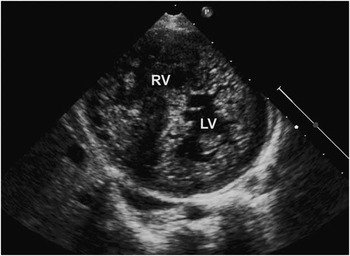
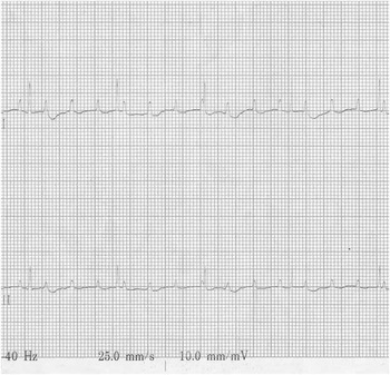
At birth, the Apgar scores were 8 at 1 and 5 minutes of age. She was self-ventilating in room air, with oxygen saturations of 98%. Her heart rate was 50 beats/minute, mean blood pressure was 40–45 mmHg, and respiratory rate 50 breaths/minute. There were normal heart sounds and a 2/6 systolic murmur at the left sternal border. The pulses were normal. There was no organomegaly. Electrocardiogram demonstrated complete heart block with a ventricular rate of 60 per minute. Echocardiography demonstrated biventricular non-compaction cardiomyopathy with a severely hypertrophic phenotype (Fig 1), multiple muscular ventricular septal defects with bidirectional shunting, patent arterial duct, and mild pulmonary valve stenosis with a peak velocity 2.6 m/second. The left ventricular ejection fraction measured 64%. She was commenced on isoprenaline infusion for complete atrioventricular block (Fig 2).
Two-dimensional echocardiography in the parasternal short-axis view demonstrating biventricular non-compaction hypertrophic cardiomyopathy.

Electrocardiogram demonstrating complete heart block with ventricular escape rate 60 beats/minute following treatment with isoprenaline infusion. LV=left ventricle; RV=right ventricle.

An epicardial VVI pacemaker was placed on day 3 of life. She tolerated the procedure well and was successfully extubated 3 days later. Extensive genetic and metabolic investigations were undertaken. On muscle biopsy, specific flux experiment demonstrated complex 1 deficiency. The skeletal muscle biopsy also showed type I muscle atrophy. Cytogenetic evaluation confirmed a normal female karyotype with a normal fluorescent in situ hybridisation for 22q11 deletion. Automated sequencing of the entire mitochondrial genome sequence demonstrated no sporadic or maternally inherited mtDNA point mutation. Specific testing for mt3243>G, mt8344A>G, and mt8993T>C/G mutations was negative, and the mitochondrial genome analysis was normal. Sequencing of the nuclear encoded gene, ACAD9, was undertaken, but no known or potentially pathogenic mutation was identified. Therefore, point mutations and small indels within the coding region of ACAD9 as a possible cause of her clinical condition were excluded. She had a normal renal and cranial ultrasound.
She was discharged on day 15 of life on carvedilol, aspirin, coenzyme Q10, and carnitine. She underwent a pacemaker change at 7 years of age. The ventricular septal defects restricted over time, and the pulmonary valvar stenosis peak velocity measured 3 m/second on the most recent echocardiogram. She has mild gross motor delay but normal cognitive development on neurological assessment. Her siblings and parents had normal echocardiogram studies.
Discussion
We report this first foetal diagnosis of biventricular non-compaction hypertrophic cardiomyopathy in association with complete congenital heart block and complex 1 mitochondrial disease. Previously, an adult patient with biventricular non-compaction and complete heart block has been described.Reference Patra, Kumar, Sadananada, Basappa and Nanjappa 4 In our case, prenatal and postnatal echocardiography confirmed ventricular non-compaction cardiomyopathy, affecting both ventricles with severe hypertrophic phenotype, with multiple small Swiss cheese ventricular septal defects and pulmonary stenosis. She had associated congenital heart block and required pacemaker placement on day 3 of life. She was a non-dysmorphic neonate with normal cytogenetic and mtDNA investigations. Muscle biopsy demonstrated complex 1 respiratory chain enzyme deficiency and type I fibre atrophy. Similarly, sequencing of ACAD9 gene, known for its association with oxidative phosphorylation disorders and cardiomyopathy, also failed to identify any pathogenic point mutations or small indels within the coding region of ACAD9. She manifested mild gross motor delay but normal cognition and no major cardiovascular events.
Isolated non-compaction cardiomyopathy with its typical echocardiographic appearance was first described by Engberding.Reference Engberding and Bender 5 Chin et alReference Chin, Perloff, Williams, Jue and Mohrmann 6 suggested the name “isolated non-compaction of the left ventricular myocardium” on the basis of the background developmental physiology. Awareness of the prevalence of this disease in the paediatric population has increased with clarification of its diagnostic criteria.Reference Pignatelli, McMahon and Dryer 2 , Reference Hoedemaekers, Caliskan and Michels 7 , Reference Finsterer 8 Liu et alReference Liu, Bai and Huang 9 suggest that lower mtDNA copy number and morphological abnormalities of mitochondria suggest mitochondrial dysfunction that may be associated with the aetiology of non-compaction cardiomyopathy. There are several known genetic mutations involving tafazzin, α-dystrobrevin, and the sarcomeric proteins.Reference Pignatelli, McMahon and Dryer 2 , Reference Hoedemaekers, Caliskan and Michels 7 , Reference Finsterer 8 The genetic heterogeneity could suggest that non-compaction cardiomyopathy is not only a primary result of embryonic non-compaction, but could be a compensatory morphological expression/result of an impaired myocardium.Reference Finsterer 8 From our case, there was clear non-compaction involvement of the right ventricle, although a strict definition of right ventricular non-compaction has not been reached, given the normal trabeculated nature of the right ventricle.
A number of metabolic conditions are associated with cardiomyopathies including respiratory chain enzyme defects. In the process of oxidative phosphorylation, molecular oxygen is consumed and adenosine triphosphate is produced, as the protons fall back down the electrochemical gradient. All 13 proteins encoded by the mitochondrial genome are subunits of these oxidative phosphorylation process complexes. Thus, mtDNA mutations alter the crucial oxidative phosphorylation process. Cardiomyopathies with underlying mitochondrial disorders continue to be underdiagnosed, as the mitochondrial mutations inheritance pattern and phenotypic expression is different and variable as compared with Mendelian inheritance pattern.Reference Fosselin 3 Several cardiomyopathies of metabolic origin, often owing to respiratory chain defects, showed a restrictive pattern or belonged to the non-classified type of cardiomyopathy.Reference Gilbert-Barness 10
In conclusion, our report describes a case of biventricular non-compaction hypertrophic cardiomyopathy with congenital heart block in association with complex 1 respiratory chain enzyme deficiency. Early diagnosis, genetic evaluation, timely pacemaker placement, and ongoing medical management in our case have successfully avoided serious cardiovascular events.
Acknowledgements
The authors are grateful to the Cardiac and Genetics Departments at Our Lady’s Hospital for Sick Children.
Financial Support
This research received no specific grant from any funding agency, commercial or not-for-profit sectors.
Conflicts of Interest
None.