Case report
We successfully removed a huge vegetation from an extremely low-birth-weight baby using a catheter. This report details this globally rare case involving a 19-day-old girl (corrected age, 29 weeks, 0 days). She was the firstborn of a set of twins, born at 26 weeks and 2 days of gestation, weighing 714 g. On day 4, the stool culture revealed methicillin-resistant Staphylococcus aureus, but she showed no signs of infection. On day 10, C-reactive protein level increased, and vancomycin, cefotaxime, and immunoglobulin were administered. Methicillin-resistant Staphylococcus aureus was detected in blood cultures on the same day. On day 14, echocardiography showed a tumour measuring 4 mm in diameter in the right atrium. Despite aggressive antibiotic treatment, the tumour gradually grew to a diameter of 6 mm and extended from the superior vena cava in a threadlike manner, forming a large aneurysm in the right atrium (Figure 1a). The aneurysm had no continuity with the cardiac structures and was separate from cardiac tissue. A peripherally inserted catheter was inserted into the right upper limb, and it was identified as an infectious vegetation that originated from the tip of the peripherally inserted catheter. Reference Marks, Zucker, Kapelushnik, Karplus and Levitas1,Reference Kobayashi, Iwatani and Izimi2 The tumour was mobile, travelling between the right atrium and ventricle (Figure 1b). We were concerned that the string-like part of the vegetation could break off, causing a pulmonary embolism and potentially fatal consequences. Additionally, we believed that the catheter could not remove the vegetation unless it was confined to the right atrium.
( a ) Vegetation (★) growing like a string from the superior vena cava, entering and exiting the right atrium and right ventricle. ( b ) Vegetation (★) extending from the right atrium into the right ventricle (transthoracic echocardiography: four-chamber view).

We determined that the vegetation presented a high risk of main pulmonary artery embolisation and that emergency extraction was necessary. However, open chest surgery using cardiopulmonary bypass was not possible, considering the low birth weight of the infant. We resorted to removing the vegetation using a snare catheter. As this was a catheter for an extremely premature infant, the parents were informed of the risks of vascular damage and pulmonary embolism due to tumour dispersion during surgery, and approval was obtained after an emergency ethical review (Kindai University Hospital Clinical Ethics Committee approval number: RR06-17). The procedure was performed only after it was comprehensively explained to the parents, and their consent was provided.
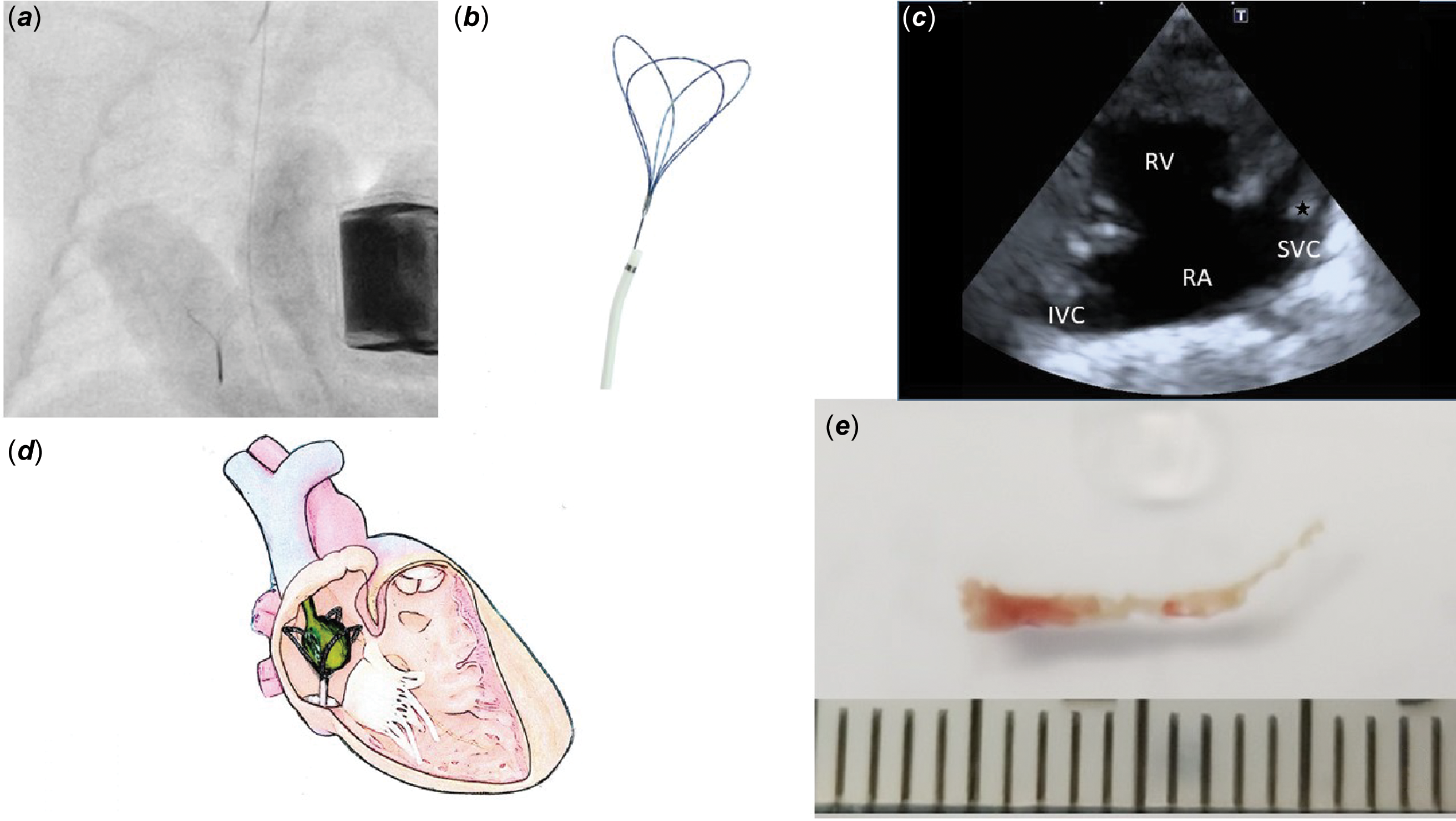
The vegetation was removed percutaneously under general anaesthesia. The femoral vein of the newborn weighing 740 g was punctured with a 22G indwelling needle, and a 4 Fr Prelude Ideal® sheath was inserted. It is difficult to capture a huge vegetation with a single-loop snare system, and there is a risk that grabbing the string-like stalk will break the stalk and allow the huge vegetation to enter the pulmonary artery. Therefore, we selected a three-loop EN Snare system. The EN Snare system (guiding catheter 3.2 Fr) is a three-loop basket system that captures foreign bodies (Figure 2a). We believed that this three-loop basket could directly capture large aneurysmal vegetations. Snares with loops measuring 4–8 mm in size were used. Reference Seow, Lin and Wong3 The EN Snare was inserted into the right atrium, where the vegetation was captured with the snare using fluoroscopy and transthoracic echocardiography as guides (Figure 2b). The snare can easily capture vegetations (Figure 2d). However, in this case, the vegetation was huge, and we determined that there was a risk of the vegetation dispersing in the heart if pulled into the 4F sheath. Therefore, we removed the vegetation together with the sheath. The chordae extending from the superior vena cava were ruptured during the recovery process (Figure 2c).
( a ) Snare used for extraction. ( b ) Transthoracic echocardiography and fluoroscopy were performed to manipulate the snare. ( c ) Ultrasound image after tumour resection (remaining vegetation margins visible in the SVC (★)) ( d ) An image of vegetation being removed with the EN snare system. ( e ) Removed vegetation. RA = right atrium; TV = tricuspid valve; RV = right ventricle; LA = left atrium; LV = left ventricle; SVC = superior vena cava; IVC = inferior vena cava.

The resected lesion was fibrous and approximately 9 mm in size (Figure 2e) and was thought to be a thrombus that had attached during the removal process. The lesion was examined histopathologically, and the patient was diagnosed with a vegetation caused by methicillin-resistant Staphylococcus aureus. Reference Marks, Zucker, Kapelushnik, Karplus and Levitas1,Reference Kobayashi, Iwatani and Izimi2 No vegetation was found in the IVC on abdominal ultrasound after catheter treatment. A CT scan was performed three months after catheterisation, but no pulmonary infarction was found. The vegetation remaining in the superior vena cava had also disappeared. With continued antibiotic treatment, the residual lesions resolved, and the patient was discharged at 163 days of age.
In extremely low-birth-weight infants with bacteraemia, attention must be paid to infective endocarditis, and catheter-based procedures are useful for removing vegetations.
Financial support
This research received no specific grant from any funding agency, commercial, or not-for-profit sectors.
Competing interests
The authors declare none.
Ethical standards
The study has been approved by the institutional committees (Kindai University Hospital Clinical Ethics Committee approval number: RR06-17).