





Non-technical Summary
Research on the Mexican amber fossil record of true hoppers has been little explored with five known species to date in the last 20 years. The present work adds three new species of planthoppers (Fulgoromorpha) grouped in a new genus. Results show schematic illustrations, high-quality pictures, line drawings, and diagnostic characters to distinguish them from other similar taxa in the tribe Nogodinini. The new Nogodinid planthopper genus is characterized by distinctive face marks, a unique wing-vein scheme, and five spines on the hind legs. In addition, a dichotomous key to separate this new Mexican planthopper taxon from the living and extinct genera in the same tribe is also provided.
Introduction
Nogodinidae Melichar, Reference Melichar1898, considered as a “higher fulgorid” family within Fulgoroidea Latreille, Reference Latreille and Latreill1807, which is composed of five subfamilies, 10 tribes, four subtribes, 81 genera, 375 extant and 31 fossil species distributed worldwide (Hamilton, Reference Hamilton2011; Dmitriev et al., Reference Dmitriev, Angelova, Anufriev, Bartlett and Blanco-Rodríguez2022-onward). Although its morphology drastically varies between subfamilies, some external macroscopic features are shared among Nogodinidae: (1) lateral carinae on the frons and sometimes in the clypeus, (2) pronotum with anterior margin projected beyond mid-length of eye, (3) absence of tubercles on the clavus, (4) hind tibia with lateral stout spines, and (5) second metatarsomere with two single apical spines on each side. Some of these characters sometimes vary, including the mesonotum extension, tegmina shape, length, and venation arrangement (Fennah, Reference Fennah1984; O’Brien and Wilson, Reference O’Brien, Wilson, Nault and Rodriguez1985; Wilson, Reference Wilson2005). Past phylogenetic studies consistently indicate the non-monophyletic status of Nogodinidae and its lineages, which reinforces the need to reevaluate its systematic position at each taxonomic level (Bucher et al., Reference Bucher, Condamine, Luo, Wang and Bourgoin2023; Deng et al., Reference Deng, Stroiński, Szwedo, Ghanavi, Yapar, Franco, Prus-Frankowska, Michalik, Wahlberg and Łukasik2024).
Within the family, the subtribe Nogodinina Melichar, Reference Melichar1898, of Nogodinini Melichar, Reference Melichar1898, is a small, but morphologically unique planthopper group, integrated by five living genera and 11 extant species, widely spreading from southeast Mexico through south of Brazil with presence in Guatemala, Belize, Honduras, El Salvador, Nicaragua, Costa Rica, Panama, Colombia, Guyana, Surinam, Ecuador, and Peru (Dmitriev et al., Reference Dmitriev, Angelova, Anufriev, Bartlett and Blanco-Rodríguez2022-onward). Nogodinini is almost exclusive to the New World with one exception, Nogodina kotoshonis Matsumura, Reference Matsumura1938, which has been reported from Taiwan (Matsumura, Reference Matsumura1938; Liang and Suwa, Reference Liang and Suwa1998).
A detailed account of the fossil record of definite Nogodinids can be found in Luo et al. (Reference Luo, Gnezdilov, Zhuo and Song2023). However, when including fossil incertae sedis taxa, the record encompasses six subfamilies, four tribes, 24 genera, and up to 36 extinct species on the list (Dmitriev et al., Reference Dmitriev, Angelova, Anufriev, Bartlett and Blanco-Rodríguez2022-onward; Luo et al., Reference Luo, Gnezdilov, Zhuo and Song2023). Within Nogodinina, three extinct species have been described: Tainosia quisqueyae Szwedo and Stroiński, Reference Szwedo and Stroiński2001, Tonacatecutlius gibsoni Stroiński and Szwedo, Reference Stroiñski and Szwedo2000, and Nogodina chiapanceca Solórzano Kraemer and Petrulevičius, Reference Solórzano Kraemer and Petrulevičius2007, the latter two extracted from Mexican amber deposits. Despite the known current worldwide fossil record, the number of specimens described in Mexican amber (two species) is still limited. Thus, the present work aims to expand the known fossil record by exploring the diversity of Auchenorrhyncha fossil inclusions in Simojovelite and contribute to the state of knowledge of Mexican amber to further classification attempts.
After exhaustive and detailed study of undetermined specimens from Mexican Early Miocene amber, a new genus, Pauchus Iscavel and Pinedo-Escatel, and three new species: P. loisae Iscavel and Pinedo-Escatel, n. gen. n. sp., P. tsakal Iscavel and Pinedo-Escatel, n. gen. n. sp., and P. nensak Iscavel and Pinedo-Escatel, n. gen. n. sp. are described and allocated into the subtribe Nogodinina. A functional dichotomous key to all New World Nogodinini genera is given. Morphology exhibited by the new taxa and similarities to other nogodinids are discussed.
Materials and methods
Locality information
The three studied fossil specimens are from the Lower Miocene, Simojovel Formation (34–16 Ma) in Simojovel Municipality, Chiapas State, Mexico (Riquelme et al., Reference Riquelme, Ortega-Flores, Estrada-Ruiz and Córdova-Tabares2025).
Specimen preparation and visualization
All specimens studied were cut and polished prior to acquisition. To reshape or enhance coronal visibility resin was cut using a hacksaw then polished with water sandpaper, polishing paste, and microfiber cloth at the Escuela Nacional de Ciencias Biológicas of the Instituto Politécnico Nacional (IPN). Inclusions were visualized under a Zeiss Stemi 508 optical microscope while immersed in pure agave honey (Sadowski et al., Reference Sadowski, Schmidt, Seyfullah, Solórzano-Kraemer, Neumann, Perrichot, Hamann, Milke and Nascimbene2021) to eliminate visual distortions caused by faceting of the amber.
Specimen image and vector processing
Scale photographs were obtained using a MonoZoom EDF 3.0 at the Laboratorio Nacional de la Biodiversidad (LANABIO) of the Instituto de Biología, Universidad Nacional Autónoma de México (IB-UNAM), using the RisingView software for photo stacking. Overall morphology of each specimen observed was vectorized in schematic diagrams using Clip Studio Paint Ex version 3.1.0.
Terminology and criteria
Terminology follows O’Brien and Wilson (Reference O’Brien, Wilson, Nault and Rodriguez1985), Wilson (Reference Wilson2005), and Gnezdilov (Reference Gnezdilov2009, Reference Gnezdilov2017) for head and thorax while Bourgoin et al. (Reference Bourgoin, Wang, Asche, Hoch, Soulier-Perkins, Stroiński, Yap and Szwedo2015) was used for tegminal vein scheme. Descriptions use standard planthopper protocol denoting coloration, head, thorax, legs, abdomen, and terminalia (e.g., Szwedo and Stroiński, Reference Szwedo and Stroiński2001) while tegmina followed Stroiński and Swedo (2000) and Solórzano Kraemer and Petrulevičius (Reference Solórzano Kraemer and Petrulevičius2007). Venation workflow used Bourgoin et al. (Reference Bourgoin, Wang, Asche, Hoch, Soulier-Perkins, Stroiński, Yap and Szwedo2015): width and length, costal border, ScP+R (+MA), ScP+RA, RA+MA, MP, CuA, CuP, Pcu, and A1.
Repositories and institutional abbreviations
The specimens studied are housed at the Colección de Paleontología of the Escuela Nacional de Ciencias Biológicas, Instituto Politécnico Nacional (IPN PAL) and the Colección Nacional de Paleontología of the Instituto de Geología, Universidad Nacional Autónoma de México (IGM).
Systematic paleontology
Family Nogodinidae Melichar, Reference Melichar1898
Subfamily Nogodininae Melichar, Reference Melichar1898
Tribe Nogodinini Melichar, Reference Melichar1898
Subtribe Nogodinina Melichar, Reference Melichar1898
Pauchus Iscavel and Pinedo-Escatel, new genus
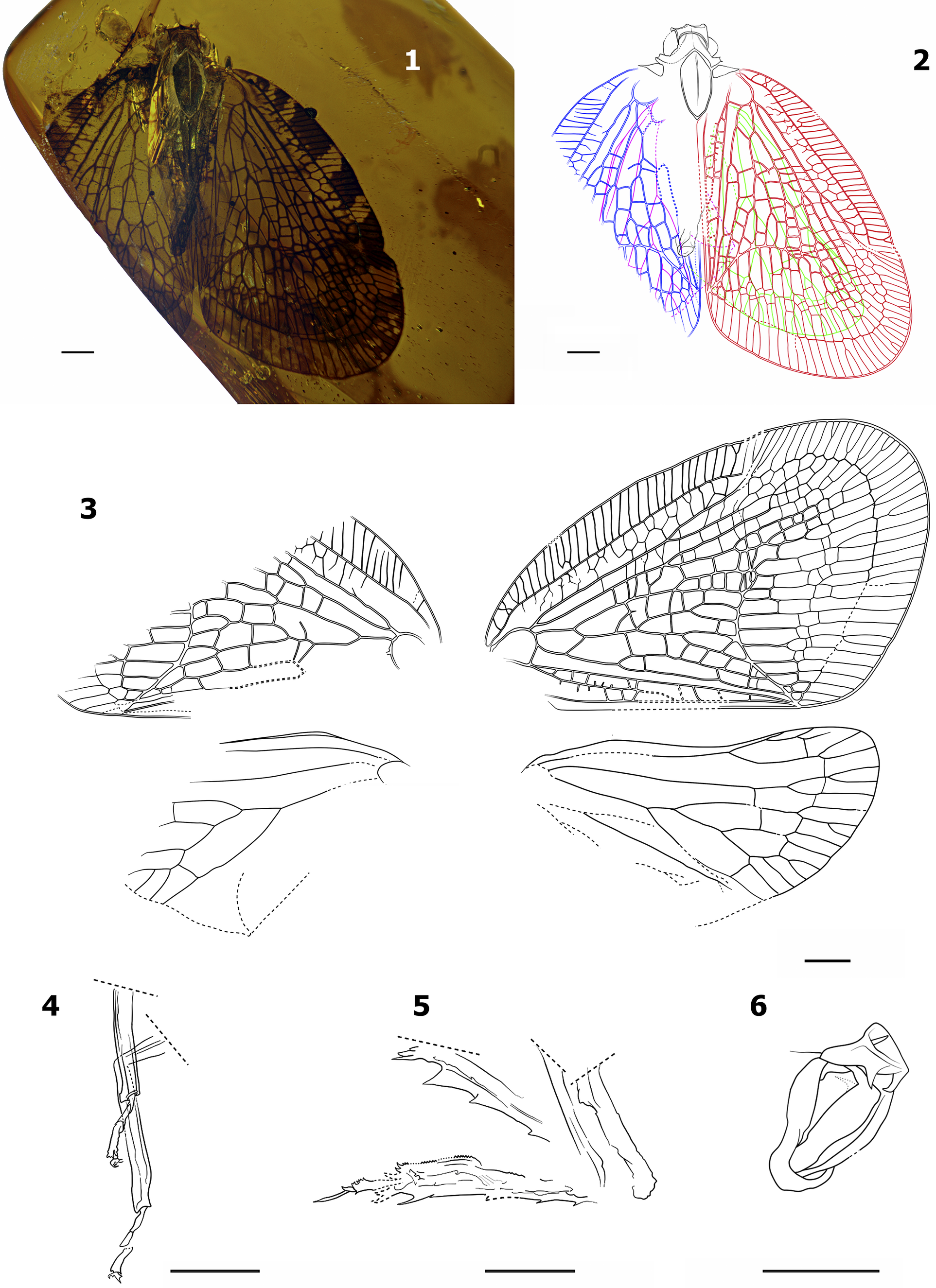
Overall habitus of Pauchus loisae Iscavel and Pinedo-Escatel n. gen. n. sp., holotype IGM 14695. (1) Specimen in resin, dorsal view; (2) head, thorax, anal tube (black), right (red) and left (blue) tegmen, left (fuchsia) and right (green) hindwing, dorsal view; (3) schematic details of tegmen and hindwings; (4) first (tibiae and tarsomeres) and second (femur, tibia, and tarsomeres) legs of the right side, ventral view; (5) femur, tibiae, and tarsomeres of both hind legs, ventral view; (6) genitalia, lateral view. All scales = 1 mm.

Figure 1. Long description
The plate consists of six panels, each with a 1 mm scale bar.
Panel 1: A photograph of the holotype specimen IGM 14695 encased in amber-colored resin, shown in dorsal view with wings spread.
Panel 2: A dorsal view line drawing of the specimen. The head, thorax, and anal tube are outlined in black. The right tegmen is red, the left tegmen is blue, the left hindwing is fuchsia, and the right hindwing is green.
Panel 3: Detailed black and white schematics of the wing venation. The top pair shows the larger, more complex tegmina with dense reticulate venation. The bottom pair shows the smaller hindwings with simpler venation patterns.
Panel 4: Ventral view line drawing of the right-side first and second legs. The first leg shows the tibiae and tarsomeres. The second leg shows the femur, tibia, and tarsomeres.
Panel 5: Ventral view line drawing of both hind legs, illustrating the femur, tibiae, and tarsomeres with visible spines along the tibial margins.
Panel 6: A lateral view line drawing of the genitalia, showing a curved, complex structure with internal sclerotized elements.
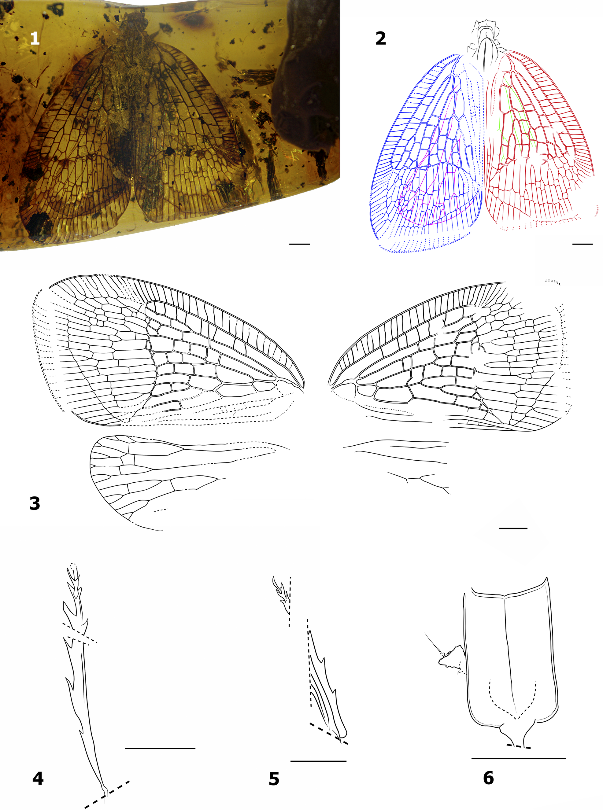
Overall habitus of Pauchus tsakal Iscavel and Pinedo-Escatel n. gen. n. sp., holotype IPN PAL 221. (1) Specimen in resin, dorsal view; (2) head and thorax (black), right (red) and left (blue) tegmen, left (fuchsia) and right (green) hindwing, dorsal view; (3) schematic details of tegmen and hindwings; (4) right hind tibiae, ventral view; (5) left hind tibiae, ventral view; (6) rostrum. All scales = 1 mm.

Figure 2. Long description
The figure consists of six panels, each with a 1 mm scale bar.
Panel 1: A photograph of the holotype specimen preserved in yellowish-brown translucent resin. The insect is shown in dorsal view with its wings spread, revealing intricate venation patterns.
Panel 2: A color-coded line drawing corresponding to the dorsal view in panel 1. The head and thorax are outlined in black. The right tegmen is red, the left tegmen is blue, the left hindwing is fuchsia, and the right hindwing is green.
Panel 3: A schematic line drawing showing the isolated details of the tegmen and hindwings. The upper pair shows the large, triangular forewings with dense, reticulated venation. The lower pair shows the smaller hindwings with simpler branching veins.
Panel 4: A line drawing of the right hind tibiae in ventral view, showing a long, slender structure with several sharp, thorn-like spines projecting along the outer edge.
Panel 5: A line drawing of the left hind tibiae in ventral view, showing a similar spined structure, though partially fragmented as indicated by dashed lines.
Panel 6: A line drawing of the rostrum, showing a rectangular base with a small, pointed lateral projection on the left side, indicated by a thin arrow.
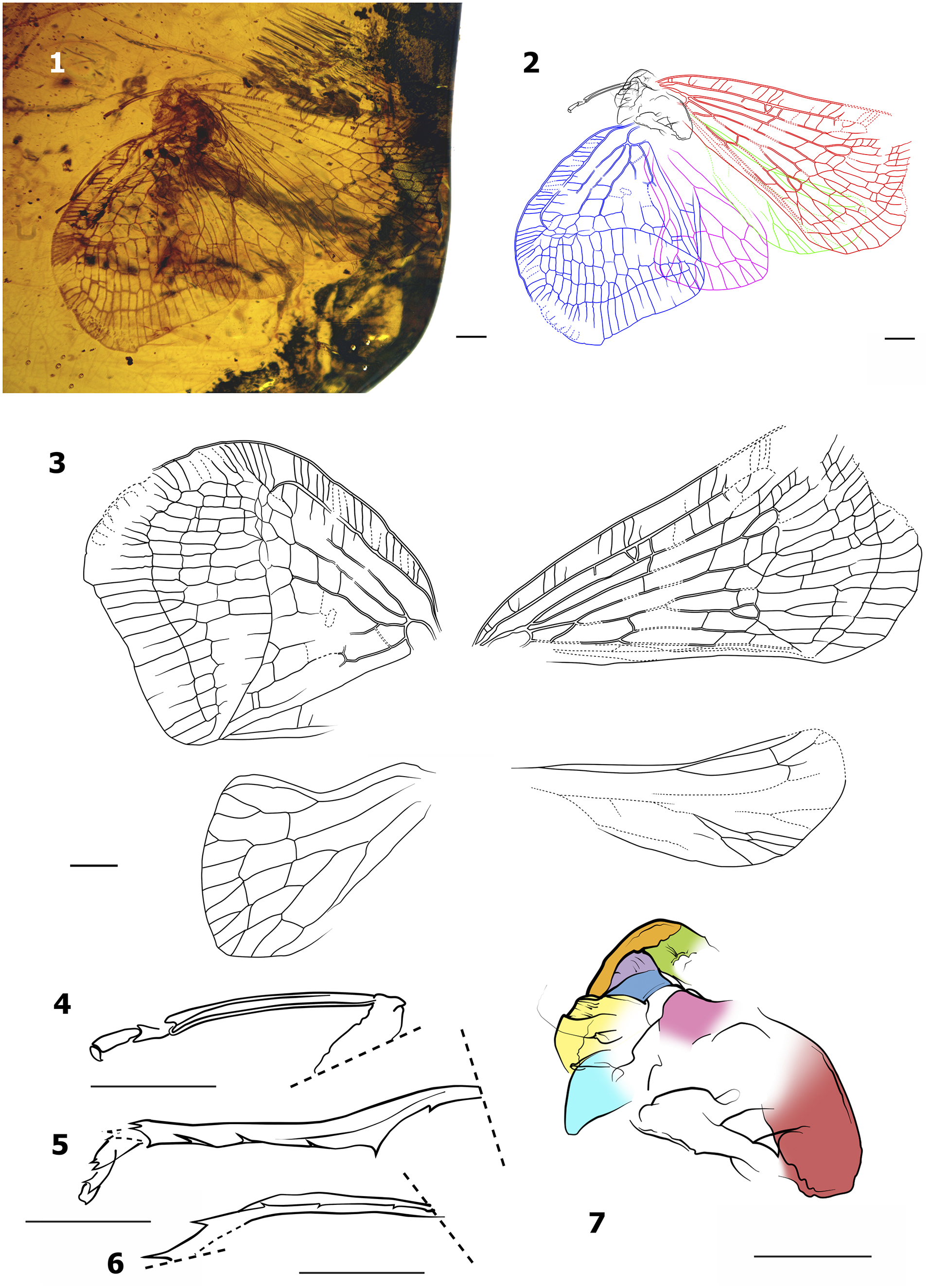
Overall habitus of Pauchus nensak Iscavel and Pinedo-Escatel n. gen. n. sp., holotype IGM 14696. (1) Specimen in resin, dorsal view; (2) head and thorax (black), right (red) and left (blue) tegmen, left (fuchsia) and right (green) hindwing, dorsal view; (3) schematic details of tegmen and hindwings; (4) femur, tibiae, and tarsomeres of the right foreleg, ventral view; (5) right hind tibiae and tarsomeres, dorsal view; (6) left hind tibiae and tarsomeres, dorsal view; (7) head and thoracic regions: vertex (green), internal side of right lateral carinae (orange), internal side of middle carinae (lilac), left half of frons (navy blue), portion of left carinae and left antennae (yellow), clypeus (cyan), pronotum (magenta), and posterior area of mesonotum (red). All scales = 1 mm.

Figure 3. Long description
A multi-panel figure detailing the morphology of a fossil insect.
* Panel 1: A photograph of the specimen preserved in yellow-orange amber, showing the dorsal view of the body and wings.
* Panel 2: A color-coded line drawing of the dorsal habitus. The head and thorax are black. The right tegmen is red, and the left tegmen is blue. The left hindwing is fuchsia, and the right hindwing is green.
* Panel 3: Four separate black-and-white line drawings showing the schematic venation of the tegmina and hindwings.
* Panel 4: A ventral view of the right foreleg showing the femur, tibiae, and tarsomeres.
* Panel 5: A dorsal view of the right hind tibiae and tarsomeres.
* Panel 6: A dorsal view of the left hind tibiae and tarsomeres.
* Panel 7: A detailed color-mapped diagram of the head and thoracic regions. The vertex is green. The internal side of the right lateral carinae is orange. The internal side of the middle carinae is lilac. The left half of the frons is navy blue. Portions of the left carinae and left antennae are yellow. The clypeus is cyan. The pronotum is magenta. The posterior area of the mesonotum is red.
Each panel includes a scale bar representing 1 mm.
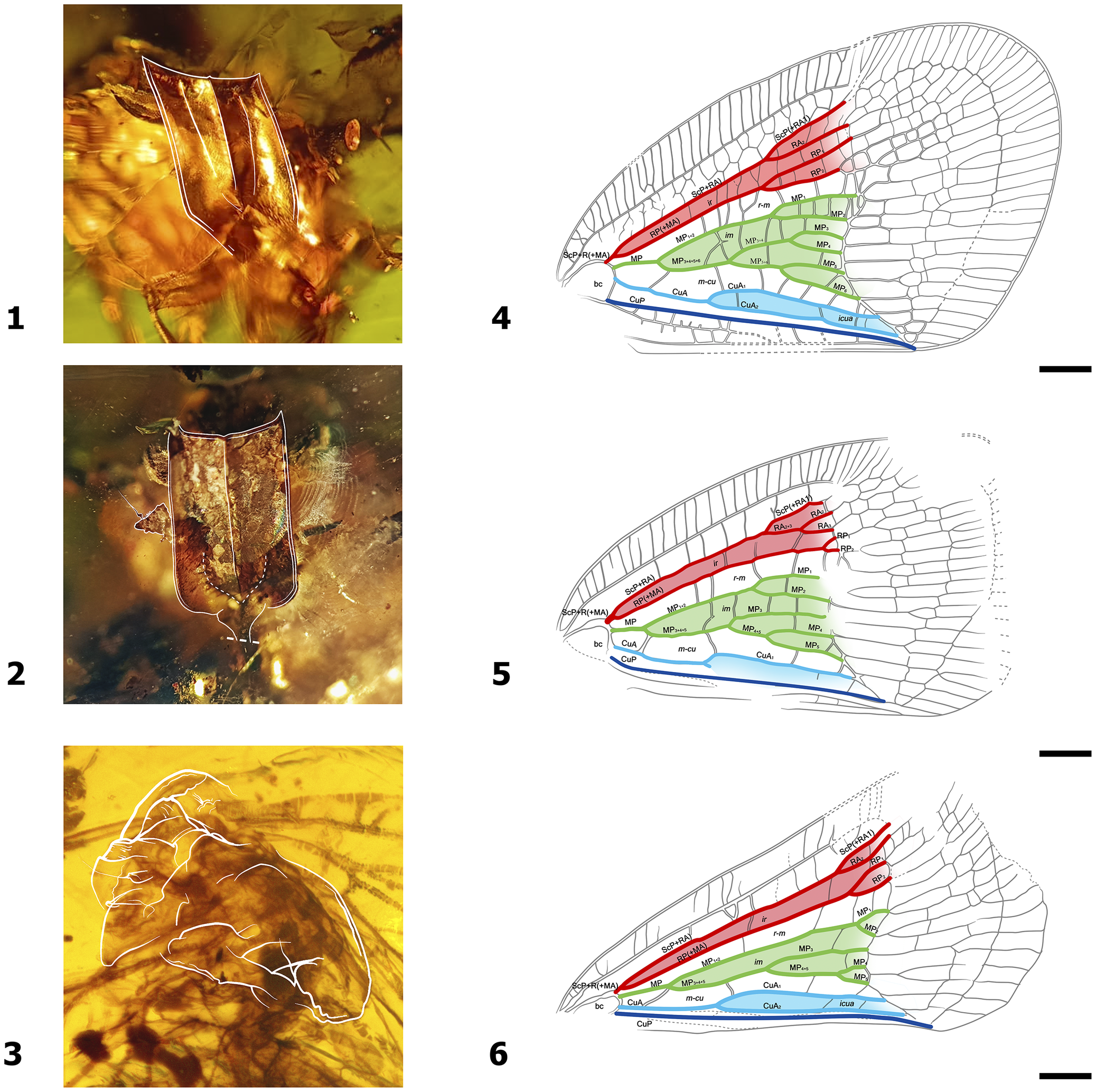
Comparative morphology of frons and right tegmen among species of Pauchus Iscavel and Pinedo-Escatel n. gen. (1, 4) Pauchus loisae Iscavel and Pinedo-Escatel n. gen. n. sp., holotype, IGM 14695; (2, 5) Pauchus tsakal Iscavel and Pinedo-Escatel n. gen. n. sp., holotype, IPN PAL 221; (3, 6) Pauchus nensak Iscavel and Pinedo-Escatel n. gen. n. sp., holotype, IGM 14696. ScP+RA(+MA) veins and C1 cells in red; MP veins and C3 cells in green; CuA veins and C5 cells in blue; CuP vein in navy blue. All bar scales = 1 mm.

Figure 4. Long description
A six-panel scientific plate organized into two columns.
Left Column (Photos 1, 2, 3):
* Panel 1: A dorsal view of the frons of Pauchus loisae preserved in amber, highlighted with a white outline showing a rectangular, shield-like structure.
* Panel 2: A dorsal view of the frons of Pauchus tsakal, outlined in white, showing a similar but slightly more elongated shield shape.
* Panel 3: A lateral view of the head and thorax of Pauchus nensak in amber, with a white outline tracing the profile of the frons and pronotum.
Right Column (Line Drawings 4, 5, 6):
* Panel 4: Right tegmen of Pauchus loisae. The venation is color-coded. The S c P plus R A plus M A veins and C 1 cells are red. The M P veins and C 3 cells are green. The C u A veins and C 5 cells are light blue. The C u P vein is a thick navy blue line at the bottom. Labels include b c, r minus m, i r, and i m.
* Panel 5: Right tegmen of Pauchus tsakal. It follows the same color scheme as panel 4 but shows a different branching pattern in the R A and R P sectors.
* Panel 6: Right tegmen of Pauchus nensak. It follows the same color scheme, showing a more condensed arrangement of the M P green cells compared to the other two species.
Each panel in the right column includes a black scale bar representing 1 mm at the bottom right.
Type species
Pauchus loisae Iscavel and Pinedo-Escatel n. sp.; here designated.
Included species
Three fossil species embedded in Mexican amber: Pauchus loisae Iscavel and Pinedo-Escatel n. gen. n. sp., IGM 14695 (Fig. 1); P. tsakal Iscavel and Pinedo-Escatel n. gen. n. sp., IPN PAL 221 (Fig. 2); and P. nensak Iscavel and Pinedo-Escatel n. gen. n. sp., IGM 14696 (Fig. 3).
Diagnosis
Pauchus n. gen. is characterized by the presence of short ScP+R(+MA) vein, unlike Nogodina Stål, Reference Stål1859, where vein is completely reduced to absence and significatively shorter than other genera in the subtribe. In addition, subparallel markings on the frons; a transverse vein in the median cell running after the first bifurcation of MP to CuA, similar to Neovarcia, Varciopsis tenguelana Fennah, Reference Fennah1945, and V. vitripennis (Lethierry, Reference Lethierry1890); transverse veins on the clavus; and five lateral spines in the hind tibia (also in Biolleyana).
Head
Overall body form slender and moderately robust. Vertex (Figs. 1.1, 1.2, 2.1, 2.2, 4.3) 2.5–3.5× wider than long; frons longer than wide; superior margin convex; lateral margin parallel, strongly carinated, and inconspicuously wider at base; short subparallel faint pair of sutures between medial and lateral carina converging near to clypeus (Fig. 4.1, 4.2); middle carina prominent; frontoclypeal suture indiscernible. Compound eyes prominent in dorsal and lateral view.
Thorax
Pronotum (Figs. 1.1, 1.2, 2.1, 2.2, 4.1, 4.2) surface depressed, surrounding margins strongly carinated, striated or rugous lengthwise; middle carina present whether complete or incomplete; anterior margin medially truncated and extended beyond middle length of eyes; subparallel convex anterolateral margins; posterior margin strongly emarginated. Mesonotum subtriangular; middle carinae conspicuous and not reaching anterior margin; carinae conspicuous and anteriorly convergent.
Legs
Hind tibia (Figs. 1.4, 1.5, 2.4, 2.5, 3.4–3.6) formula 5+5; interior margin serrated (discernible only on P. loisae n. gen. n. sp.).
Tegmina
Macropterous (Figs. 1.1–1.3, 2.1–2.3, 3.1–3.3, 4.4–4.6) with apex beyond terminalia, longer than wide, surface membranous, venation highly reticulated; shape triangularly obtuse with rounded apex; mostly hyaline with colored stripes across the costal and apical area; corium with pterostigma. Tegulae 1.6–2.7× longer than wide in dorsal view. Costal area as wide or wider than post-costal cell, usually with 28–38 transverse veins and six pigmented stripes from base to pterostigma (except P. nensak n. gen. n. sp.); small, pigmented spot near pterostigma at the end of RA or RP. Post-costal cell extends partially through length of tegmina. Basal cell oblong with subsequent emergence of ScP+R(+MA), MP, and CuA veins. ScP+R(+MA) vein greatly reduced, forks into ScP+RA and RP(+MA) immediately after emerging from basal cell and split around the halfway length of the tegmina. MP vein 4× longer than ScP+R(+MA), forks 4–5× before nodal line. CuA vein 9.5× longer than ScP+R(+MA), forks once before nodal line. Apical border of the tegmina may have irregular pigmented spots that fade towards the edge.
Abdomen
Conic and narrowing to apex; significantly shorter than total length of the tegmina; usually fading from black to brown.
Terminalia
Anal tube conical, projected beyond genital capsule, 2× longer than wide, 1.3× longer than gonostyles in lateral view, with two teeth-like ventral projections pointing downwards. Gonostyles elongated, 3.3× longer than the maximum width; sharply bent into a hook pointing upwards towards the apex (similar to Biolleyana spp.), hook-like projection 0.4× longer than gonostyle total length; no visible bristles (Fig. 1.6).
Etymology
The name of the new genus is based on the Tzotzil word “pauch” which means “amber”. Tzotzil is a variation belonging to the Mayan language family commonly spoken in the Highlands of Chiapas. Gender neutral.
Measurements
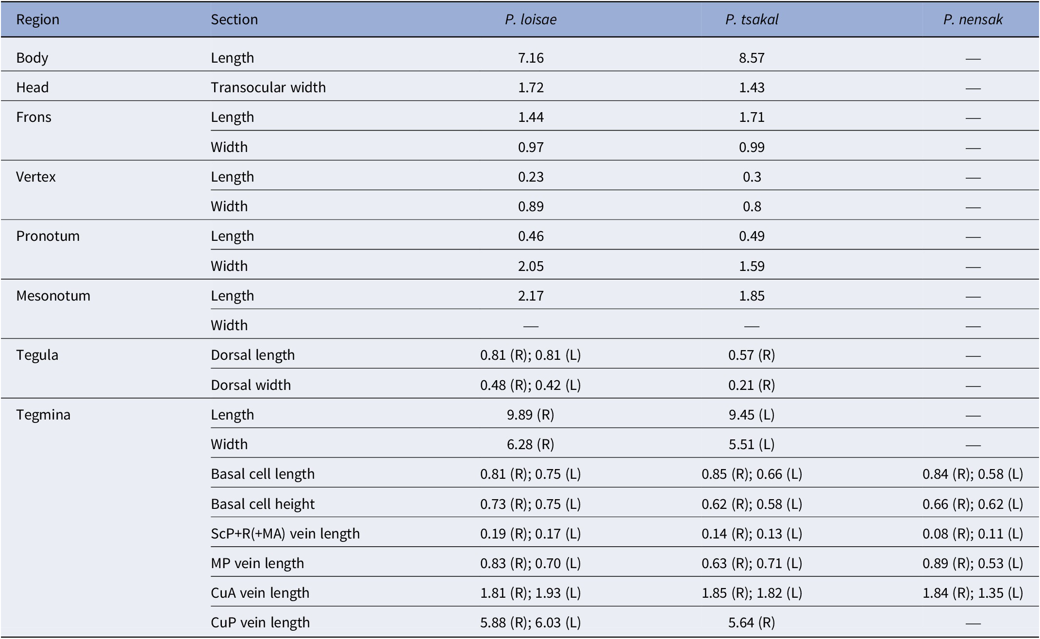
Bodies 7.16–8.57 mm, details for species as shown in Table 1.
Overall body measurement of all species of Pauchus Iscavel and Pinedo-Escatel n. gen. in millimeters.

Table 1. Long description
The table lists measurements in millimeters for P. loisae, P. tsakal, and P. nensak across various anatomical regions.
* Body: Length is 7.16 for P. loisae and 8.57 for P. tsakal.
* Head: Transocular width is 1.72 for P. loisae and 1.43 for P. tsakal.
* Frons: Length is 1.44 for P. loisae and 1.71 for P. tsakal. Width is 0.97 for P. loisae and 0.99 for P. tsakal.
* Vertex: Length is 0.23 for P. loisae and 0.3 for P. tsakal. Width is 0.89 for P. loisae and 0.8 for P. tsakal.
* Pronotum: Length is 0.46 for P. loisae and 0.49 for P. tsakal. Width is 2.05 for P. loisae and 1.59 for P. tsakal.
* Mesonotum: Length is 2.17 for P. loisae and 1.85 for P. tsakal.
* Tegula: Dorsal length for P. loisae is 0.81 for both Right and Left; for P. tsakal it is 0.57 Right. Dorsal width for P. loisae is 0.48 Right and 0.42 Left; for P. tsakal it is 0.21 Right.
* Tegmina: Length is 9.89 Right for P. loisae and 9.45 Left for P. tsakal. Width is 6.28 Right for P. loisae and 5.51 Left for P. tsakal. Basal cell length for P. loisae is 0.81 Right and 0.75 Left; for P. tsakal is 0.85 Right and 0.66 Left; for P. nensak is 0.84 Right and 0.58 Left. Basal cell height for P. loisae is 0.73 Right and 0.75 Left; for P. tsakal is 0.62 Right and 0.58 Left; for P. nensak is 0.66 Right and 0.62 Left. S c P plus R plus M A vein length for P. loisae is 0.19 Right and 0.17 Left; for P. tsakal is 0.14 Right and 0.13 Left; for P. nensak is 0.08 Right and 0.11 Left. M P vein length for P. loisae is 0.83 Right and 0.70 Left; for P. tsakal is 0.63 Right and 0.71 Left; for P. nensak is 0.89 Right and 0.53 Left. C u A vein length for P. loisae is 1.81 Right and 1.93 Left; for P. tsakal is 1.85 Right and 1.82 Left; for P. nensak is 1.84 Right and 1.35 Left. C u P vein length for P. loisae is 5.88 Right and 6.03 Left; for P. tsakal is 5.64 Right.
Remarks
Pauchus n. gen. bears slight similarities to Tonacatecutlius Stroiński and Szwedo, 2000, but largely differs by the presence of aforementioned subparallel small, faint pair of sutures on the frons, the noticeable extension of the ScP+R(+MA) vein, and the robust body shape. Morphology also shares traits resembling in part Tainosia Szwedo and Stroiński, Reference Szwedo and Stroiński2001. However, from the latter, second pair of carinae on mesonotum absent, transverse veins on the clavus, and fewer costal margin pigmented spots are clearly distinct in form. Features on pronotum, mesonotum, abdomen, genitalia, and hind tibia of P. nensak n. gen. n. sp. are difficult to interpret based on the translucent nature of specimen, nevertheless major traits mirror synapomorphies with P. loisae n. gen. n. sp. and P. tsakal n. gen. n. sp.
Key to species of Pauchus new genus
-
1. Costal margins of tegmen with 5–6 clearly pigmented stripes…… 2
-
—. Costal margins of tegmen without pigmented stripes, only pigmented veins = P. nensak n. gen. n. sp.
-
-
2. Transverse venation in post-costal cell. Slender head and thorax complexion = P. tsakal n. gen. n. sp.
-
—. Dense reticulate venation in post-costal cell. Wider head and thorax complexion = P. loisae n. gen. n. sp.
-
Pauchus loisae Iscavel and Pinedo-Escatel, new species
Holotype
Male, IGM 14695; Mexico: Chiapas, Simojovel de Allende, Minas de Montecristo.
Diagnosis
Vertex medially narrower than P. tsakal Iscavel and Pinedo-Escatel n. gen. n. sp. Post-costal cell venation reticulated. Transverse veins on costal cell from base to pterostigma: 13 discernable on left tegmen and right tegmen with 38.
Occurrence
Inclusion in Mexican amber (23–16 Ma), adult specimen.
Description
Vertex (Figs. 1.1, 1.2, 4.1) 3.5× wider than long; posterior margin inconspicuously carinated; frons approximately 1.3× longer than wide; eyes oblong and well visible from dorsal view.
Thorax
Pronotum (Fig. 1.1, 1.2) wider than long; surface striated lengthwise; middle carina emerges from anterior and posterior margin but not converging in the middle. Mesonotum disc 2.8× longer than wide.
Tegmina
Left tegula 1.9× longer than wide; right tegula 1.6× longer than wide. Transversal venation pigmented through costal margin; post-costal cell with dense reticulated venation (Figs. 1.2, 1.3, 4.4).
Right tegmen
Complete scheme (Figs. 1.2, 1.3, 4.4); 1.5× longer than wide. Costal border hyaline with six well-defined pigmented spots. ScP+R(+MA) forks into ScP+RA and RP(+MA) near the basal cell; ScP+RA forks into ScP(+RA1) and RA2, and RP(+MA) into RP1 and RP2, both bifurcations taking place at 2/5 of the total tegmen length. Seven transversal inter-radial veins (ir) between ScP+RA and RP(+MA); six ir veins between RA2 and RP1; nine transverse radial-medial (r-m) veins before the nodal line. MP vein forks into MP1+2 and MP3+4+5+6 at 1/5 of the total tegmen length; MP1+2 forks into MP1 and MP2 near 2/5 of the total tegmen length; MP3+4+5+6 forks into MP3+4 and MP5+6 at 1/3 the length of the tegmen; MP3+4 fork into MP3 and MP4, and MP5+6 into MP5 and MP6 between 2/5 and 1/2 of the tegmen length. Six transverse veins inter-medial (im) between MP1 and MP2; two im veins between MP1+2 and MP3+4+5+6, two between MP1+2 and MP3+4, one between MP2 and MP3+4, and one between MP2 and MP3; one im vein between MP3+4 and MP5+6, and one between MP4 and MP5; eight medial-cubital transverse (m-cu) veins. Main CuA veins extend up to 3/10 of the total length of the tegmen, then fork into CuA1 and CuA2. Four inter-CuA (icua) transverse veins between CuA1 and CuA2. CuP runs straight through 2/3 of the total length of tegmen. Pcu and A1 fuse into Pcu+A1 near 2/5 of the total length of tegmen. Total of eight transverse veins, five complete and three incomplete between CuP and Pcu; four complete transverse veins between CuP and Pcu+A1; six transverse veins, three complete and three incomplete between A1 and Pcu.
Left tegmen
Incomplete scheme (Fig. 1.2, 1.3); cut subparallel from costal posterior margin to halfway to the post-costal cell; venation roughly symmetrical to right tegmen. Four visible pigmented stripes along the costal border. ScP+R(+MA) forks into ScP+RA and RP(+MA) near the basal cell; RA(+MA) splits into RP1 and RP2 closer to the base than on the right tegmen. A single ir vein between ScP+RA and RP(+MA) discernible; another single ir vein is visible between RP1 and RP2; four r-m veins preserved. MP forks into MP1+2 and MP3+4+5+6 in a similar fashion to the right tegmen; subsequent bifurcations follow the exact same pattern described on the right tegmen. One im vein between MP1 and MP2; single arched transverse vein emerging from MP3+4+5+6 towards MP1+2 but not reaching it; two im veins between MP2 and MP3+4; one im vein between MP2 and MP3; one im vein between MP3+4 and MP5+6; one im between MP4 and MP5. Five m-cu veins. Main CuA mostly indiscernible, then forks into CuA1 and CuA2. Three visible inter-CuA (icua) transverse veins between CuA1 and CuA2. CuP, Pcu, A1, and Pcu+A1 veins indiscernible; clavus area diffuse to elucidate.
Abdomen
Abdomen is 0.3× shorter than total length of the tegmina; tergum and sclerites barely distinguishable; bent towards the left (Fig. 1.1, 1.2).
Terminalia
As stated in genus description (Fig. 1.6).
Etymology
This species is named in honor of Lois Breimeier O’Brien for her major contributions to Fulgoromorpha systematics.
Condition
Specimen well preserved and complete; head, pronotum, and right tegmina clearly visible; frons and venter opaque.
Coloration
General ground color fuscous: tegmina translucent with spots along the costal cell and apical region; body moderately robust (Fig. 1.1).
Measurements
Total body length 7.16 mm; additional structures as shown in Table 1.
Remarks
This species differs from congers in body size, number of costal spots, and supernumerary distal region of tegmen.
Pauchus tsakal Iscavel and Pinedo-Escatel, new species
Holotype
Male, IPN PAL 221; Mexico: Chiapas, Simojovel de Allende, Minas de Montecristo.
Diagnosis
Vertex medially wider than P. loisae Iscavel and Pinedo-Escatel n. gen. n. sp.; transverse venation throughout the post-costal cell; costal area with 5–6 pigmented stripes; coloration streaks through tegmen venation.
Occurrence
Inclusion in Mexican amber (23–16 Ma), adult specimen, organism complete.
Description
Vertex (Figs. 2.1, 2.2, 4.2) 2.5× wider than long; posterior margin convex and carinated. Frons 1.6× longer than wide. Clypeus and mouth pieces mostly indiscernible. Right antenna visible in dorsal view and scape discernible with basal projection, pedicelum trapezoidal apically widened, and flagellum widened at the base.
Thorax
Pronotum (Fig. 2.1, 2.2) longer than wide; texture slightly rugose; median carinae emerges from anterior margin; mesonotum disc 2.5× longer than wide.
Tegmina
Right tegula 2.7× longer than wide and left tegula indiscernible. Transverse venation pigmented near stripes; stripes along the costal border (Figs. 2.1–2.3, 4.2, 4.5).
Right tegmen
Almost complete. Five subtly pigmented stripes through costal border. A single dark spot on the intersection of the nodal line, RP1 and RP2. Post-costal cell with seven transverse veins. ScP+R(+MA) forks into ScP+RA and RP(+MA); ScP+RA forks into ScP(+RA1) and RA2+3 at 2/5 the assumed total length of the tegmen; RA2+3 forks into RA2 and RA3 halfway through the tegmen. RP(+MA) forks past the middle of the tegmen into RP1 and RP2. Two ir veins between ScP+RA and RP(+MA); one ir vein between RA3 and RP(+MA). Two r-m veins between RP(+MA) and MP1+2; two r-m veins between RP(+MA) and MP1; one r-m vein between RP2 and MP1. MP forks into MP1+2 and MP3+4+5 at 1/5 the total length of the tegmen; MP1+2 forks into MP1 and MP2 at 2/5 the total length of the tegmen; MP3+4+5 forks into MP3 and MP4+5 near 1/3 the length of the tegmen; MP4+5 forks into MP4 and MP5 past 2/5 the length of the tegmen. One i-m vein between MP1+2 and MP3+4+5; one i-m vein between MP1+2 and MP3; two i-m veins between MP2 and MP3; one i-m vein between MP3 and MP4; and one i-m vein between MP5 and the arch of transverse veins by the nodal line. One m-cu vein between CuA and MP3+4+5; one m-cu vein between MP3+4+5 and what seems to be CuA1 and another between MP4+5 and the potential CuA1. Two barely distinguishable transverse veins near the apical end of the clavus (Figs. 2.1–2.3, 4.5).
Left tegmen
Complete; 1.7× longer than wide. Six subtly pigmented stripes through costal border. Post-costal cell with nine transverse veins. ScP+R(+MA) forks into ScP+RA and RP(+MA); ScP+RA forks into ScP(+RA1) and RA2 halfway through the total length of the tegmen; RP(+MA) forks past the middle of the tegmen into RP1 and RP2. One ir vein between ScP+RA and RP(+MA); one ir vein between ScP(+RA1) and RA2. One r-m RP(+MA) and MP1+2 and three r-m RP(+MA) and MP1. Low visibility in the lower half of the medial-cubital area suggests fusion between the basal half of the MP4+5 and the second fourth section of CuA1. MP forks into MP1+2 and MP3+4+5 at 1/5 the total length of the tegmen; MP1+2 forks into MP1 and MP2 at 1/3 the total length of the tegmen; MP3+4+5 forks into MP3 and MP4+5 before 1/3 the length of the tegmen; MP4+5 fuses briefly with CuA1 before forking into MP4 and MP5 past 2/5 the length of the tegmen. One i-m vein between MP1+2 and MP3; two i-m veins between MP2 and MP3; one i-m vein between MP3 and MP4; one i-m vein between MP4 and MP5. CuA vein poorly visible, assumed fork into CuA1 and CuA2 at the same distance as MP3+4+5 forks into MP3 and MP4+5 before 1/3 the length of the tegmen. A single icua vein between CuA1 and CuA2 (Fig. 2.1–2.3).
Abdomen
Abdomen is 0.1× shorter than total length of the tegmina; tergum and sclerites barely distinguishable due to low visibility around clavus area (Fig. 2.1, 2.2).
Terminalia
As stated in genus description.
Etymology
Epithet is rooted from the Tzotzil word “ ts’akal ” meaning “whole/complete”, referencing to the good state of specimen preservation within the resin.
Condition
Specimen mostly well preserved and complete; head, frons, pronotum, right and left tegmina visible whereas venter is opaque.
Coloration
General ground color ochreous; hyaline tegmina with striped edges. Body slender; head, thorax, and tegmina visible in dorsal view; frons and right antenna visible from coronal view (Fig. 2.1).
Measurements
Total body length 8.57 mm; additional visible structures listed in Table 1.
Remarks
Numerous flat, bubble-like structures surround the organism, specially near the clavus area in both tegmens. Visible frons morphology bears resemblance to the trapezoidal frons described in Tonacatecutlius gibsoni Stroiński and Szwedo, 2000.
Pauchus nensak Iscavel and Pinedo-Escatel, new species
Holotype
Male, IGM 14696; Mexico: Chiapas, Simojovel de Allende, Minas de Montecristo.
Diagnosis
Costal area without pigmented stripes (opposite as displayed in P. loisae n. gen. n. sp. and P. tsakal n. gen. n. sp.), transverse veins within costal area show color variations resembling striped pattern in the rest of the genus.
Occurrence
Inclusion in Mexican amber (23–16 Ma), adult specimen, organism complete.
Description
Partially distorted (Figs. 3.1, 3.7, 4.3); seemingly dislocated from proper anatomical position, twisted counterclockwise and separated from thorax; vertex visible; frons with prominent middle and lateral carinae, subparallel markings on the frons indiscernible; clypeus visible; both antennae visible, scape and pedicelum enlarged, cylindrical.
Tegmina
Indiscernible tegula. Fully extended and partially distorted; longer than wide. Costal area inconspicuously wider than post-costal cell; transverse venation on costal area pigmented forming six pigmented stripes; apical edge of tegmen hyaline (Figs. 3.1–3.3, 4.6).
Right tegmen
Ten discernible transverse veins in post-costal cell. ScP+RA and RP(+MA) fork into ScP(+RA1) and RA2, and RP1 and RP2 respectively near the 2/3 of the total tegmen length. Two ir veins between ScP+RA and RP(+MA) and one between RA2 and RP1. Two r-m veins between RP(+MA) and MP1+2; one r-m vein between RA2 and MP1+2 ; two r-m veins between RA2 and MP1. MP forks into MP1+2 and MP3+4+5 by 1/5 the total length of the tegmen; MP1+2 forks into MP1 and MP2 at past half the length of the tegmen; MP3+4+5 forks into MP3 and MP4+5 before half of tegmen length; MP4+5 forks into MP4 and MP5 past half the length of the tegmen. One i-m vein between MP1+2 and MP3; one i-m vein between MP3 and MP4. One m-cu vein between CuA and the first fork of MP; one m-cu vein between MP3+4+5 and CuA1; one m-cu between MP4+5 and CuA1; and one m-cu between MP5 and the arch of transverse veins by the nodal line. CuA vein forks into CuA1 and CuA2 near 1/3 the total length of the tegmen. Two icua veins between CuA1 and CuA2; past 1/2 the total length of the tegmen. Clavus region poorly visible; a single inconspicuous transverse vein between CuP and Pcu (Figs. 3.1–3.3, 4.6).
Left tegmen
Seven transverse veins discernible in post-costal cell. ScP+RA and RP(+MA) fork into ScP(+RA1) and RA2, and RP1 and RP2, respectively near the halfway total length of the tegmen. One i-r vein between RA2 and RP(+MA); and one i-r vein between RA2 and RP1. Two r-m veins between RP(+MA) and MP1+2; one r-m vein RP(+MA) and MP1. MP forks into MP1+2 and MP3+4+5; MP1+2 forks into MP1 and MP2 before the midlength of the tegmen. MP3+4+5 poorly visible, fork into MP3 and MP4+5 indiscernible; MP4+5 forks into MP4 and MP5 near halfway through the tegmen length. CuA vein forks into CuA1 and CuA2 near 1/3 total length of the tegmen. Two icua veins between CuA1 and CuA2; past 1/3 the total length of the tegmen (Fig. 3.1–3.3).
Abdomen
Indiscernible.
Terminalia
Indiscernible.
Etymology
Epithet formed based on the Tzotzil words “nen” meaning “glass”, and “sak” meaning “clear”, referencing to the translucent appearance of the specimen studied.
Condition
Specimen partially damaged and incomplete; right and left tegmina clearly visible but head, pronotum, thorax, and abdomen are strongly compressed and deformed.
Coloration
General translucent specimen, ground color indiscernible, tegmina with stripe-like patterns over translucent veins. Head and thorax vaguely discernible; abdomen mostly indiscernible (Fig. 3.1).
Measurements
Total body length indiscernible; other well-defined structures as shown in Table 1.
Remarks
Organism displays obvious degradation on thoracic and abdominal areas; pigmentation fully absent from exoskeleton; head presumably deformed and dislocated. Pigmented vein scheme pattern resembles costal spots similar to those found on P. loisae n. gen. n. sp. and P. tsakal n. gen. n. sp., but the costal area lacks pigmentation.
Key to fossil and extant New World Nogodinini genera
-
1. Tegmina hyaline, with pigmented stripes on corium or costal area forming clear windows. Frons distinctly longer than wide (except Nogodina) = Subtribe Nogodinina ……………….. 2
-
—. Tegmina opaque, with clear windows on corium or costal area. Frons subquadrate = Subtribe Vutinina (Vutina)
-
-
2. Tegmina with pigmented stripes through corium forming three hyaline windows; multiple stripes on costal margin or with pigmented veins forming pale stripes ……………….. 3
-
—. Tegmina with uniform hyaline corium; costal margin hyaline or with narrow stripes on pterostigma ……………….. 4
-
-
3. Three veins of similar length emerging from basal cell: ScP+R(+MA), MP, and CuA. Post-costal cell with transverse veins. Shape of frons subrectangular (always longer than wide) with lateral, middle, and extra subparallel pair of carina next to central. Hind tibia with five lateral spines = Biolleyana.
-
—. Four veins of different length emerging from basal cell: SCP+RA, RP(+MA), MP, and CuA. Post-costal cell with transverse or reticulated veins. Shape of frons subquadrate (similar width and length) with lateral and middle carinae. Hind tibia with four lateral spines = Nogodina.
-
-
4. Costal margin with multiple pigmented stripes. Numerous veins on costal margin prior to pterostigma (more than 16) ……………….. 5
-
—. Costal margin hyaline, just pterostigma. Few transverse veins on costal margin prior to pterostigma (fewer than 16) ……………….. 7
-
-
5. Five spines on hind tibia. With transverse veins on clavus. Transverse vein from CuA to first fork of MP splitting median cell in half ……………….. 6
-
—. Three spines on hind tibia. No transverse veins on clavus. No transverse vein from CuA to first fork of MP splitting median cell in half = †Tainosia
-
-
6. ScP+R(+MA) vein of similar length to MP and CuA. Frons without subparallel markings between medial and lateral carina = †Tonacatecutlius
-
—. ScP+R(+MA) significantly shorter than MP and CuA. Frons with subparallel markings between medial and lateral carina = †Pauchus n. gen.
-
-
7. Tegmina elongated, almost subrectangular, more than 2× as long as wide. Mesonotum as wide as the transocular width of the head = Orthothyreus
-
—. Tegmina distinctly subtriangular, up to 1.5× longer than wide. Mesonotum wider than the transocular width of the head ……………….. 8
-
-
8. Transverse vein from CuA to MP splitting median cell present = Neovarcia
-
—. Transverse vein from CuA to MP splitting median cell absent = Varciopsis (except V. tenguelana and V. vitripennis)
-
Discussion
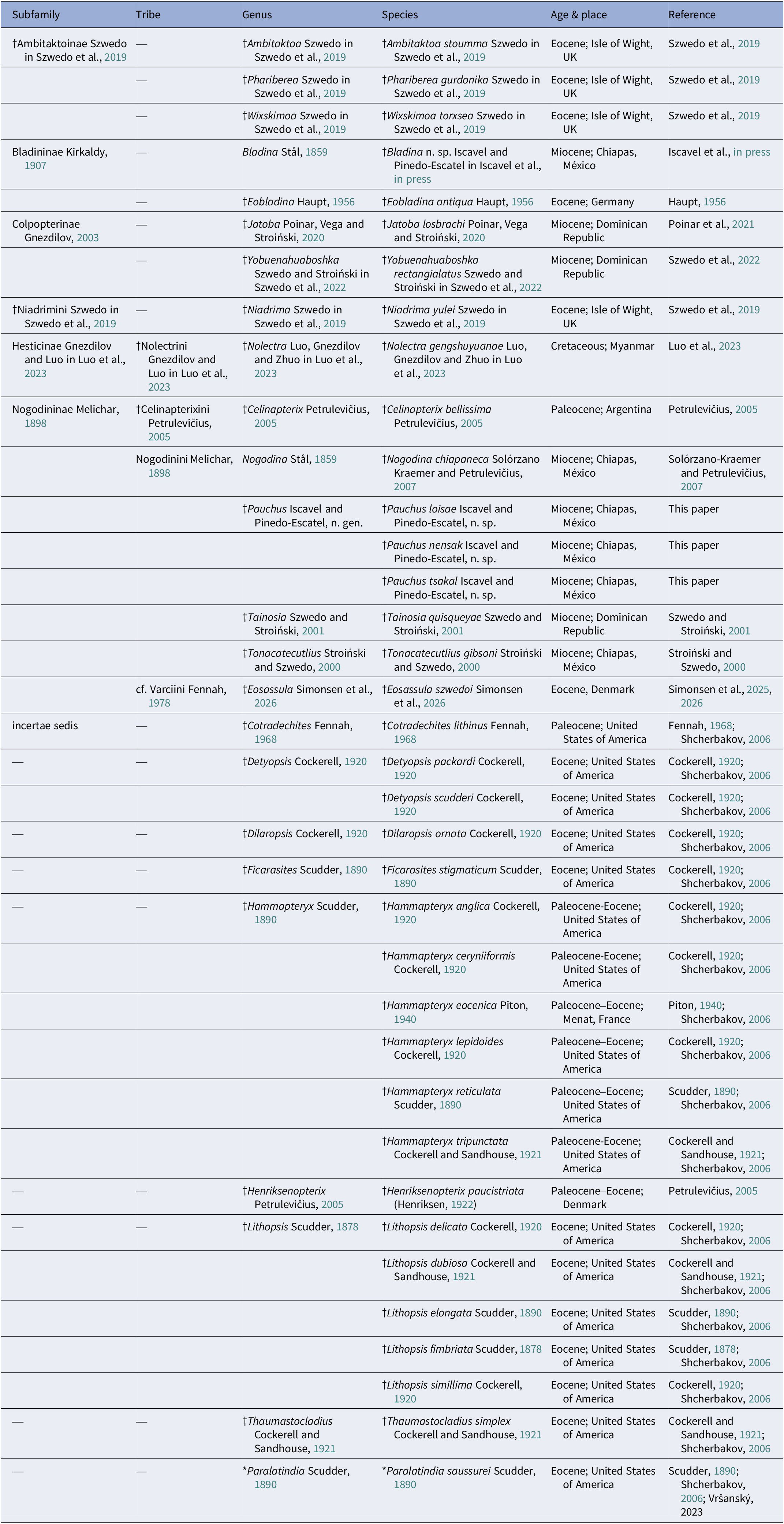
Although a cautionary approach is recommended when relying on tegmina for the study of fossil material because tegmen structure and venation represent some of the best, if not the only, preserved anatomical structures for various identified fossil Nogodinidae (e.g., Scudder, Reference Scudder1890; Cockerell, Reference Cockerell1920; Cockerell and Sandhouse, Reference Cockerell and Sandhouse1921; Haupt, Reference Haupt1956, etc.), the observation and comparison of tegmina morphology, vein arrangement, and color patterns in exceptionally preserved specimens become the main strategies when delimiting fossil taxa and defining characteristic boundaries of the family (Stroiński and Szwedo, Reference Stroiñski and Szwedo2000; Szwedo and Stroiński, Reference Szwedo and Stroiński2001). Some fossil material (Table 2) exhibits anatomical features (e.g., tegmen) that can be directly compared to living relatives using homologous traits, becoming one of the most essential resources to date for redefining a non-monophyletic group such as Nogodinidae from other similar fulgorid taxa (Stroiński and Szwedo, Reference Stroiński and Szwedo2025).
Updated record of Nogodinidae fossils from Luo et al. (Reference Luo, Gnezdilov, Zhuo and Song2023). *Taxa included in Shcherbakov (Reference Shcherbakov2006) and Vršanský et al. (Reference Vršanský, Palková, Vršanská, Koubová and Hinkelman2023) as incertae sedis.

Table 2. Long description
The table is organized into six columns.
* The first column, Subfamily, includes groups such as Ambitaktoinae, Bladininae, Colpopterinae, Niadrimini, Hesticinae, Nogodininae, and incertae sedis.
* The second column, Tribe, lists specific tribes like Nolectrini, Celinapterixini, Nogodinini, and cf. Varciini, though many entries are marked with a dash.
* The third and fourth columns list Genus and Species, including Ambitaktoa stoumma, Phariberea gurdonika, Wixskimoa torxsea, Bladina n. sp., Eobladina antiqua, Jatoba losbrachi, Yobuenahuaboshka rectangialatus, Niadrima yulei, Nolectra gengshuyuanae, Celinapterix bellissima, Nogodina chiapaneca, and three new species of Pauchus (loisae, nensak, and tsakal).
* The fifth column, Age and place, details geological periods and locations, ranging from the Cretaceous in Myanmar to the Miocene in Mexico and the Dominican Republic, the Eocene in the U K, Germany, and Denmark, and the Paleocene in Argentina and the U S A.
* The final column provides academic references for each record, including Szwedo et al. 2019, Luo et al. 2023, and several citations to Shcherbakov 2006.
Most characters used in the current description of the new genus can be found among modern and fossil taxa Nogodinini, such as similar markings on the frons towards the frontoclypeal suture, which are found on the extant species Varciopsis trigutta (Walker, Reference Walker1858) and Biolleyana costalis (Fowler, Reference Fowler1900); colored patterns along the costal edge of tegmen not dissimilar to those in Biolleyana Distant, Reference Distant1909, and Nogodina Stål, Reference Stål1859; number of lateral spines on the hind tibia in the new genus is widely shared with all known species of Biolleyana and the fossil species Tonacatecutlius gibsoni; most Nogodinina species, except for fossil Tainosia quisqueyae, possess transverse veins on the claval area (Stroiñski and Szwedo, Reference Stroiñski and Szwedo2000; Szwedo and Stroiñski, Reference Szwedo and Stroiński2001). The distinctive synapomorphy, short length of ScP+R(+MA) vein, distinguishes the new genus from other described Nogodinina; found either completely absent or fully developed and elongated in close relatives. The overall aforementioned traits create a unique combination of features that solidify its unique placement within Nogodinini.
The herein described Pauchus n. gen. and its three new species contribute to the ever-growing fossil record of the New World Nogodinini and increase known numbers of exclusive Mexican Auchenorrhyncha fauna, still unarticulated and largely undocumented. This contribution along with the previously described fossils in the same subtribe, may potentially serve as a keystone to redefine the backbone to promote validity of some groups supported with additional pipelines compiled in future phylogenetic analyses focused on this family, and reposition any non-related taxa into more appropriate families.
Data availability statement
All vouchers of specimens studied are available at the museums cited in the materials and methods section.
Acknowledgments
We thank the Colección de Paleontología of the Escuela Nacional de Ciencias Biológicas, Instituto Politécnico Nacional and the Colección Nacional de Paleontología of the Instituto de Geología, UNAM, for loaning the specimens described in this work. The first author R. Iscavel, born “R.I. Cañete-Velázquez”, is grateful to the Secretaría de Ciencia, Humanidades, Tecnología e Innovación (SECIHTI), and to the Posgrado en Ciencias Biológicas of the UNAM, for the MSc scholarship (CVU: 2008743). Thanks to C. Mayorga (CNIN, Instituto de Biología, UNAM) who kindly helped process specimens to accomplish this work. Thanks to B. Mendoza Garfias for her assistance at LANABIO (Laboratorio Nacional de Biodiversidad), Instituto de Biología, UNAM. Thanks to curator D.I. Rendon Mera from the Natural History Museum, London, for her support and for giving us access to primary type Nogodinid material. The first author also wholeheartedly thanks E.M. Velázquez Gómez, E.X. Cañete Velázquez, and R. Cañete Ibáñez for their never-ending love and support throughout their life; and to all supportive friends at the CNIN, Instituto de Biología, UNAM. This research was supported in full by PAPIIT-DGAPA-UNAM (grant IA203825). Collection of specimens IGU 14695 and IGU 14696 were made possible through UNAM-DGAPA-PAPIIT (grant IN207314).
Author contribution
Conceptualization (RI and JAPE), methodology (RI), original draft preparation (RI and JAPE), writing (RI, JAPE, EER, and JMJM), and funding acquisition (JAPE).
Funding
This research was supported by the UNAM-DGAPA-PAPIIT (grant IA203825).
Competing interests
The authors declare no competing interests.

 Open access

