Scimitar syndrome is a rare CHD composed of a partial anomalous pulmonary venous connection from the right lung to the inferior vena cava via a scimitar vein. The birth prevalence of scimitar syndrome is approximately 1 in 50,000 live births. Reference Gudjonsson and Brown1 Scimitar syndrome is often associated with varying degrees of right lung hypoplasia and patients with scimitar syndrome often suffer from chronic lung parenchymal disease, asthma, and recurrent respiratory infections. Reference Wang, Kalfa and Rosenbaum2 There is also an increased prevalence of right pulmonary artery hypoplasia, atrial and ventricular septal defects, coarctation of the aorta, tetralogy of Fallot, and other congenital heart lesions including single-ventricle lesions. Reference Gudjonsson and Brown1,Reference Wang, Kalfa and Rosenbaum2 Patients with scimitar syndrome and multiple congenital anomalies can be challenging to manage due to their multiple organ system pathologies.
Although some forms of CHD have been linked to various genetic syndromes, the mechanisms that contribute to scimitar syndrome remains elusive. Reference van Nisselrooij, Lugthart and Clur3–Reference Evans, Acherman, Ciccolo, Castillo and Restrepo9 Studies have demonstrated an association between scimitar syndrome and extracardiac anomalies including congenital diaphragmatic hernia, imperforate anus, and variants of VACTERL association. Reference Ashida, Itoh and Naruko10–Reference Lloyd, Rowe, Erickson, Paxton, South and Alashari14
Although pathogenic variants in specific genes can clearly cause CHDs, the genetic factors contributing to most cases of scimitar syndrome remain unidentified. Reference van Nisselrooij, Lugthart and Clur3–Reference Evans, Acherman, Ciccolo, Castillo and Restrepo9 In this study, we sought to determine the frequency and efficacy of genetic testing in patients with scimitar syndrome from a single institution. We also use the results of clinically based genetic testing to identify genes and pathways whose alteration may lead to scimitar syndrome development. We hypothesiszed that genetic testing would be performed in a minority of patients with scimitar syndrome but would have a considerable rate of positive findings.
Methods
This retrospective cohort study included all patients evaluated at Texas Children’s Hospital (TCH) from January 1987 to July 2019 diagnosed with Scimitar syndrome. Patients were first identified from institutional echocardiographic and clinical databases. All patient charts were reviewed to confirm an anatomic diagnosis of scimitar syndrome. A portion of these patients have been used in a prior publication from our institution, focusing on different aspects than our study and not consisting of our entire cohort. Patients were only included if they had been seen by a provider and had sufficient data for chart review. Coexisting diagnoses were also identified and extracardiac defects were classified as: asthma (any diagnosis of asthma, irrespective of medical management), right lung hypoplasia (if documented on imaging), tracheal stenosis, frequent respiratory infections (if indicated as such by the provider), pulmonary sequestration (if indicated on cross-sectional imaging), any gastrointestinal symptoms (poor feeding and failure to thrive), or status post-nasogastric feeding requirement or gastrointestinal tube placement, liver dysfunction, omphalocele, pyloric stenosis or imperforate anus. Developmental delay, intellectual disability, and psychiatric diagnosis (attention-deficit hyperactivity disorder, autism spectrum disorder, anxiety disorder, and obsessive–compulsive disorder) were also identified. Cardiac abnormalities were identified by review of echocardiograms, MRI, and CT and reported, with the exclusion of patent foramen ovale and small patent ductus arteriosus.
All clinically obtained genetic testing was reviewed, including chromosome analysis, chromosomal microarray analyses, and whole-exome sequencing. Chromosome analysis and/or single gene or panel sequence testing in isolation were not performed on any patients in this cohort. Positive findings on genetic testing were labelled by the third-party laboratory as “abnormal” or indicative of a pathologic change. All copy number variants reported on chromosomal microarray analysis were recorded and results were described. Variants identified by whole-exome sequencing were interpreted according to the American College of Medical Genetics and Genomics guidelines. Reference Richards, Aziz and Bale15 Studies that revealed “pathogenic” or “likely pathogenic” variants in genes associated with the patient’s phenotype were considered positive if their inheritance pattern was also consistent the proposed diagnosis. Variants of unknown significance found in genes that associated with the patient’s phenotype were also recorded and results described.
Descriptive analyses included the assessment of categorical variables expressed as counts and percentages. This study was approved by the Baylor College of Medicine Institutional Review Board.
Results
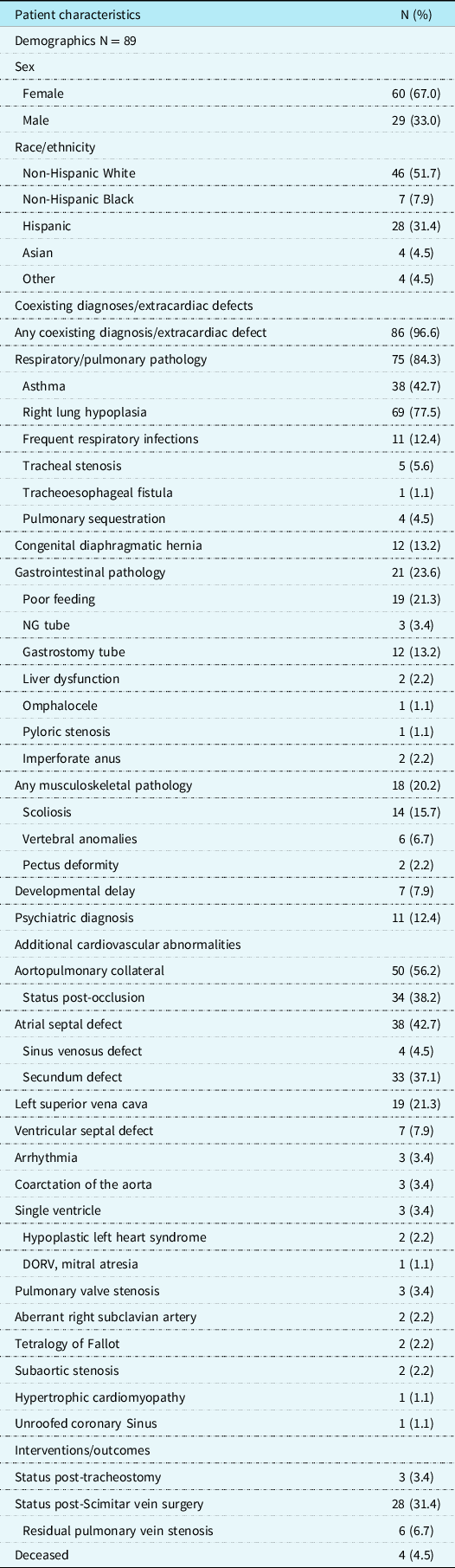
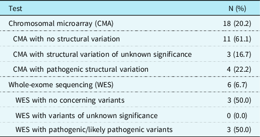
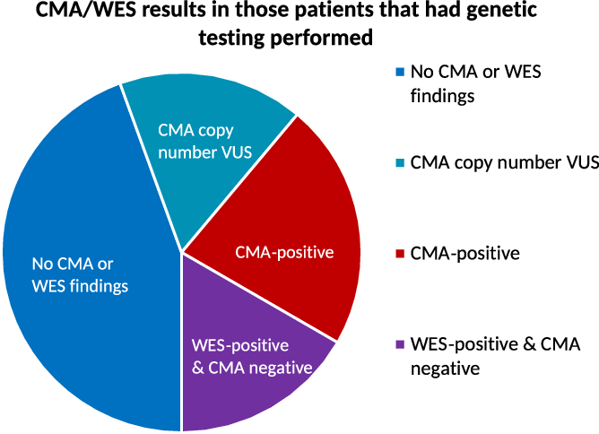
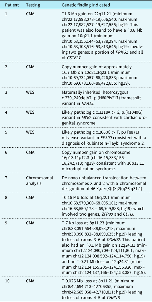
Ninety-eight patients with scimitar syndrome were identified. Eighty-nine of these patients met study criteria (Table 1). The majority of patients were female (60/89, 67.0%). Of these 89 patients, 18 patients (20.2%) had chromosome analysis or chromosomal microarray analysis testing performed and six (7%) had whole-exome sequencing performed in addition to chromosomal microarray analysis (Table 2). No genetic tests were performed prior to 2000, and the majority of genetic testing was performed since 2010, with 16 of 18 chromosomal microarray analyses performed after 2010 and all whole-exome sequencing performed after 2010 (supplemental Figure 1). Of the individuals for whom chromosomal microarray analysis was obtained, 4 of 18 (22.2%) revealed clearly pathogenic copy number variants (Patients 1, 2, 6, and 7). Three patients had positive genetic findings delineated by whole-exome sequencing. Hence, a genetic diagnosis was made in 7 of 18 (38.9%) of patients in whom genetic testing was obtained (Fig 1).
Patient characteristics

DORV = double-outlet right ventricle; NG = nasogastric.
Genetic testing results in patients with SS

Pie graph indicating the distribution of chromosomal microarray (CMA) and/or whole-exome sequencing (WES) results on patients with SS. No CMA or WES findings were noted in 8 of 18 patients (44%). CMA revealed copy number variants of unknown significance (VUS) in 3 of 18 (17%). CMA was positive in 4 of 18 (22%) and WES was positive, with negative CMA, in 3 of 18 (17%).

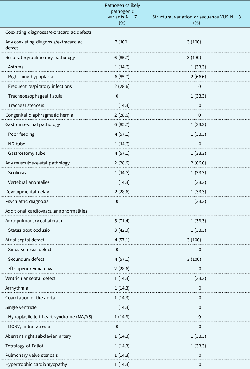
Extracardiac defects and coexisting diagnoses are described in Table 1, and at least one of these was identified in a total of 86 of 89 patients (96.6%). Among the entire cohort, 43% of patients were diagnosed with asthma compared to 14% of the genetic-positive subgroup. Four patients were found to have pulmonary sequestration. Congenital diaphragmatic hernia was identified in 13% of the entire cohort and 29% in the genetic-positive subgroup. Any gastrointestinal pathology was identified in only 24% of the entire cohort versus 86% of the genetic-positive subgroup. All patients that had positive genetic findings had some type of extracardiac finding (Table 3) and their genetic findings are summarised in Table 4.
Cardiac and extracardiac findings in patients with pathogenic/likely pathogenic or VUS on genetic testing

AS = aortic stenosis; DORV = double-outlet right ventricle; MA = mitral atresia; NG = nasogastric.
All values as n (%).
Summary of genetic testing results

CMA = chromosomal microarray; WES = whole-exome sequencing.
Four patients were deceased, none of which had genetic testing performed. One patient had VACTERL syndrome with a transitional atrioventricular septal defect, subaortic stenosis, and coarctation of the aorta. No further information surrounding the death was available. A second patient had hypoplastic left heart syndrome (mitral and aortic atresia) and underwent stage 1 palliation with Norwood/Sano repair and then stage 2 palliation with Glenn repair. The patient had poor PO feeding, intestinal malrotation, and pulmonary sequestration but was eventually discharged from the hospital following Glenn palliation. No further information surrounding the death was available. A third patient had a secundum atrial septal defect, right-sided congenital diaphragmatic hernia, hemivertebrae, and a sacral anomaly. She had severe lung hypoplasia and oxygen requirement and was managed with home hospice care. A fourth patient had a secundum atrial septal defect as well as horseshoe lung, tracheal stenosis requiring surgical repair and subsequent tracheal stent, chiari malformation, and pulmonary hypertension and died due to pulmonary hypertensive crisis.
Clinical and molecular summaries
Seven patients (Patient 1–7) had a definitive molecular diagnosis made through genetic testing. Three patients (Patients 8–10) had copy number variants identified on chromosomal microarray analysis and that were of variants of unknown significance. None of these patients had whole-exome sequencing performed. Patient 11 did not have genetic testing but was diagnosed on a clinical basis with Marfan syndrome.
Patient 1
Patient 1 had scimitar syndrome with a congenital diaphragmatic hernia, right lung hypoplasia, right pulmonary artery narrowing, dextroposition of the heart, secundum atrial septal defect, and aortic arch elongation. Chromosomal microarray analysis demonstrated ˜1.6 Mb gain on 22q11.21 (minimum chr22:17,998,078–19,606,540; maximum chr22:17,982,527–19,627,555; hg19). The proximal portion of the DiGeorge critical region was involved in the duplication, but the TBX1 gene is located outside of the duplication. Duplications involving the DiGeorge critical region have a variable phenotype with significant clinical overlap to 22q11.2 deletion syndrome that has been described in association with total anomalous pulmonary venous return. Reference De La Rochebrochard, Joly-Hélas and Goldenberg16,Reference Hasten, McDonald-McGinn and Crowley17
This patient was also found to have a ˜0.6 Mb gain on 10q21.1 (minimum chr10:53,155,144–53,788,294, maximum chr10:53,108,516–53,813,645; hg19) involving two genes; a portion of PRKG1 and all of CSTF2T. Missense variants in PRKG1 are associated with autosomal-dominant aortic aneurysm, familial thoracic 8 through a gain-of-function mechanism. Reference Guo, Regalado and Casteel18 It is possible that PRKG1 may be disrupted by this gain, but data from normal individuals catalogued in the gnomAD database (https://gnomad.broadinstitute.org/) suggest that haploinsufficiency of PRKG1 is unlikely to be associated with a significant phenotype. Similarly, a gain of CSTF2T is not currently associated with a known phenotype. Hence, is it unlikely that this change contributed to the development of scimitar syndrome in this patient.
Patient 2
Patient 2 had scimitar syndrome with right lung hypoplasia as well as secundum atrial septal defect, coarctation of the aorta, and pulmonary valve stenosis. This patient also had extracardiac findings including congenital diaphragmatic hernia, osteopenia and rib fractures, failure to thrive with nasogastric feeding tube requirements, and nystagmus. Chromosomal microarray analysis demonstrated a copy number gain of approximately 16.7 Mb on 10q21.3q23.1 (minimum chr10:69,734,057–86,426,833; maximum chr10:69,678,160–86,472,655; hg19) that overlaps the region associated with 10q22.3-q23.2 deletion syndrome. Duplications overlapping that which was seen in this individual have been reported in patients with CHD, dysmorphisms, and delays in speech and motor development. Reference Tang, Yang and Xie19,Reference Erdogan, Belloso and Gabau20
Patient 3
Patient 3 had scimitar syndrome with right lung hypoplasia as well as tetralogy of Fallot, an aortopulmonary collateral to the right side requiring device occlusion, a secundum atrial septal defect, and hypertrophic cardiomyopathy. Of note, the patient developed progressive severe biventricular hypertrophy and underwent orthotopic heart transplant with reimplantation of the scimitar vein at 22 months of age. The reimplanted scimitar vein developed stenosis requiring transcatheter angioplasty and stenting of the vein. The patient had a normal chromosomal microarray analysis. Trio whole-exome sequencing revealed a maternally inherited, heterozygous c.239_240delAT, p.(H80Rfs*17) frameshift variant in NAA15. This gene encodes an acetyltransferase subunit and has a role in maintaining cell proliferation. Reference Sugiura, Adams and Corriveau21 Changes in this gene have been identified in separate patients with complex CHDs, hypertrophic cardiomyopathy, and developmental delay. Reference Ritter, Berger and Deardorff22 Variants in this gene have also been described in patients with isolated congenital diaphragmatic hernia. Reference Longoni, Pober and High23 No report was available on whether the mother was symptomatic.
Patient 4
Patient 4 had scimitar syndrome and right lung hypoplasia, with hypoplastic left heart syndrome with mitral atresia/aortic stenosis, left ventricular sinusoids, and severe coarctation. The patient underwent single-ventricle palliation consisting of a Norwood procedure and, subsequently, a Glenn palliation. Extracardiac findings for this patient included feeding intolerance requiring gastrostomy tube placement, global developmental delay, and a para-oesophageal hernia. Chromosomal microarray analysis was normal. Whole-exome sequencing revealed a likely pathologic c.3118A > G, p.(R1040G) variant in MYRF consistent with cardiac urogenital syndrome. Reference Qi, Yu and Zhou24–Reference Rossetti, Glinton and Yuan26 This patient has previously been reported in the literature. Reference Rossetti, Glinton and Yuan26
Patient 5
Patient 5 had scimitar syndrome, an aberrant right subclavian artery, and a left superior vena cava. The patient also had extracardiac findings of failure to thrive, global developmental delay, right lung hypoplasia, congenital diaphragmatic hernia, and a laryngeal cleft. His chromosomal microarray analysis was normal. Whole-exome sequencing revealed a likely pathologic c.2660C > T, p.(T8871) missense variant in EP300 consistent with a diagnosis of Rubinstein–Taybi syndrome 2, which has a known association with CHDs. Reference Stevens and Bhakta27
Patient 6
Patient 6 had scimitar syndrome with an atrial septal defect and a history of supraventricular tachycardia along with pyloric stenosis. Chromosomal microarray analysis revealed a gain on chromosome 16p13.11p12.3 (chr16:15,333,155–18,242,713; hg19) consistent with 16p13.11 microduplication syndrome. Individuals with this syndrome can have CHDs Reference Warburton, Ronemus and Kline28,Reference El Khattabi, Heide and Caberg29 as well as aortopathy. Reference Kuang, Guo and Prakash30,Reference Erhart, Brandt and Straub31 Developmental delay, seizure disorder, autism spectrum disorder, and speech and learning disorders have also been seen in patients with 16q13.11 microduplications.
Patient 7
Patient 7 had scimitar syndrome with perimembranous and muscular ventricular septal defects, a history of poor feeding, developmental delay and intellectual disability. A chromosome analysis revealed a de novo unbalanced translocation between chromosomes X and 2 with a chromosomal designation of 46,X,der(X)t(X;2)(q26;q31.1). Additional studies demonstrated preferential inactivation of the abnormal X chromosome, with no inactivation of the translocated material from chromosome 2. This results in the presence of three active copies of the distal long arm of chromosome 2 in most cells.
Patient 8
Patient 8 had scimitar syndrome with tetralogy of Fallot as well as ectrodactyly, right lung hypoplasia, scoliosis, thoracic vertebrae anomalies and developmental delay. Chromosomal microarray analysis revealed a ˜0.16 Mb loss at 16q22.1 (minimum chr16:68,579,360–68,695,101; maximum chr16:68,550,178 – 68,709,888; hg19) which involved two genes, ZFP90 and CDH3, neither of which have been associated with CHDs.
Patient 9
Patient 9 had scimitar syndrome with a large atrial septal defect, aberrant right subclavian artery, and pulmonary hypertension requiring extracorporeal membrane oxygenation. This patient also had a tracheoesophageal fistula requiring surgical repair, right lung hypoplasia, necrotising enterocolitis requiring medical management, intraventricular hemorrhage noted at birth, and adrenal insufficiency. Chromosomal microarray analysis revealed a ˜ 7 kb loss at 8p11.23 (minimum chr8:38,091,564–38,098,218; maximum chr8:38,090,832–38,099,625; hg19) leading to loss of exons 3–6 of DDHD2, which is not known to be associated with CHDs. This patient also had an ˜0.1 Mb gain on 12q24.31 (minimum chr12:124,090,709–124,111,801; maximum chr12:124,008,592–124,114,750; hg19) and an ˜ 0.21 Mb loss on 12q24.31 (minimum chr12:124,155,205–124,156,920; maximum chr12:124,137,166–124,158,087; hg19), both of which carry no association with CHDs.
Patient 10
Patient 10 had scimitar syndrome, a perimembranous ventricular septal defect, a secundum atrial septal defect, scoliosis, asthma, and ADHD. This patient also had a tracheoesophageal fistula requiring surgical repair, necrotising enterocolitis requiring medical management, intraventricular hemorrhage noted at birth, and adrenal insufficiency. Chromosomal microarray analysis revealed an ˜ 0.026 Mb loss at 8p11.21 (minimum chr8:42,694,713–42706855; maximum chr8:42,685,068–42,710,811; hg19) leading to loss of exons 4–5 of CHRNB3, which has not been associated with a human disorder and carries no association with CHD.
Additionally, Patient 11 was clinically diagnosed with Marfan syndrome. The patient has a family history of clinical Marfan syndrome in multiple siblings, aunts/uncles, and cousins that all carry the clinical diagnosis of Marfan syndrome. The patient had the following findings on evaluation: aortic root dilation with most recent measurement of 4.0 cm (z-score for age + 5.0 years), ectopia lentis, positive thumb and wrist sign. Of note, no genetic testing has been performed on this patient and specifically FBN1 sequencing has not yet been performed. The patient is being medically managed for Marfan syndrome with atenolol and losartan. She has an extracardiac history of spontaneous pneumothorax requiring apical bleb resection. She has not required surgical intervention on her aortic root.
Discussion
We present a cohort of 89 patients with scimitar syndrome and describe the frequency and findings of genetic testing, describing the genetic variants as well as the extracardiac findings seen in these patients. This is largest single-centre cohort of patients with scimitar syndrome described to date. Our findings correlate with similar literature, describing a 2:1 female-to-male preponderance. Reference Gudjonsson and Brown1 The percentage of patients with scimitar syndrome who had genetic testing was relatively low at only 20% of patients in the cohort (18 of 89). However, genetic testing provided a molecular diagnosis in 39% of the individuals tested (7 of 18). Variants of unknown significance were identified in another three individuals that had genetic testing performed and one individual was clinically diagnosed with Marfan syndrome.
Among the genes implicated in this study, EP300 and NAA15 may lead to involved changes related to different phases of cell growth and proliferation, including transcription coactivators and post-translational acetylation processing. Reference Gayther, Batley and Linger32,Reference Sugiura, Patel and Corriveau33 MYRF has been shown to play a role in oligodendrocyte cell proliferation, Reference Koenning, Jackson and Hay34 and we can assume that MYRF plays an important developmental role outside of the central nervous system as well.
Patient 1 had duplication of the 22q11.21 region, previously reported to be associated with total anomalous pulmonary venous return. However, duplications of this region have not been previously reported in individuals with scimitar syndrome. Reference Cao, Liu and Liu35 Altered dosage of the TBX1 gene has been implicated in the commonly noted outflow tract abnormalities in both 22q11.2 deletion syndrome and 22q11.2 duplication syndrome. Reference De La Rochebrochard, Joly-Hélas and Goldenberg16,Reference Hasten, McDonald-McGinn and Crowley17 However, it is important to note that the TBX1 gene was not involved in our patient’s duplication. This suggests that increased copy number of genes other than TBX1 are responsible for the heart defects seen in this patient.
Patient 7 had an unbalanced chromosomal translocation between chromosome X and 2 that effectively resulted in trisomy of the terminal portion of the q arm of chromosome 2 extending to band 2q31.1. This large region is likely to harbour several genes that play a role in cardiac development. Although CHD can be seen in Turner syndrome, losses of material distal to Xq25 rarely give rise to phenotypes associated with Turner syndrome beyond secondary amenorrhoea or premature menopause. Reference Therman and Susman36 In keeping with this observation, this patient had no stigmata or findings consistent with Turner syndrome. Of note, the patient population was predominantly female, calling into question if other sex chromosome variants could be implicated.
Patient 6 had a 16p13.11 microduplication, which has been associated with CHD as well as multiple neuropsychiatric anomalies and developmental delay, as well as hypermobility and various other musculoskeletal changes. Reference Warburton, Ronemus and Kline28–Reference Kuang, Guo and Prakash30,Reference Nagamani, Erez and Bader37 At present, there is no clear mechanistic link between the genetic findings described and cardiac embryogenesis or the development of scimitar syndrome.
The copy number variants classified as variants of unknown significance involved several different cellular pathways. For example, Patient 8 had a loss at 16q22.1 that involved CDH3. Autosomal recessive variants in CDH3, located within the 16q22.1 region, are associated with ectodermal dysplasia, ectrodactyly, and macular dystrophy (OMIM# 225,280) and hypotrichosis, congenital, with juvenile macular dystrophy (OMIM# 601,553). This gene has not been associated with CHD in the literature to date.
Extracardiac findings were seen in a large majority of our cohort, and in all of the patients with positive genetic findings and copy number variants of unknown significance. The most common extracardiac findings throughout the cohort were asthma and right lung hypoplasia, gastrointestinal, and skeletal pathologies. Four patients were found to have pulmonary sequestration, a rare but known finding amongst scimitar syndrome patients. Reference Kayawake, Motoyama and Date38,Reference Gonzalez, Bize, Ris and Krueger39
Our study must be considered in the light of certain limitations. First, this is a retrospective study, which limits the applicability of our results and introduces the risk of selection bias. This was a single-centre study, and the decision to perform genetic testing was likely altered by institutional practices as well as intrinsic sampling bias towards those patients with extracardiac findings. We relied on chart review for description of extracardiac findings, which places great emphasis on accurate documentation as the sole source for description of extracardiac findings and instances may have been missed due to this limitation. Right lung hypoplasia was seen in a large percentage of patients, but this and other extracardiac findings may be susceptible to reporting bias. Given the retrospective nature of the study, we also cannot describe what individual aspects drove each clinician to obtain genetic testing on some patients with scimitar syndrome and not others. It is possible that providers performed more genetic tests on patients with scimitar syndrome and other extracardiac abnormalities, as compared to those without additional findings. We also note that genetic testing was performed on a relatively small percentage of our cohort. Many of these limitations could be addressed in future studies performed on a prospective basis. Future studies may also consider analysis of the 2:1 female preponderance, which to date has been described but its mechanism remains unknown.
In conclusion, our study highlights the ability of genetic testing to identify a molecular diagnosis in a significant percentage of scimitar syndrome patients (7/18, 39%). Since all of these individuals had extracardiac defects, we conclude that genetic testing should be performed in all individuals with scimitar syndrome who have extracardiac defects. If a specific genetic syndrome is not suspected, chromosomal microarray analysis should be performed as the first-tier test. If a molecular diagnosis is not identified on chromosomal microarray analysis, whole-exome sequencing should then be performed. This testing will allow the clinician to better counsel families going forward and prepare the provider for extracardiac pathologies.
Supplementary material
To view supplementary material for this article, please visit https://doi.org/10.1017/S1047951121002535
Acknowledgements
The Frequency and Efficacy of Genetic Testing in Individuals with Scimitar Syndrome – none.
Financial support
Research reported in this publication was supported by the National Institutes of Health under award number R01HD093660.
Conflicts of interest
None.
Ethical standards
The authors assert that all procedures contributing to this work comply with the ethical standards of the relevant national guidelines on human experimentation and with the Helsinki Declaration of 1975, as revised in 2008, and have been approved by the institutional committees at Baylor College of Medicine.