Advanced technical equipment along with the introduction of foetal echocardiography as an integral part of prenatal ultrasound examination has significantly improved antenatal detection of congenital heart disease, which occurs with an incidence of about 4–13 per 1000 live births. Nevertheless, cardiac anomalies are among the most frequently overlooked diagnoses during prenatal ultrasound, and they remain a leading cause of infant mortality causing nearly 20% of neonatal deaths and up to 50% of infant deaths.Reference Ferencz, Rubin and McCarter1–Reference Cuneo, Curran, Davis and Elrad3 Recent studies have demonstrated a positive impact of prenatal diagnosis on mortality and morbidity rates in specific groups of congenital heart disease.Reference Bonnet, Coltri and Butera4–Reference Franklin, Burch, Manning, Sleeman, Gould and Archer6
Recently, the International Society of Ultrasound in Obstetrics and Gynecology published a consensus statement regarding the foetal echocardiogram, and in 2006 the society published guidelines for performing the basic and extended basic cardiac scan. Foetal echocardiography should be performed at any time during the second trimester when cardiac anatomical details are adequately visible.7, 8 The members of the “International Society of Ultrasound in Obstetrics and Gynecology foetal echocardiography task force group” advise that mothers with a slightly increased risk of, for example, 2% as opposed to the normal risk of 0.8% should be electively scheduled for a detailed cardiac scan between 18 and 22 weeks of gestation. This includes mothers with a family history of congenital heart disease, maternal diabetes, exposure to teratogenic drugs, and foetuses with an increased nuchal translucency. They advocate that a scan may be performed before 14 weeks of gestation, particularly for anxious women, or in cases with a nuchal translucency above 3.5 millimetres.7, 8
In contrast, there are strong arguments for early foetal echocardiography. Recently, Vogel and co-workers have shown that nearly half of the prenatally diagnosed foetuses with congenital heart disease were found to have increased nuchal thickness in the first or early-second trimester ultrasound.Reference Vogel, Sharland and McElhinney9 Earlier diagnosis allows a safer termination of pregnancy or a longer time period for karyotyping or genetic counselling of parents. Confirmation of normal cardiac anatomy for parents at risk of having a child with congenital heart disease diminishes their concerns early in gestation per pregnancy. Initiation of pharmacological therapy, or in selected anomalies foetal cardiac intervention, could influence the outcome by improving cardiac function or even curing the disease. In recent years, data have been published, stating the reliable accuracy of early foetal echocardiography.Reference Rasiah, Publicover, Ewer, Khan, Kilby and Zamora10–Reference Bronshtein, Zimmer, Milo, Ho, Lorber and Gerlis13
Our study presents detection rates of early foetal echocardiography and subsequent cumulative detection rate during gestation in a single tertiary centre over a long time period.
Materials and methods
We reviewed 3221 singleton pregnancies examined in a tertiary referral centre for prenatal medicine between 1993 and 2007 with an early anomaly scan including foetal echocardiography. Nuchal translucency measurement was carried out according to international guidelines, and values above 95th percentile were considered abnormal.Reference Pandya, Snijders, Johnson, De Lourdes Brizot and Nicolaides14 In general, foetal karyotyping was obtained by chorionic villous sampling, but on completion of 15 weeks of gestation by amniocentesis.
This study focused on foetuses with congenital cardiac defects detected after an initial unsuspicious echocardiographic examination. The first diagnosis of a congenital heart disease in a child was made either in subsequent ultrasound scans during pregnancy or in the neonatal period. The study group consisted of a mixed high- and low-risk population and in some cases have been published.Reference Smrcek, Berg and Geipel15 The majority of examined women underwent a routine nuchal translucency screening to exclude foetal anomalies caused by maternal age or on maternal request (37.1%). In 15.2%, increased nuchal translucency, and in 10.5% unspecified foetal anomalies detected by other institutions were the reason for referral to our unit; 13.4% of the women had gynaecological problems; 2.8% were referred to our unit with suspected intrauterine growth restriction. Reasons for referral due to problems in former pregnancies are classified as follows: foetal anomalies (8.6%), intrauterine foetal death (2.9%), and chromosomal defects (3.7%). In 3.8%, in vitro fertilisation/intra-cytoplasmatic sperm injection was performed, and in 2.0% parents had a positive family history for foetal anomalies.
During the study period, an anatomical survey and foetal echocardiography was performed in a standardised method. Two-dimensional foetal echocardiography was followed by colour Doppler flow mapping, spectral Doppler interrogation, and, if necessary, continuous wave Doppler sonography. Colour M-mode echocardiography was applied for exact timing of atrioventricular valve regurgitation. Foetal echocardiography was carried out by a segmental approach using standardised anatomical transverse and longitudinal planes with the trans-abdominal and the trans-vaginal routes.7, 8, Reference Gembruch16, Reference Yagel, Cohen and Achiron17
In 64.2% of all examinations in the first trimester, only trans-abdominal ultrasound was carried out. In 35.8%, the trans-vaginal approach was performed additionally.
Doppler evaluation of the umbilical arteries, the ductus venosus, and the umbilical vein was performed whenever possible. All Doppler measurements were obtained during foetal quiescence and apnoea. Ductus venosus blood flow was considered abnormal when flow during atrial contraction, A wave, was absent or reversed. The transmitting power levels for all Doppler modes were less than 50 milliwatts per square centimetre spatial-peak time-averaged intensity and the principles of as low as reasonably achievable were followed.
For all examinations, Acuson 128XP, Siemens Medical Solutions, Erlangen, Germany (until 1999), ATL HDI 5000, Philips, Solingen, Germany (until 2004), or Voluson 730 Expert Pro, GE Healthcare, Waukesha, Wisconsin, United States of America (until 2007) machines were used, equipped with 5, 7.5, or 9 mega hertz sector or curved array probes.
The medical records, including reports, prints, and videotapes of the ultrasound examinations, autopsy reports, when available, and neonatal records, were evaluated.
Isolated intracardiac shunt malformations were not classified as a major congenital cardiac defect and were excluded from the final analysis.
Results
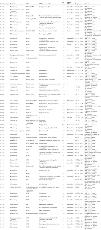
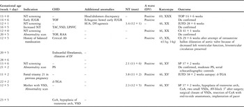
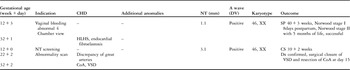
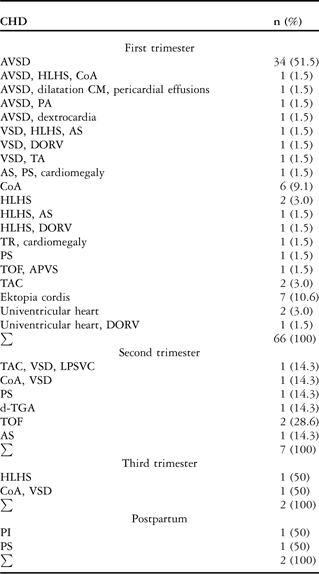
During the study period, 77 foetuses were diagnosed prenatally or postnatally with a congenital cardiac anomaly (22 of 1000, 2.2%). The first trimester echocardiography showed 66 (85.7%) malformations, that is, 22 cases at 11 plus 0 to 11 plus 6 weeks, 23 cases at 12 plus 0 to 12 plus 6 weeks, 21 cases at 13 plus 0 to 13 plus 6 weeks. At mid-gestation, seven (9.1%) cases of coronary heart disease, which were not diagnosed in the first trimester, were detected by trans-abdominal ultrasound, with a detection rate of 94.8%. Examination in the third trimester showed two (2.6%) previously undetected cardiac malformations, resulting in an overall prenatal detection rate of 97.4% (75/77) in this series. Postnatal echocardiography added another two cases (2.6%) of congenital heart disease (Table 1a–d).
Characteristics of 77 cases with CHD over gestational age and postnatally (first trimester).

APVS = absent pulmonary valve syndrome; AS = aortic stenosis; AVSD = atrioventricular septal defect; AV-block = atrioventricular block; CHD = congenital heart disease; CM = cardiomyopathy; CoA = coarctation of the aorta; CS = caesarean delivery; DORV = double outlet right ventricle; DV = ductus venosus; Dx = diagnosis; HLHS = hypoplastic left heart syndrome; IVF = in vitro fertilisation; ICSI = intra-cytoplasmatic sperm injection; NIHF = non immune hydrops fetalis; SP = spontaneous partus; SUA = single umbilical artery; TA = tricuspid atresia; PS = pulmonary stenosis; SP = spontaneous partus; TAC = common arterial trunk; TOF = tetralogy of Fallot; TOP = termination of pregnancy; TR = tricuspid insufficiency; IUFD = intra uterine fetal death; VSD = ventricular septal defect
Characteristics of 77 cases with CHD over gestational age and postnatally (second trimester).

AS = aortic stenosis; CHD = congenital heart disease; CoA = coarctation of the aorta; CS = caesarean delivery; DV = ductus venosus; Dx = diagnosis; IUGR = intra uterine growth restriction; LPSVC = left persistent superior vena cava; LV = left ventricle; PS = pulmonary stenosis; RAA = right aortic arch; SP = spontaneous partus; TAC = common arterial trunk; TOF = tetralogy of Fallot; TOP = termination of pregnancy; VSD = ventricular septal defect
Characteristics of 77 cases with CHD over gestational age and postnatally (third trimester).

CHD = congenital heart disease; CoA = coarctation of the aorta; CS = caesarean delivery; DV = ductus venosus; Dx = diagnosis; HLHS = hypoplastic left heart syndrome; SP = spontaneous partus; VSD = ventricular septal defect
Characteristics of 77 cases with CHD over gestational age and postnatally (postpartum)

CHD = congenital heart disease; CS = caesarean delivery; DV = ductus venosus; Dx = diagnosis; PI = pulmonary insufficiency; PS = pulmonary stenosis; SP = spontaneous partus
Diagnoses comprised a broad variety of congenital heart diseases within the first trimester including left- and right heart anomalies and abnormalities of the outflow tracts, with and without intracardiac shunt malformations. Table 1a–d, outlines the types of malformations and includes detailed information for each case. Table 2 shows congenital heart disease and its time of diagnosis. In the second trimester, diagnosis of congenital heart disease included one coarctation of the aorta at 23 plus 5 weeks of gestation and one case of a common arterial trunk and left-persistent superior vena cava at 16 plus 5 weeks of gestation, both combined with a ventricular septal defect. One case with pulmonary stenosis and another with aortic stenosis were diagnosed at mid-gestation. There was one case of tetralogy of Fallot and triploidy with early intrauterine growth restriction at 14 plus 6 weeks. In these cases, suspicion of congenital heart disease was raised in the first trimester and the subsequent diagnosis was made at the time of karyotyping. Another diagnosis of tetralogy of Fallot was made at 20 plus 5 weeks of gestation. Nuchal translucency measurement was within the normal range and ductus venosus blood flow velocity waveforms were normal. One case with complete transposition was diagnosed at 23 weeks of gestation. Nuchal translucency measurement was 3.8 millimetres at 11 plus 2 weeks of gestation and blood flow velocities in the ductus venosus were positive during atrial contraction – A wave. Unexplained intrauterine foetal death occurred at 38 plus 2 weeks of gestation. There was one case of coarctation of the aorta with a ventricular septal defect and left-to-right shunt of blood flow across the septal defect in the third trimester. The diagnosis of hypoplastic left heart was confirmed in one foetus at 32 + 1 weeks of gestation, and in this case nuchal translucency measurement and ductus venosus A wave velocities were normal during the first and second trimester screening.
Foetal cardiac anomalies detected during first, second, and third trimester echocardiography and postnatally
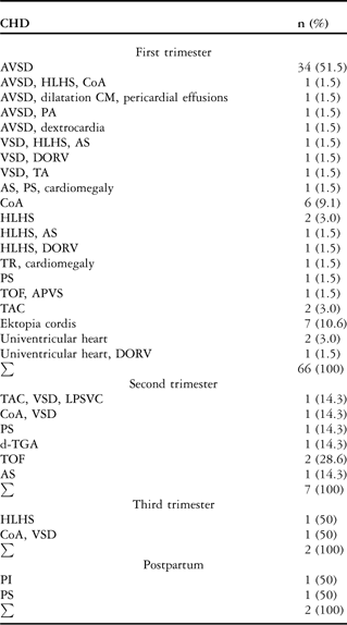
APVS = absent pulmonary valve syndrome; AS = aortic stenosis; AVSD = atrioventricular septal defect; CHD = congenital heart disease; CM = cardiomyopathy; CoA = coarctation of the aorta; DORV = double outlet right ventricle; HLHS = hypoplastic left heart syndrome; LPSVC = left persistent superior vena cava; PA = pulmonary atresia; PS = pulmonary stenosis; TA = tricuspid atresia; TAC = common arterial trunk; TGA = transposition of the great arteries; TOF = tetralogy of Fallot; TR = tricuspid insufficiency; VSD = ventricular septal defect
Discussion
The vast majority of cardiac anomalies can be detected at 11–14 weeks of gestation through trans-vaginal and trans-abdominal foetal echocardiography.Reference Rasiah, Publicover, Ewer, Khan, Kilby and Zamora10, Reference Smrcek, Berg and Geipel15, Reference Yagel, Weissman and Rotstein18 This study presents data of a single tertiary referral centre: during gestation, correct diagnosis of congenital heart disease was made in more than 95% of affected foetuses. But even with a systematic echocardiographic examination by experienced health-care professionals during the first half of pregnancy, a proportion of congenital heart disease may remain undetected. One reason may be that some congenital heart diseases cause only mild alterations in the first trimester, which are not easily detectable by echocardiographic ultrasound. Those alterations may become more apparent in later gestation, or after birth, and can only be detected at that time. Limited resolution and erroneous diagnosis can also be a reason for unsuccessful diagnosis.Reference Smrcek, Berg and Geipel15, Reference Yagel, Weissman and Rotstein18–Reference Allan21 A normal echocardiographic survey in the first trimester does not exclude alterations in chamber size or morphology. Discrepancies in great artery proportion or small ventricular septal defects become apparent in later foetal or neonatal life. From our series, coarctation of the aorta, hypoplastic left heart resulting from aortic stenosis, pulmonary and aortic stenosis, and Tetralogy of Fallot may serve as examples for this theory. The following – that is, 4 of 77 (5.2%) – cardiac anomalies remained undetected until the third trimester or neonatal period: mild pulmonary stenosis, insufficiency of the pulmonary valve, coarctation of the aorta, and hypoplastic left heart.
In a retrospective series, Yagel et al evaluated detection rates of early echocardiography for congenital heart disease in a mixed risk population with two-dimensional ultrasound using the trans-vaginal and trans-abdominal approaches.Reference Yagel, Weissman and Rotstein18 Colour flow mapping or spectral Doppler was only performed in cases of suspected congenital heart disease. They reported a 64% detection rate of trans-vaginal foetal echocardiography performed between 13 and 16 weeks of gestation and subsequently a 17% detection rate via trans-abdominal examination at mid-gestation. They compared this result with the result of a group of foetuses examined for the first time at mid-gestation using trans-abdominal ultrasound only. In this group, the detection rate was 78%. Subsequently 4% in study of yagel, 7% in the other study, where fetuses were evaluated not until mid-trimester. In each group, 15% of congenital heart disease was diagnosed postnatally. Reasons for the slightly higher detection rate in our series, compared with the results of Yagel and co-workers at 13–16 weeks gestation, may be the routine use of colour flow and spectral Doppler interrogation in our echocardiographic set-up and improved equipment, as our study took place after the mentioned study by Yagel and co-workers. Smrcek and co-workers reported slightly lower detection rates during the first trimester with an identical set-up. However, the sample of congenital heart diseases comprised in this series was too small (44 cases) to draw definitive conclusions.
The types of cardiac lesions missed at the first trimester were comparable to our findings; however, with regard to a larger population examined by Yagel et al, lesions such as cardiac rhabdomyoma or ventricular aneurysm, which might show a late onset appearance, have not been observed in our series.Reference Smrcek, Berg and Geipel15, Reference Yagel, Weissman and Rotstein18 In both series, as well as in ours, there seems to be a comparable number of cardiac lesions of about 10%, which are – despite the advances in resolution and image processing – not detectable in the first or early-second trimester echocardiography, but at mid-gestation, and another 5% in later gestation or postnatally. These findings underline the theory of varying appearance of foetal cardiac pathology.Reference Allan, Sharland and Tynan19–Reference Allan21 One of the possible causes for delayed diagnosis of congenital heart disease may be the limitation in resolution because of foetal orientation, size of the foetal heart, or technology used.
There is now sufficient evidence in literature that some cardiac lesions show an in utero progression such as ventricular outflow tract obstructions and major vessel stenosis.Reference Allan, Sharland and Tynan19–Reference Hornberger, Sanders, Rein, Spevak, Parness and Colan27 Several studies have shown a reduced growth of left heart structures in case of severe left ventricular outflow tract obstruction identified in the mid-trimester. This has also been shown in the animal model.Reference Fishman, Hof, Rudolph and Heymann28 In cases with aortic stenosis, the appearance of the foetal heart will depend on the degree of obstruction, and the alteration may not be obvious until later in gestation. This also applies to stenosis of the pulmonary valve. Foetal hearts with a milder outflow tract obstruction in the mid-trimester may nevertheless show normal growth of the ventricle, although abnormal growth of the semilunar valve may result in increasing stenosis by the time of birth or in the neonatal period. If aortic or pulmonary stenoses are severe, a dilated and poorly contracting ventricle with evidence of increased echogenicity of the ventricular walls suggestive of endocardial fibroelastosis might be detected. Critical stenosis of the aortic valve may show a progression to hypoplastic left heart syndrome in later gestation, because of impaired growth of the left ventricle. Coarctation of the aorta is another example of in utero progression of arch hypoplasia with an unsuspicious aortic arch in early foetal life developing into an arch with significant hypoplasia later in gestation with more pronounced discrepancies in chamber and great artery size. Those alterations may be subtle and not necessarily detectable at the time of first echocardiographic examination. Our cases with aortic stenosis or ventricle discrepancy followed by the diagnosis of coarctation of the aorta may serve as examples for this phenomenon.Reference Marasini, DeCaro, Pongiglione, Ribaldone and Caponetto25, Reference Hornberger, Sanders, Rein, Spevak, Parness and Colan27, Reference Hornberger, Sahn, Kleinman, Copel and Silverman29 A minority of cases of hypoplasia of the left heart is located at the severe end of the spectrum of coarctation of the aorta, where marked hypoplasia of all components of the left heart might develop during gestation, but the mitral and aortic valves remain patent. Hypoplasia of the pulmonary arteries and dilatation of the aorta, respectively, may also have progressive character in case of Tetralogy of Fallot during gestation, resulting in more pronounced discrepancy of great artery size and marked malalignment of the great vessels, which may be missed at the first trimester evaluation.Reference Hornberger, Sanders and Sahn30 In this context, it is important to underline the use of colour flow mapping and spectral Doppler interrogation depicting turbulent blood flow with high velocities above the valves in case of outflow tract obstruction and retrograde blood flow in the ascending aorta or transverse arch, or in the pulmonary trunk in case of aortic or pulmonary atresia. In our series, we diagnosed the following coronary heart diseases after the completion of the first trimester: two cases of Tetralogy of Fallot, both cases in the second trimester, two cases of coarctation of the aorta, one in the second and one in the third trimester, one aortic stenosis and pulmonary stenosis, both in the second trimester, one pulmonary stenosis after birth, and one hypoplastic left heart in the third trimester. We carefully reviewed the images in our cases of delayed diagnosis of outflow tract obstruction and we could not detect any abnormality during examination. In addition, one has to take into account that in cases with outflow tract obstructions abnormal flow patterns may be evident in advanced stages of the disease only, because of foetal intracardiac shunts preventing early development of pressure gradients between chambers. Endocardial fibroelastosis in the absence of viral infection and hypertrophic cardiomyopathy are further examples of cardiac pathology becoming apparent later during gestation, as in our series, or even postnatally.
Of those cardiac pathologies that do not alter from a normal appearance in the first trimester – for example atrioventricular septal defect, single ventricle, double-outlet right ventricle, transposition of great arteries, common arterial trunk, Ebstein anomaly, and heterotaxy syndrome – we missed one complete transposition of great arteries at 11 plus 2 weeks of gestation, increased nuchal translucency 3.8 millimetres, and one common arterial trunk at 12 plus 1 weeks of gestation, increased nuchal translucency 3.4 millimetres. This common arterial trunk – in combination with a ventricular septal defect and left-persistent superior vena cava – was subsequently diagnosed at 16 plus 5 weeks. At the initial examination, a single umbilical artery – in combination with a ductus venosus agenesis – was diagnosed, and chorionic villous sampling was performed showing a normal karyotype.
Recently, Smrcek et alReference Smrcek, Berg, Geipel, Fimmers, Diedrich and Gembruch31 studied the feasibility of early foetal echocardiography between 10 and 15 weeks of gestation in 123 normal singleton pregnancies using modern high-end ultrasound equipment. The authors evaluated the appearance of the atrioventricular and ventriculoarterial connections and incorporated the appearance of the venoatrial connections by using trans-vaginal and trans-abdominal B-mode and colour Doppler sonography. Complete evaluation of the foetal heart including the pulmonary veins was impossible at 10 weeks, the total success rate increased from 45% at 11 weeks to 90% between 12 and 14 weeks and to 100% at 15 weeks of gestation. Between 10 and 13 weeks, the trans-vaginal approach exceeded the trans-abdominal sonography. From 13 plus 0 to 14 plus 0 weeks, the trans-abdominal approach was sufficient in over 80% and 90% of cases, respectively. At 14 weeks, both methods had similar success. At 15 weeks, trans-abdominal sonography allowed adequate visualisation of all cardiac structures. When visualisation of pulmonary veins was excluded, the rate of successful survey by using a combined trans-vaginal and trans-abdominal approach including colour Doppler examination increased from 67% in the 10 completed week of gestation to 100% in the 11th week of gestation. These data demonstrate the increase in successful detection of cardiac structures along with technical improvement.Reference Smrcek, Berg, Geipel, Fimmers, Diedrich and Gembruch31
The rate of association between increased nuchal translucency and congenital cardiac defect is well known. It varies in different reports from 17% to 56%. Increased nuchal translucency is not connected to any particular type of congenital heart disease with the exception of atrioventricular septal defects, which are associated with increased nuchal translucency above 2.5 millimetres in the recent series of Vogel et al.Reference Vogel, Sharland and McElhinney9, Reference Hyett, Perdu, Sharland, Snijders and Nicolaides32–Reference Mavrides, Cobian-Sanchez and Tekay34
The routine measurement of nuchal translucency in the first trimester screening is an expeditious tool that may be a reliable indicator for coronary heart diseases in cases of increased translucency. As a large proportion of affected foetuses in this series were aneuploid, our results do not add new information to that issue.
The Doppler flow profile in the ductus venosus has received much attention as an indicator of foetal cardiac function in past years. An abnormal profile can be associated with increased nuchal translucency, chromosomal abnormalities, and congenital cardiac defects.Reference Matias, Gomes, Flack, Montenegro and Nicolaides35, Reference Matias, Huggon, Areias, Montenegro and Nicolaides36 In our series,  of foetuses with congenital heart disease in the first trimester had absent – or reversed flow during A wave in the Ductus venosus. As stated before, there was a large proportion of aneuploid foetuses, precluding any conclusion to be drawn from this information.
There are three main limitations of successful early foetal echocardiography:
Skills of the examining physician, quality of technical equipment, and possible in utero progression of coronary heart diseases later in pregnancy.
Sub-specialised physicians using high-end ultrasound machines with high-frequency trans-vaginal probes are mandatory. Suboptimal visualisation because of limited resolution or foetal size and position also contributes to erroneous diagnosis. If a congenital heart disease is suspected during foetal sonographic examination, referral to a centre for prenatal medicine should be discussed for thorough second evaluation by sub-specialized physicians. If this re-evaluation confirms alterations of the foetal cardiac development, the risk of unnecessary interventions because of false-positive results – such as chorionic villous sample/amniocentesis or even termination of pregnancy – is reduced.
The third limitation of early foetal echocardiography is the possible in utero development of congenital heart diseases: Cardiac embryogenesis occurs in the first 7 weeks of pregnancy. From 10 weeks on, the foetal heart has reached its definite anatomic form in intrauterine life. The final process of closure of the foetal shunts takes place after birth, including the separation of the systemic and pulmonary circulation. With better knowledge and increasing experience in prenatal cardiology, it has become apparent that many cardiac lesions have initially developed within the first 6 or 7 weeks of gestation. But detectable effects caused by those lesions may be missing or may only exist in a very mild way at the time of the first trimester screening. They may become apparent in the second or third trimester or even postnatally.
Therefore, when counselling parents, it is important to state that normal echocardiographic appearance of the foetal heart cannot completely rule out the diagnosis of structural cardiac defects in later gestation or even postnatally.
It has to be acknowledged that this series consists of cases representing a high-risk population at a tertiary referral centre. Therefore, our recommendations apply for comparable institutions only.
Only 20% of congenital heart disease in the foetus occur in high-risk pregnancies; therefore, routine screening of all pregnancies is necessary. The results presented strongly advocate the use of early trans-vaginal and trans-abdominal standardised sequential foetal echocardiography as part of early anomaly scanning.
Early foetal echocardiography performed by experienced personnel using high-end ultrasound equipment reached detection rates of 85% of congenital heart disease in this series, with a further 10% at mid-gestation. There was only one delayed diagnosis of a cardiac lesion detectable in early foetal echocardiography in our series. Approximately 97% of the cumulative antenatal detection rate of congenital heart disease was identified by sequential standardised foetal echocardiography. The possible evolvement of many cardiac abnormalities during gestation has to be kept in mind, and therefore early foetal echocardiography should be followed by mid-trimester echocardiography in all ongoing pregnancies.