Modern surgical treatment of anomalous origin of the left coronary artery from the pulmonary trunk aims at creation of a dual coronary arterial system. Among the variety of surgical options for such treatment, aortic re-implantation is the preferred option.Reference Dodge-Khatami, Mavroudis and Backer1–Reference Schwartz, Jonas and Colan4 This can be difficult to perform when the anomalous arterial orifice is greatly separated from the aorta, and in those presenting late with diminished elasticity of the vessels, and extensive formation of collateral vessels around the pulmonary sinuses.Reference Dodge-Khatami, Mavroudis and Backer1–Reference Chan, Hare and Buxton10 In these situations, it has been proposed to interpose a free segment of the subclavian artery;Reference Arciniegas E Farooki, Harkmi and Green11 to create an intrapulmonary tunnel;Reference Takeuchi, Imamura and Katsumoto6 or to prolong flaps of pulmonary arterial wall with or without anastomosis to the right subclavian artery.Reference Turley, Szarnicki, Flachsbart, Richter, Popper and Tarnoff3, Reference Vigneswaran, Campbell, Pappas, Wiggins, Wolfe and Clarke9, Reference Sese and Imoto12–Reference Katsumata and Westaby15 We present here our experience with the modification of the trapdoor technique, wherein the anomalous left coronary artery is detached from the pulmonary arterial sinus, and combined autogenous aortic and pulmonary arterial flaps are used to augment its length.
Criterions for decision-making and selection of patients
In our prospective study, we evaluated outcomes after aortic re-implantation of the anomalous left coronary artery from the pulmonary trunk having elongated the main stem of the artery using autogenous aortic and pulmonary arterial flaps. We made the decision to use the technique having assessed the site of origin of the anomalous left coronary artery. All patients with anomalous left coronary artery arising from the right posterior wall or sinus of the pulmonary trunk, or from the anterior pulmonary sinus, were candidates for direct coronary re-implantation, and we excluded such patients from this analysis, excluding also patients in whom the repair was made by creating an aorto-pulmonary window and inserting an intrapulmonary baffle, the so-called Takeuchi procedure.
Thus, there were nine forces driving our decision-making for anatomic repair of anomalous left coronary artery from the pulmonary trunk:
• The desire to obtain improved operative exposure and direct access to the anomalous left coronary artery and its neo-aortic location.
• The desire to re-implant the anomalous left coronary artery in the aortic sinus, which could offer haemodynamic advantages.
• The desire to avoid injury to the aortic valvar apparatus by performing the procedure under direct visualization away from the components of the valve.
• The desire to create a dual coronary arterial system irrespective of the location of the anomalous orifice within the pulmonary trunk.
• The desire to extend the left coronary artery by interposing a tube created from the autologous arterial wall between the neoaortic orifice and the anomalous artery, thereby avoiding tension, torsion, distortion of the coronary arterial button, and allowing full potential for subsequent growth of the newly constructed structure.
• The desire to minimize dissection around the proximal part of the coronary artery.
• The desire to minimize the use of pericardial augmentation and maximize the use of endothelialized autogenous arterial wall flaps to avoid thrombosis.
• The desire to place the extended coronary artery posterior to the pulmonary trunk to avoid extrinsic mechanical compression, and finally,
• The desire to apply the procedure in all age groups, and in all anatomical variants.
Patients and methods
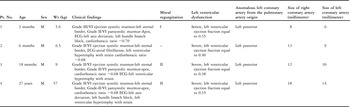
Between January, 1999, and December, 2006, we encountered four male patients, aged 3 months, 6 months, 18 months, and 27 years respectively, in whom we reimplanted an anomalous left coronary artery arising from the left posterior wall of the pulmonary trunk or sinus using the surgical technique described herein. Consent was taken for anonymous analysis of data, and we obtained approval from our institutional ethics committee. The procedures were performed by a single surgeon, thus achieving uniformity in the surgical protocol.
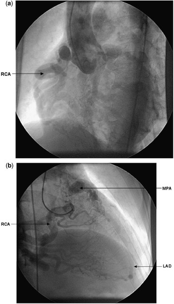
The preoperative characteristics and haemodynamic variables for the 4 patients are presented in Table 1. The diagnosis was established preoperatively by echocardiography, cardiac catherterization, and angiocardiography in all. Electrocardiography demonstrated evidence of myocardial ischaemia in the anterolateral leads. Cross-sectional echocardiography demonstrated regional abnormalities of wall motion involving the anteroapical areas, with reduced ejection fraction ranging from 0.35 to 0.40, with a median of 0.38, and central mitral regurgitation graded at levels 1 or 2 in our first, third, and fourth patients. Cardiac catheterization showed a step-up in arterial saturation of oxygen in the pulmonary trunk, and the ratio of pulmonary to systemic flows varied from 1 to 1.4 to 1 to 1.6. Coronary arteriography revealed an ectatic and enlarged right coronary artery supplying the left coronary artery through collateral vessels. The right coronary artery and its collateral vessels were unusually large and tortuous (Fig. 1a, b).
Demographic data, cardiac anatomy and the intraoperative details of the patients.

(a, b) Preoperative selective right coronary arteriogram showing a dilated tortuous, right coronary artery (RCA) with extensive collaterals draining into the left anterior descending coronary artery (LAD) which opens into the pulmonary trunk (MPA). Aortic root injection shows (a) absence of left coronary artery.

Left ventriculography confirmed these findings. Surgical correction was performed in all patients as described below.
Operative technique
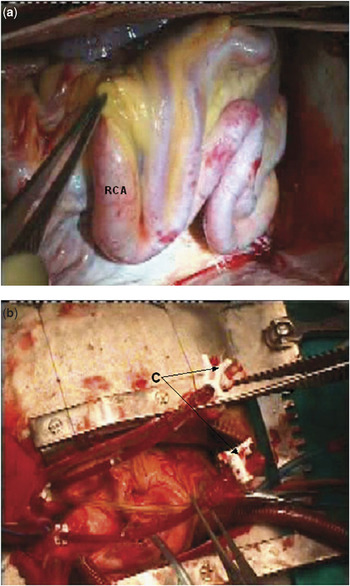
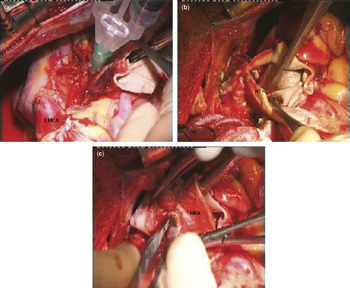
All operations were performed through a median sternotomy. The coronary arteries were grossly dilated and tortuous (Fig. 2a). A continuous thrill was palpable at the root of the pulmonary trunk. The ascending aorta and pulmonary trunk were separated from each other, and the right and left pulmonary arteries dissected and freed up to their first lobar branch. The operations were performed with moderately hypothermic cardiopulmonary bypass, at 28 degrees Celsius, through angled venous cannulas inserted into the superior and inferior caval veins, and with distal aortic cannulation. The aorta and pulmonary trunk were individually cross clamped. and cold hyperkalemic blood cardioplegic solution was infused simultaneously into both vessels at a pressure of 80 millimetres of mercury for 3 minutes to achieve optimal myocardial protection (Fig. 2a, b). This technique is important in maintaining high pressure within the proximal pulmonary trunk, avoiding run-off from the orifice of the anomalous left coronary artery. The left heart was vented through the right superior pulmonary vein. The location of the anomalous artery, its relationship to the sinuses and the zones of apposition between the valvar leaflets, and its pattern of branching, were all examined.
(a, b) Operative techniques used for aortic re-implantation of the anomalous left coronary artery from the pulmonary trunk. Note the dilated, tortuous right coronary artery [(RCA), b] and simultaneous infusion of the cardioplegic solution through two cardioplegic cannulas (C) into both the aorta and pulmonary trunk. The ascending aortic, superior and inferior caval venous cannulas are not shown.

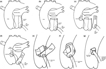
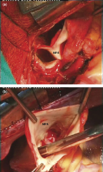
The pulmonary trunk was then transected between stay sutures just below its bifurcation so as to fashion a long arterial cuff to cover a bridging aortic flap to the anomalous coronary artery. On opening the pulmonary trunk, the orifice of the anomalous left coronary artery was observed to vary in diameter from 6 to 14 millimetres. The orifice was located posterolaterally and on the left side in all 4 patients (Figs 3a–g and 4a, b).
(a–g) Diagrammatic representation of the techniques used to repair anomalous origin of the left coronary artery from the pulmonary trunk. (a, b) After routine cannulation of the ascending aorta and caval veins and cross-clamping of the aorta, the pulmonary trunk was opened transversely just below the bifurcation and completely transected. (c) A vertical flap of pulmonary arterial wall of the respective sinus of Valsalva containing the anomalous left coronary artery at its bottom was mobilized avoiding injury to the pulmonary valvar leaflets. The dotted line on the aorta represents the creation of an anteriorly based, obliquely directed, rectangular flap of the aortic wall 10 to 15 millimeters above the sinutubular junction. (d, e and f) These demonstrate the step-bystep method of suturing of the pulmonary arterial flap to the aortic opening. The right inferomedial margin of the pulmonary arterial flap was sutured along with posterior lip of the neoaortic orifice. The suturing continues inferiorly along the base of the left coronary arterial orifice. The next point of suturing starts on the left inferolateral portion of the pulmonary arterial flap. The two adjacent edges of the left inferolateral margin of the pulmonary arterial flaps were sutured with each other with the left coronary artery orifice at its fulcrum. Subsequently, the redundant superior edge of the pulmonary arterial flap was folded anteriorly over the left coronary sinus. Finally, the anteriorly based, obliquely directed, small rectangular aortic flap was sutured over the residual gap to obtain a trap door effect. (g) The implanted left main coronary artery elongated to its neoaortic site. Note the absence of narrowing, flattening, kinking, wasting, tension, torsion or saccular configuration of the neoaortic tube.

(a, b) The pulmonary trunk (MPA) is mobilized and transected just below the bifurcation to allow sufficient length of the pulmonary arterial flap to create an extended left main stem.

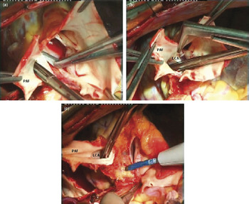
Because extra length was required to ensure a tension-free communication with the ascending aorta, the coronary arterial button was mobilized along with a vertical flap of pulmonary arterial wall from the respective sinus of Valsalva, taking care to avoid injury to the leaflets of the pulmonary valve, according to the technique described by Katsumata and Westaby.Reference Katsumata and Westaby15 The orifice of the anomalous left coronary artery was situated low in the lateral part of the left posterior pulmonary sinus in all patients. The proximal part of the left coronary artery was mobilized away from the pulmonary trunk over a distance of about 1 centimetre (Figs 3c and 5a).
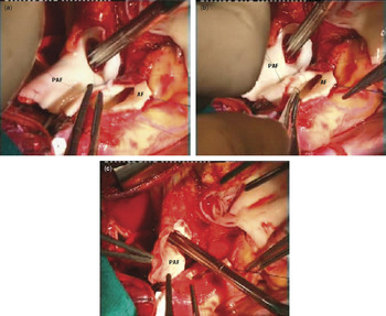
Subsequently, an anteriorly based, obliquely directed, rectangular flap of the aortic wall was created, approximately 10 to 15 millimetres above the sinutubular junction, starting from the posterior aspect towards the left lateral side and continuing to the anterior aspect of the aorta, thereby maintaining anterior continuity of the aortic flap (Figs 3c, d, 5a–c and 6a–c). While selecting the site of re-implantation of the anomalous coronary artery, we took care not to damage or distort the aortic valve, to implant the anomalous left coronary artery in the appropriate sinus, to elongate the main stem of the artery by combining the obliquely positioned, rectangular aortic flap with a vertical pulmonary arterial flap so as to obtain a trap-door effect, and avoiding any narrowing, flattening, tension, torsion or saccular formation, to direct the extended main stem of the anomalous artery posterior to the pulmonary trunk in anatomical position to avoid extrinsic compression, to implant the artery above the sinutubular junction to ensure absence of kinking, external waisting of the left coronary artery or its branches, and to design appropriate length of flaps so that the aortic re-implantation could be performed without any tension (Figs 3c, d, 5a–c and 6a–c).
(a–c) Mobilization of the left coronary button along with a vertical flap of pulmonary arterial wall of the respective sinus of valsalva (PAF) avoiding injury to the pulmonary valvar leaflets.

(a–c) Creation of an anteriorly based, obliquely directed rectangular flap of the aortic wall (AF), 10–15 mm above the sinutubular junction. The aortic (AF) and pulmonary arterial flaps (PAF) are sutured using a trapdoor technique to make a tubular extension for anomalous left coronary artery.

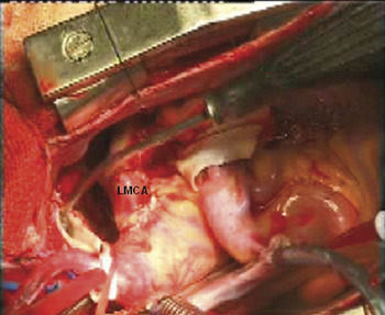
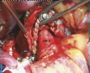
The isolated segment of the pulmonary trunk containing the origin of the anomalous artery at its bottom was approximated to the obliquely positioned neo-aortic window. The right inferomedial margin of the pulmonary arterial flap was then sutured along the posterior lip of the neo-aortic orifice (Figs 3d and 5a–c). Subsequently, the redundant superior edge of the pulmonary arterial flap was folded anteriorly over the left coronary sinus. The side edges of the left inferolateral portion of the pulmonary arterial flaps were sutured around the left coronary arterial orifice to form an extension tube of pulmonary arterial tissue, thus lengthening the left coronary artery (Figs 3d, f and 5a–c). The anteriorly based, obliquely positioned, small rectangular flap of the aortic wall was sutured next over the residual gap to produce a trap door effect (Figs 3f and 5a–c). Thus, an obliquely positioned, extended left main stem was created, ensuring a tubular configuration with the orifice of the left coronary artery at its fulcrum (Figs 3g and 7). The defect created in the pulmonary trunk and its sinus was repaired with an autogenous pericardial patch, and the transected ends of the pulmonary trunk were directly sutured using 6-0 monofilament polypropylene suture (Ethicon, Inc, Somerville, NJ) (Figs 8a–c and 9).
The completed extended left main stem, translocating the left coronary artery (LMCA) to its neo-aortic orifice.

(a–c) Repair of the defect in the pulmonary trunk with a pericardial patch, and direct end-to-end suturing of the two ends of the transected pulmonary trunk (MPA).

Shows the newly implanted extended left main coronary artery (LMCA) posterior to the pulmonary trunk without extrinsic mechanical compression.

Intraoperative transoesophageal echocardiography after cardiopulmonary bypass showed a well-opened, well-filled, left coronary arterial system from the ascending aorta, absence of aortic regurgitation, and no obstruction within the right ventricular outflow tract.
Results
Early results
There was no operative death. All patients were extubated on the first postoperative day. All patients were routinely started on dopamine at dosage of 5 to 10 micrograms per kilogram per minute, sodium nitroprusside at dosage of 0.5 micrograms per kilogram per minute and milrinone at dosage of 50 micrograms per kilogram intravenous bolus followed by 0.375 to 0.75 micrograms per kilogram per minute either isolated or in combination. Postoperatively, they were maintained on amiodarone at a dosage of 5 to 15 micrograms per kilogram per minute for 48 to 72 hours as a prophylactic against ventricular tachycardia.
All patients had normal renal function and were administered oral inhibitors of acetylcholinesterase prior to weaning from inotropic agents. Postoperatively, digoxin, diuretics, inhibitors of acetylcholinesterase and amiodarone were weaned at varying time intervals.
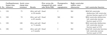
The median period of cardiopulmonary bypass was 190 minutes, with a range from 150 to 196 minutes, and the median time for aortic cross-clamping was 90 minutes, with a range from 75 to 116 minutes. Mitral valvar reconstructive procedures, a left ventricular assist device, or extracorporeal membrane oxygenation were not required for any patient.
Late results
Thus far, there has been no late death. All survivors were periodically evaluated every 6 months by our cardiologists and surgeons. Their records were reviewed for all pertinent pre-and-postoperative information, including Doppler echocardiography. Postoperatively, our fourth patient consented to investigation for myocardial perfusion by means of a stress thallium investigation, positron emission tomographic scan, cardiac catheterization, and angiocardiography. Overall, the patients have been followed for periods ranging from 9 months to 96 months, with a median of 74 months. All are in the first functional class of the system devised by the New York Heart Association at their last follow-up visit, with sinus rhythm, good biventricular function, and without the need for any medications. There was progression of R waves in leads V1 through V3 on the electrocardiogram.
Postoperatively, the left ventricular ejection fraction as measured echocardiographically ranged from 50% to 60%, with a median value of 52%, compared to the preoperative values, which ranged from 35% to 40%, with a median of 38%. These differences are statistically significant, with a value for p equal to 0.04. Normalization of ejection fraction, improvement in mitral regurgitation and abnormalities of mural motion occurred within 5 to 6 months of repair in all patients There was no evidence of obstruction to the right ventricular outflow tract in any patient (Table 2).
Intraoperative and postoperative details of the patients with anomalous left coronary artery from the pulmonary trunk who underwent aortic reimplantation by this new technique.

Stress-test Thallium-201 myocardial perfusion and single photon emission computed tomography & fluorine-18 fluoro-deoxy-glucose metabolic study
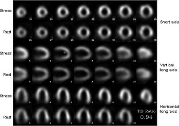
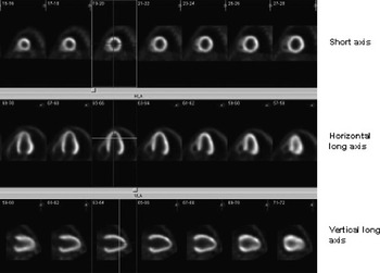
Postoperatively, our fourth patient, a 27-year-old, non-diabetic male, underwent thallium-201 stress-test myocardial perfusion single photon emission computed tomography. On a separate day, a positron emission tomographic study was performed using a dedicated system (Biograph, Siemens). The tests revealed no abnormalities in perfusion (Figs 10 and 11).
In our 4th Patient, a 27-years-old, non-diabetic male, a stress-rest thallium-201 myocardial perfusion scan reveals a dilated left ventricular cavity. Both the stress and rest images show normal perfusion to the left ventricular myocardium, and no evidence of stress-induced ischaemia.

A positron emission tomography myocardial glucose metabolism study, done on a separate day in our 4th patient, reveals normal uptake, indicating viable left ventricular myocardium.

Cardiac catheterization
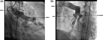
Postoperative angiography in the aortic root demonstrated simultaneous opacification of both coronary arteries. The left coronary artery was widely patent, with good flow and without any narrowing of kinking (Fig. 12a, b). The left ventriculogram performed 24 months after the operation demonstrated both improved contractility and mild mitral regurgitation.
(a, b) Postoperative selective left coronary arteriogram in our 4th patient showing a well-filled left coronary artery (LMCA) tube without any distortion.

Discussion
Creation of a dual coronary arterial system in the preferred option for surgical repair in patients with anomalous origin of the left coronary artery from the pulmonary trunk.Reference Dodge-Khatami, Mavroudis and Backer1–Reference El-Said, Ruzyllo and Williams17 It eliminates the “steal” phenomenon, and restores physiological antegrade flow to the ischaemic left ventricular myocardium. Available surgical procedures include direct coronary arterial re-implantation, the intrapulmonary tunnel repair, prolongation of flaps of pulmonary arterial wall with or without anastomosis to the right subclavian artery, left sulclavian artery-to-left coronary artery anastomosis, and ligation with saphenous venous interposition, and anastomosis of the left internal thoracic artery to the left anterior interventricular artery.Reference Dodge-Khatami, Mavroudis and Backer1–Reference Singh, Carli, Sullivan, Leonen and Morrow18
Direct implantation of the left coronary artery into the ascending aorta is the preferred option,Reference Vouhe, Baillot-Vemant and Trinquet2, Reference Alexi-Meskishvili, Hetzer and Weng7, Reference Vigneswaran, Campbell, Pappas, Wiggins, Wolfe and Clarke9, Reference Neirotti, Nijveld and Ithuralde16–Reference Backer, Stout and Zales19 and is usually performed on the left postero-lateral wall of the ascending aorta from outside. For optimal exposure of this aspect of the aorta, two ingenious techniques have been described, namely transection of the pulmonary trunk,Reference Grace, Angelini and Cooley20 and an anterior aortic approach, anastomosing the excised button of pulmonary trunk from within the lumen of the aorta under direct visualization. This technique allows the anastomosis to be performed from within the aorta, and can be achieved with low early mortality and excellent long-term outcome.Reference Laks, Ardehali, Grant, Allada and Angeles21
Direct reimplantation of the anomalous left coronary artery may be technically more difficult and hazardous, if the anomalous orifice is located in the non-facing or left posterior sinus, and in older patients with well developed collaterals around the pulmonary sinuses.Reference Dodge-Khatami, Mavroudis and Backer1–Reference Chan, Hare and Buxton10 If the distance between the aorta and the anomalous left coronary artery is longer, it would require extensive dissection around the proximal part of the coronary artery to reach the aorta. Although some authors have reported successful coronary re-implantation in adults with suitable anatomy,Reference Selzman, Zimmerman and Campbell5 a subset of adults have diminished vessel elasticity for mobilization, with the potential for tearing, and resultant catastrophic bleeding.Reference Turley, Szarnicki, Flachsbart, Richter, Popper and Tarnoff3, Reference Alexi-Meskishvili, Hetzer and Weng7, Reference Moodie, Fyfe and Gill8, Reference Chan, Hare and Buxton10
There are six basic principles involved for the establishment of an ideal dual coronary arterial system. These principles are:
• detachment of the anomalous left coronary artery from the pulmonary arterial sinus
• reattachment of the cuff of left coronary artery onto a neo-aortic orifice avoiding excessive mobilization, tension or torsion of the pedicle, and injury to the aortic and pulmonary valvar apparatus
• elongation of the main stem of the anomalous coronary artery in patients with insufficient native tissue, or lack of length due to distance of the orifice to the aorta
• placement of the newly re-constructed and elongated coronary arterial tube in an anatomic position avoiding an intrapulmonary route, and thus avoiding the potential creation of obstruction to the right ventricular outflow tract
• usage of endothelialized, viable, autogenous arterial tissue for construction of the elongated arterial tube to prevent thrombosis and allowing further growth in children
• to minimize the use of pericardium for arterial reconstruction, allowing growth of the pulmonary trunk and minimizing the chances of late obstruction to the right ventricular outflow tract.
The distance between the orifice of the anomalous left coronary artery and the aorta is the key factor for appropriate transfer.Reference Smith, Arnold and Anderson22 Analysis of the published literature documents three techniques of arterial elongation to account for lack of arterial length. The techniques include interposition of a segment of subclavian artery,Reference Arciniegas E Farooki, Harkmi and Green11 creation of an aortopulmonary window with an intrapulmonary baffle,Reference Takeuchi, Imamura and Katsumoto6 and prolongation of flaps of pulmonary arterial wall with or without anastomosis to the right subclavian artery.Reference Turley, Szarnicki, Flachsbart, Richter, Popper and Tarnoff3, Reference Vigneswaran, Campbell, Pappas, Wiggins, Wolfe and Clarke9, Reference Sese and Imoto12–Reference Katsumata and Westaby15
The reported patency rate of free subclavian arterial interposition is 80% at 11 months.Reference Arciniegas E Farooki, Harkmi and Green11 Although an intrapulmonary tunnel can be performed irrespective of the distance between the orifice of the anomalous artery and the aorta by utilizing the autologous pulmonary artery tissue, the technique has been associated with pulmonary valvar dysfunction, baffle obstruction, supravalvar pulmonary arterial stenosis, and creation of fistulas from the tunnel to the lumen of the pulmonary trunk.Reference Vouhe, Baillot-Vemant and Trinquet2, Reference Turley, Szarnicki, Flachsbart, Richter, Popper and Tarnoff3, Reference Vigneswaran, Campbell, Pappas, Wiggins, Wolfe and Clarke9, Reference Chan, Hare and Buxton10 Additionally, creating the baffle may pose technical difficulties when the anomalous left coronary artery is located immediately adjacent to a pulmonary valvar leaflet.Reference Smith, Arnold and Anderson22 Although it has been extensively used in children, there are only a few reported cases of this procedure in adults.Reference Alexi-Meskishvili, Hetzer and Weng7, Reference Kattach, Anastasiadis, Jin and Dillai23 Kattach and colleaguesReference Kattach, Anastasiadis, Jin and Dillai23 performed the tunnel repair in an adult, but also created an anastomosis for the left internal thoracic artery to the anomalous artery to ensure adequate coronary arterial perfusion should the tunnel fail.
To overcome the limitations of these procedures, a variety of techniques have been described to elongate the main stem of the left coronary artery,Reference Turley, Szarnicki, Flachsbart, Richter, Popper and Tarnoff3, Reference Sese and Imoto12–Reference Neirotti, Nijveld and Ithuralde16 each having inherent advantages and disadvantages.Reference Turley, Szarnicki, Flachsbart, Richter, Popper and Tarnoff3, Reference Sese and Imoto12–Reference Katsumata and Westaby15 Our modification of the trap-door technique involves detachment of the anomalous left coronary artery from the pulmonary arterial sinus, elongation using autogenous, viable, endothelialized aortic and pulmonary arterial flaps, side-to-side anastomosis of the aortic and pulmonary arterial flaps, creation of a neo-aortic ostium, translocation of the anomalous left coronary artery and addresses all the issues discussed above, avoiding their potential shortfalls.
The other available surgical option, of course, is to ligate the origin of the anomalous left coronary artery to avoid eventual competitive flow, combined with a conduit to bypass the left coronary artery. The conduits that have been clinically used are a saphenous vein graft, the subclavian artery, and the left internal thoracic artery.Reference Turley, Szarnicki, Flachsbart, Richter, Popper and Tarnoff3, Reference Chan, Hare and Buxton10, Reference El-Said, Ruzyllo and Williams17, Reference Kitamura, Kawachi and Nishii24 The utility of the saphenous vein grafts in infants and small children is limited by the small caliber and poor quality of the vein, as well as the prevalence of late occlusion.Reference El-Said, Ruzyllo and Williams17
Although use of the left internal thoracic artery has supplanted saphenous vein grafts, its size in infants and children is a potential problem, and there are few mid-term or long-term results. Some investigators, nonetheless, have recommended the use of bilateral left internal thoracic arteries as the procedure of choice in adults.Reference Chan, Hare and Buxton10, Reference Kitamura, Kawachi and Nishii24
Anastomosing the subclavian artery to the left coronary artery has been used in several series.Reference Backer, Stout and Zales19 The reported potential problems with the use of subclavian artery are size mismatch, technical difficulty in performing the anastomosis on a beating heart via left thoracotomy, kinking of the subclavian artery at its origin, and anastomotic stenosis.Reference Vouhe, Baillot-Vemant and Trinquet2, Reference Backer, Stout and Zales19
Conclusions
The technique we have described provides a simple and effective alternative for anatomic repair of anomalous origin of the left coronary artery from the left posterior wall of the pulmonary trunk or sinus. The resulting absence of tension, torsion, or traction, and use of viable endothelialized autogenous arterial flaps, helps to avoid thrombosis. Further investigations on a larger number of patients, and a longer follow-up, are needed to confirm the excellent early results.