Introduction
In the 1970s, Dr Robin Cotton devised laryngotracheal reconstruction to manage children with laryngotracheal stenosis.Reference Cotton1 Since then, the emphasis has been on relieving airway obstruction and allowing a higher rate of tracheostomy decannulation. There are now adolescents who no longer require their tracheostomies. A review of the current literature has shown that 69 per cent of children have dysphonia following reconstruction,Reference Sell and McCurtain2 and this is now a primary concern for many of these children.
We present three paediatric patients who presented with complex airway stenosis and dysphonia. All underwent phonosurgical intervention in a step-by-step fashion. This allowed reassessment of the voice after each intervention. In this way, we minimised the risks of possible airway obstruction, whilst still achieving the best possible vocal outcome. Using what we have learnt from these cases, we discuss a suggested algorithm for use in such paediatric patients who present with marked dysphonia following laryngotracheal reconstruction.
Case reports
Case one
A seven-year-old girl presented with significant dysphonia. She had a right-sided congenital laryngeal cyst, for which she had undergone multiple endoscopic aspiration and debulking procedures until the age of three years. She had no other significant medical history or symptoms.
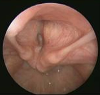
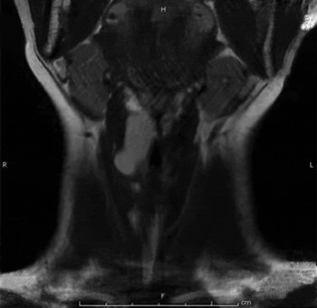
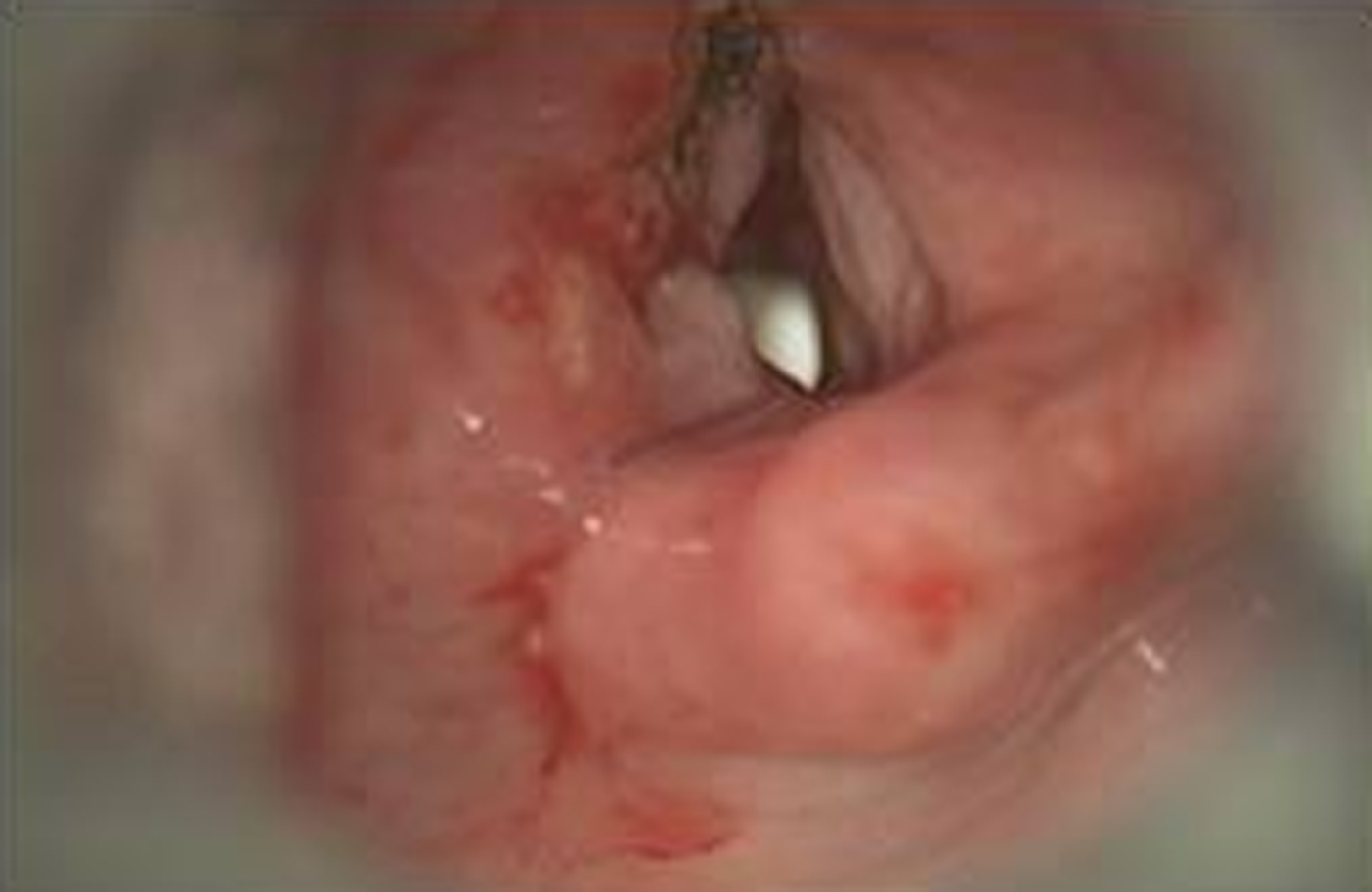
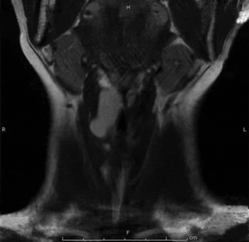
Fibre-optic laryngoscopy revealed a supraglottic mass effacing the right ventricle and obscuring the right true vocal fold. Movement of the left true vocal fold was normal (Figure 1). Magnetic resonance imaging (MRI) revealed a 1 × 2.5 cm proteinaceous cystic lesion lateral to the right vocal fold, suggestive of a congenital cyst (Figure 2).
Supraglottic mass obscuring right true vocal fold.
Coronal, T2-weighted magnetic resonance imaging scan of neck showing right-sided congenital laryngeal cyst. H = head; R = right; L = left; F = feet
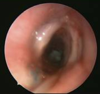
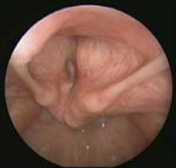
The patient had already undergone voice therapy, and its effects had plateaued. Further treatment options included watchful waiting, endoscopic removal of the cyst, or a tracheostomy with an open laryngofissure for access. A combined endoscopic and open approach was felt to be the best means to excise the cyst. Excision was successful; the vocal folds remained intact and there was no need for a tracheostomy (Figure 3).
Post excision of right-sided congenital laryngeal cyst, showing a patent airway.
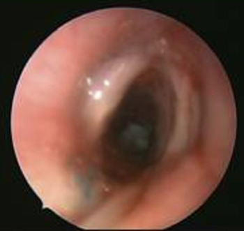
Follow-up MRI at six months post-operatively revealed no recurrence of the cyst. Laryngoscopy showed adequate lateralisation of the right vocal fold, with good movement of the left vocal fold. Objectively, the patient's voice was still hoarse, but she had no communication difficulties anymore. If, at subsequent follow up, she would like further treatment for her hoarseness, we would consider undertaking a vocal fold medialisation procedure.
Case two
A 12-year-old girl with bilateral vocal fold paralysis presented with marked dysphonia. She was born with neuromuscular weakness and remained intubated until four months of age, after which she received a tracheostomy.
The patient had previously undergone bilateral vocal fold Gore-Tex® (breathable membrane) medialisation. Post-operatively, she noticed her voice had diminished. She presented to us for a second opinion, her primary goal being to improve her voice and airway.
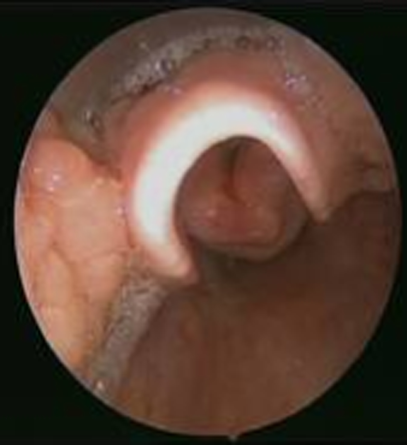
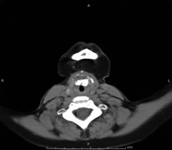
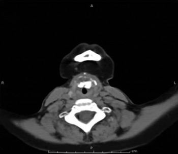
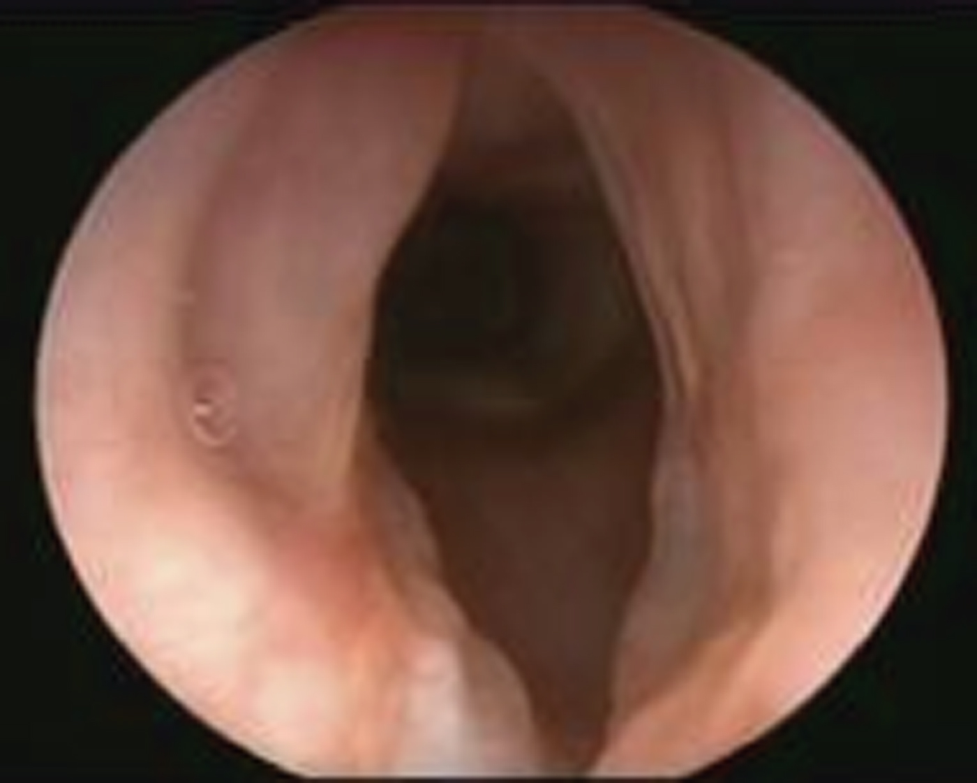
Direct laryngoscopy revealed marked anterior subglottic stenosis, with webbing and right vocal fold granuloma. There was no observed true vocal fold motion, and the right arytenoid was displaced anteriorly and medially (Figure 4). Computed tomography scanning confirmed Gore-Tex infiltration of the entire space around the anterior commissure (Figure 5).
Marked glottic stenosis observed on direct laryngoscopy.
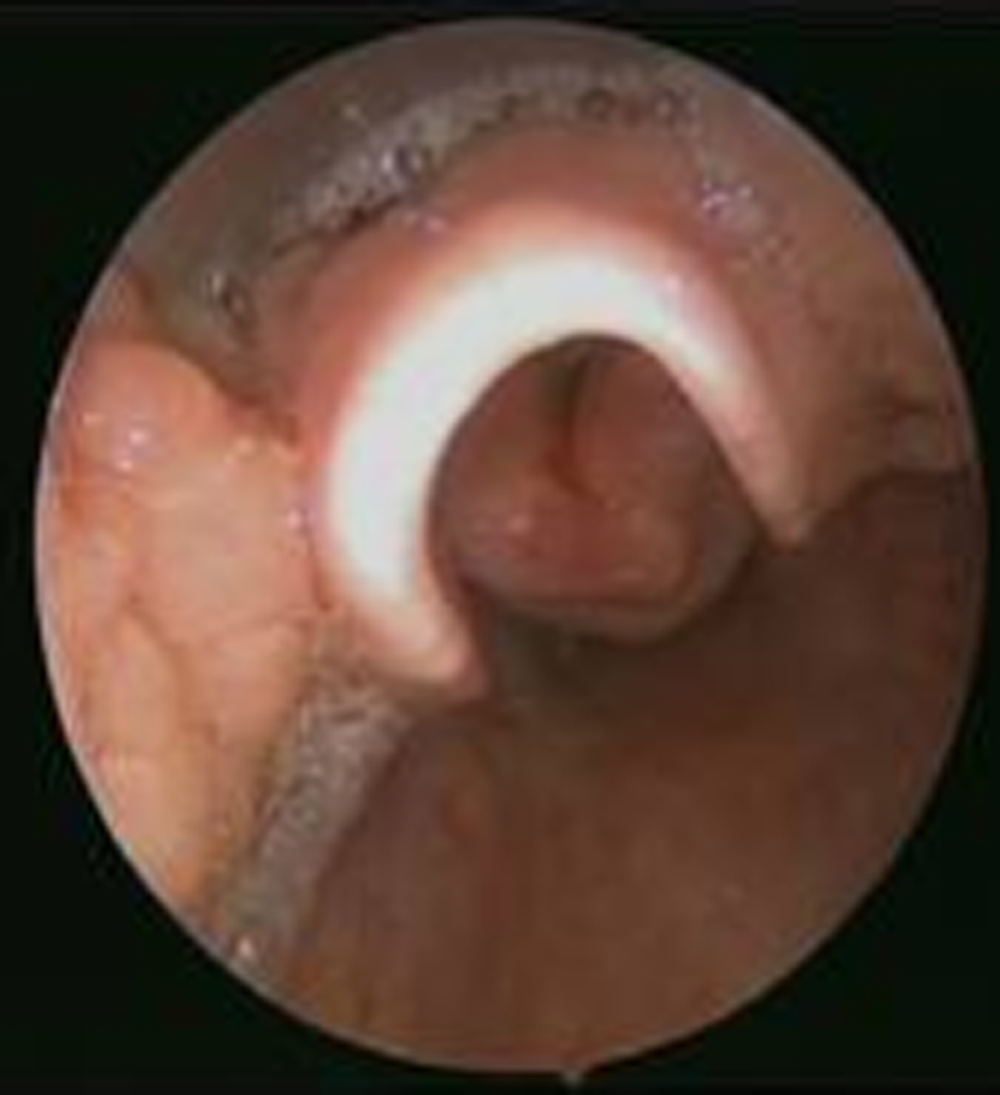
Axial computed tomography image of neck (without contrast) indicating infiltration of Gore-Tex at the anterior space, inferior to the anterior commissure. A = anterior; R = right; L = left; P = posterior
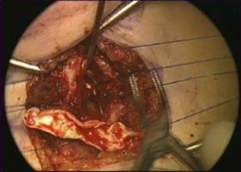
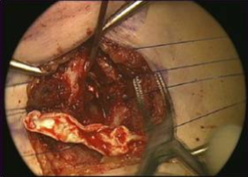
Endoscopic repair would not provide adequate visualisation; thus, an open airway approach was utilised to remove the remaining Gore-Tex and correct the stenosis. An anterior cricoid split was performed and the vocal folds were split at the anterior commissure. A tracheal T-tube stent was placed and the true vocal folds were then re-approximated (Figure 6).
Re-approximation of the true vocal folds and removal of the remaining Gore-Tex.
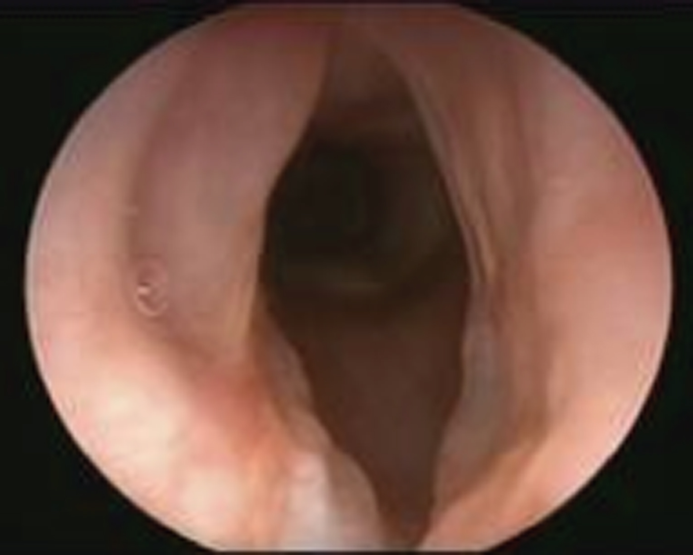
The T-tube remained in place for three months. At follow up, the stenosis had resolved, and the airway was well healed and patent (Figure 7). Open removal of the T-tube was carried out and a tracheostomy tube was temporarily placed. At a recent review, the patient was still mildly dysphonic, but her voice was much stronger. She was able to tolerate capping and we are now considering a trial of tracheostomy decannulation.
Wide glottal aperture observed, with a well-healed glottic airway.
Case three
A 13-year-old girl presented with worsening dysphonia and increasing breathlessness. She was born at 25 weeks gestation. A tracheostomy had been placed for subglottic stenosis secondary to prolonged intubation. She underwent laryngotracheal reconstruction at the age of five years, with removal of the tracheostomy.
Fibre-optic laryngoscopy showed prolapse of the left arytenoid into the airway, and the supraglottic structures were observed to be her phonatory source. We discussed with the patient and her family that in order to evaluate her vocal folds, the prolapsing left arytenoid would have to be removed, but by interfering with her current phonatory source her voice might first worsen before we could make it better. The patient and her family agreed to proceed.
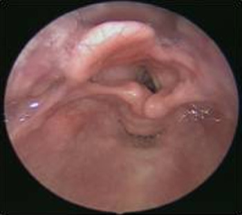
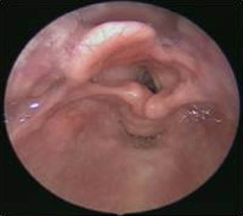
The patient then underwent operative bronchoscopy. This showed multi-level airway stenosis and left-sided cricoarytenoid joint dislocation (Figure 8). Laser supraglottoplasty was performed to the left arytenoid and subglottis to improve patency of the airway.
Prolapsed left arytenoid obscuring the vocal folds.
Despite performing the supraglottoplasty, the patient's dysphonia persisted. It appeared that she had too large a glottal gap to achieve proper glottal closure. Before attempting further surgery, we discussed a three-step open approach with re-evaluation after each step.
The first procedure consisted of open left arytenoid repositioning surgery and laryngotracheal reconstruction. A rib graft was placed in the anterior cricoid to correct the subglottic stenosis. Follow-up examination showed that there was no marked change in voice or arytenoid position.
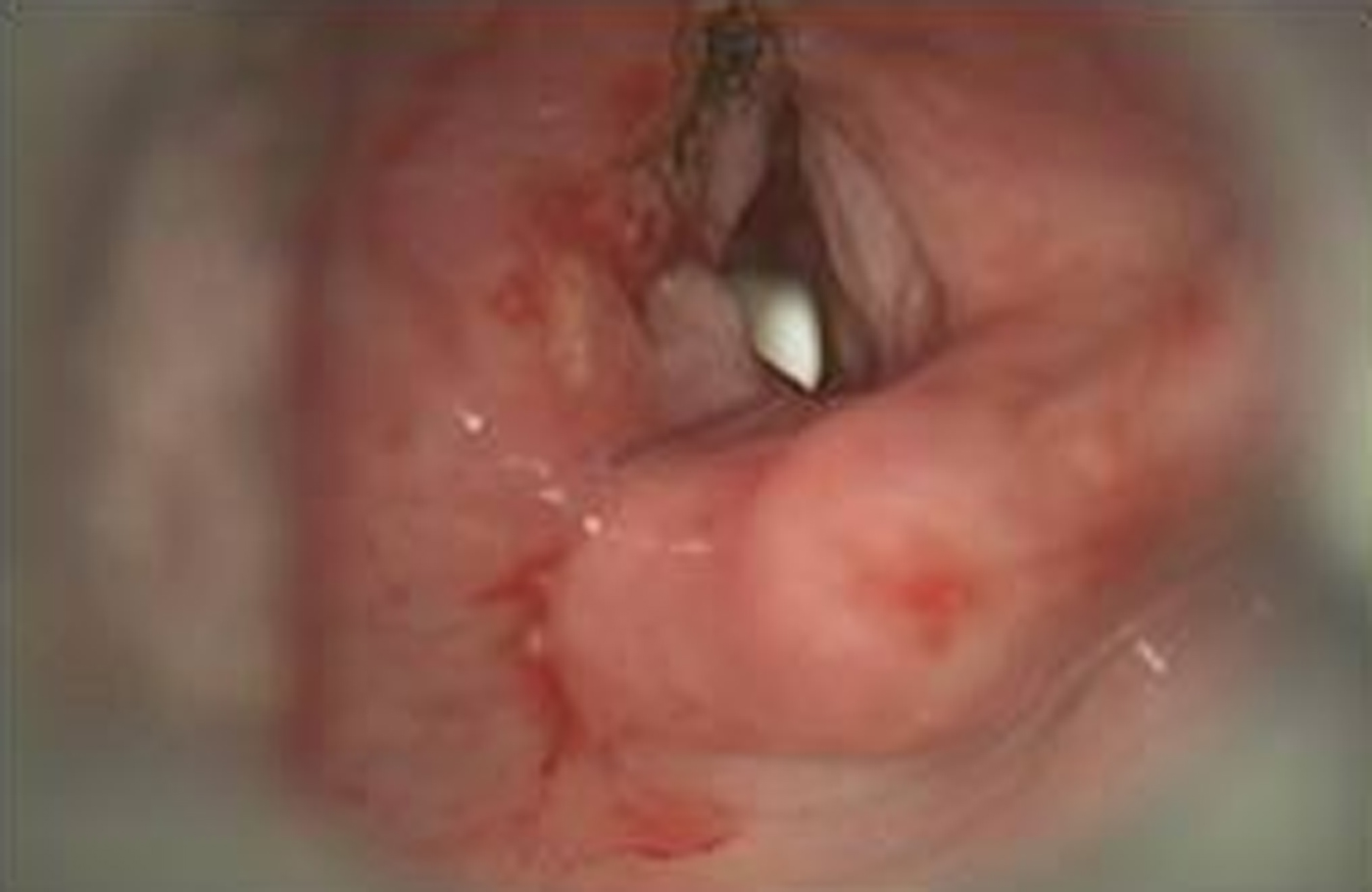
The second step was carried out two weeks later in order to reattach the left vocal fold to the left arytenoid. Supraglottoplasty was performed and the left true vocal fold was re-approximated to the vocal process (Figure 9). Post-operative review again showed no marked change in the patient's voice, but it did reveal a patent supraglottic airway.
Vocal fold re-approximation.
The third step, carried out two weeks later, comprised medialisation thyroplasty. The tracheostomy was subsequently capped and the patient was decannulated. She no longer experiences breathlessness and her airway status was markedly improved. Objectively, her voice had significantly improved and she was happy with the results.
Discussion
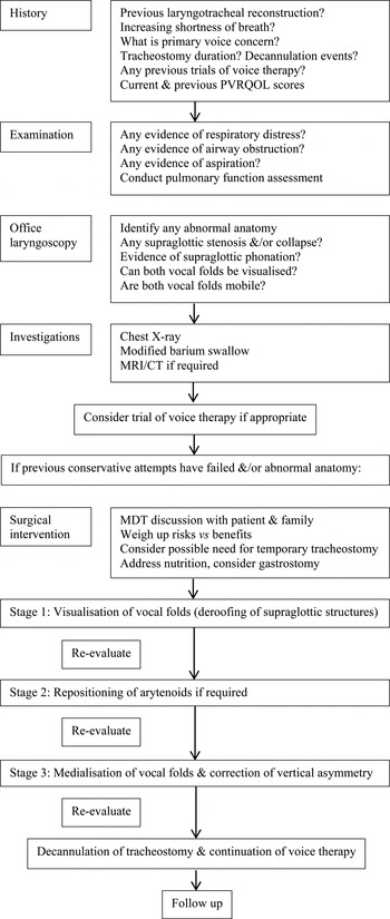
There is extensive literature on the abnormalities that can arise following laryngotracheal reconstruction.Reference Clary, Pengilly, Bailey, Jones, Albert and Comins3 However, there is no literature on the phonosurgical strategies that should be used to address dysphonia in these cases. From our three cases, the importance of a planned staged approach is highlighted. The anatomical and physiological principles of voice production should be carefully evaluated prior to intervention. We have suggested a management algorithm that could be implemented in children with dysphonia following laryngotracheal reconstruction, or in those with dysphonia and complex airway obstruction (Figure 10).
A phonosurgical reconstruction management algorithm for children who have undergone laryngotracheal reconstruction. PVRQOL = Paediatric Voice-Related Quality of Life survey; MRI = magnetic resonance imaging; CT = computed tomography; MDT = multidisciplinary team

Any child with dysphonia following laryngotracheal reconstruction needs to undergo a pre-operative assessment. In addition, each parent and child should complete the Paediatric Voice-Related Quality of Life survey. This helps us to assess the patient's current vocal and airway status, and the impact it has on daily functioning, as dysphonia can have a significant psychological impact on the child.Reference Tirado, Chadha, Allegro, Forte and Campisi4, Reference Zalzal, Loomis and Fisher5
Examination should include laryngoscopy and stroboscopy to define the anatomy. Abnormalities to be aware of following laryngotracheal reconstruction include supraglottic vibration, altered vocal fold integrity and mobility, and anterior commissure blunting.Reference Clary, Pengilly, Bailey, Jones, Albert and Comins3, Reference Bailey, Clary, Pengilly and Albert6
It is important to identify the child's phonatory source. There is often poor glottal closure in children who have undergone laryngotracheal reconstruction, and air escapes through the glottis during phonation. Thus, to prevent air escaping, there is compensatory motion of the arytenoids cartilages onto the epiglottis, which forms a supraglottic sphincter, as in case three.
Baseline voice analysis should be conducted. This allows one to assess whether voice therapy should be considered. Voice therapy techniques can minimise supraglottic compression and reduce compensatory behaviours.Reference Zur and Hartnick7
We need to highlight to families that the focus is not only to establish an adequate airway, but also to achieve a safe swallow and a type of functional voicing that is a significant improvement from their current state.Reference Baker, Kelchner, Weinrich, Lee, Willging and Cotton8 The child and parents should be made aware that a temporary tracheostomy might be required. The risks need to be weighed up against the benefit of improving the child's dysphonia and breathing difficulties.Reference Prescott9, Reference Varghese and Cheng10 If the parents and child agree to go ahead, meticulous planning of stepwise phonosurgical intervention is needed.
Post-operative evaluation should involve planning of tracheostomy decannulation (if necessary), further acoustic and perceptual voice analysis, and office-based laryngoscopy and stroboscopy. The resulting vocal quality may not be entirely normal; at this point voice therapy intervention is the next step, as now the vocal anatomy has been optimised.
• Many children suffer from dysphonia following laryngotracheal reconstruction
• Risk factors for severe dysphonia include multiple prior surgical procedures, high-grade subglottic stenosis and longer tracheostomy decannulation time
• Affected patients can have acoustic, perceptual and anatomical voice and larynx abnormalities
• Phonosurgery should involve thorough pre-operative investigation, including detailed voice analysis and phonatory source identification
• Surgery should be conducted in a stepwise fashion, using open and endoscopic techniques, with voice reassessment after each intervention
• A management strategy for paediatric patients with complex airways is suggested
These children require comprehensive and close long-term follow up, as it may take time before an improvement is observed.
Conclusion
Dysphonia following laryngotracheal reconstruction is common. The resulting differences in voice quality can significantly impact a child's ability to effectively communicate and socialise. More efforts need to be made to address the issue of dysphonia in these children.
When considering surgical intervention, the potential need for a temporary tracheostomy has to be weighed up against the benefits of improved phonation, and this should be a part of the informed consent process when counselling patients and families. Our cases series shows that by undertaking a thorough pre-operative assessment and by conducting surgical intervention in a stepwise manner, with a combination of open and endoscopic techniques, we can minimise the degree of dysphonia in such complex cases. Hopefully, future research and the use of bio-injectables will help to minimise vocal fold scarring post-operatively, and correct any residual dysphonia.