Introduction
Cerebrospinal fluid (CSF) is a clear, colourless fluid that is mainly produced in the choroid plexuses of the brain ventricles. The fluid lies in the subarachnoid space of the brain and spinal cord. It assists in normal cerebral blood flow and autoregulation, and provides mechanical support and immunological protection to the brain. It contains a very small amount of protein and differs from blood in its electrolyte content.Reference Standring 1 On rare occasions, a CSF leak can occur. When the cause is not apparent, this event is considered spontaneous.Reference Mokri 2 It can manifest as clear fluid otorrhoea or rhinorrhea, with non-specific symptoms such as low pressure headaches, nausea, tinnitus, blurred or double vision, change in hearing, and cognitive decline.Reference Graff-Radford and Schievink 3 , Reference Soler and Schlosser 4 A CSF leak represents a critical condition, with increased risks of intracranial infections and brain herniation.Reference Kou, Allen and Isaacson 5 , Reference Battal and Castillo 6
Spontaneous CSF otorrhoea has previously been reported in the literature. In the majority of cases, the CSF leak is unilateral from a defect in the skull base.Reference Rao and Redleaf 7 – Reference Patil, Trinidade, Yung and Donnelly 25 Our literature review revealed six cases of bilateral CSF otorrhoea.Reference Ferguson, Wilkins, Hudson and Farmer 26 – Reference Rotilio, Andrioli, Scanarini, Zuccarello and Fiore 30 The initial symptom of CSF leak was non-synchronous and unilateral. It was several months following surgical repair of the original symptomatic ear that CSF leak was apparent in the contralateral ear.Reference Ferguson, Wilkins, Hudson and Farmer 26 , Reference Lundy, Graham, Kartush and LaRouere 28 – Reference Rotilio, Andrioli, Scanarini, Zuccarello and Fiore 30 We report a very rare case of bilateral and concurrent CSF otorrhoea in an adult patient with no known predisposing factors. The diagnostic dilemmas encountered and final management of this rare pathology are discussed.
Case report
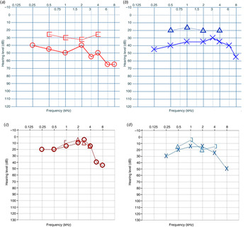
A 44-year-old female presented in the ENT department of a district general hospital with a 2-year history of bilateral fluctuating hearing loss and nasal obstruction. The patient had a background of diabetes type II, hypertension and hypercholesterolaemia. There was no history of trauma, surgical operations, cancer, middle-ear infections or congenital ear defects. Initial examination revealed bilateral hypertrophic inferior turbinates, bilaterally retracted tympanic membranes and a small adenoidal remnant at the roof of the nasopharynx. A pure tone audiogram showed bilateral mild-to-moderate conductive hearing loss (Figure 1), and tympanograms had a flat trace bilaterally.
(a) Right ear and (b) left ear pre-operative pure tone audiograms showing symmetrical mild-to-moderate conductive hearing loss bilaterally. (c) Right ear and (d) left ear post-operative pure tone audiograms with average air conduction hearing thresholds of 20 dB on the right and 25 dB on the left. [ = Bone conduction (masked) right ear; ○ = air conduction (unmasked) right ear; ▵ = bone conduction (unmasked); × = air conduction (unmasked) left ear; ] = bone conduction (masked) left ear

The patient was diagnosed with bilateral otitis media with effusion. A watch-and-wait period was initiated, and the patient was advised to perform autoinflation exercises and use a topical steroid nasal spray on a daily basis. At the two-month review, the patient reported an improvement in hearing and nasal obstruction following daily use of the nasal spray. However, she stated that symptoms returned when the spray was withheld. Her pure tone audiogram showed a 10 dB improvement on both sides. She had persistent type B tympanograms. A decision was taken to continue conservative treatment, and further follow up was arranged. After a further three months, the patient was still troubled with bilateral hearing loss. The pure tone audiogram showed bilateral moderate conductive hearing loss. Hence, a decision was taken to insert grommets bilaterally.
The patient underwent bilateral insertion of Shah grommets. Thin clear fluid was noted in both middle-ear clefts, which appeared to be under pressure. Biopsies of the nasopharynx were taken, which were subsequently reported as benign tissue growth. At the post-operative follow-up appointment, her hearing had improved, but there was an ongoing clear discharge from both ears through the patent grommets. This was treated as an infection with a 10-day course of ciprofloxacin drops.
The patient was seen on two more occasions with persistent otorrhoea through the grommets; this was treated each time as acute otitis media. She had no vertigo or otalgia. At follow up one month later, a blocked grommet was noted on the right side with the presence of middle-ear effusion, and a patent grommet was observed on the left side, with fluid actively discharging. A decision was made to replace the grommets in the operating theatre and perform adenoidectomy to remove the excess adenoidal tissue. The patient underwent adenoidectomy and bilateral re-insertion of grommets with an uneventful recovery. Following this, she was seen twice with persistent otorrhoea each time, treated as an infection. All swabs taken were negative, and each time the patient was well in herself.
Two years following her initial presentation, a review of the operating notes from the first grommet insertion, which noted that middle-ear fluid was under pressure following myringotomy, raised the suspicion of bilateral ongoing CSF leak.
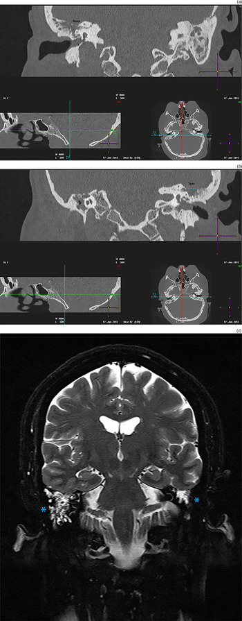
A sample was collected and a computed tomography (CT) scan was arranged. The collected sample was positive for tau protein. A fine-cut temporal bone CT scan and a subsequent magnetic resonance imaging (MRI) scan revealed a large dehiscence of the tegmen tympani of nearly 1 cm on the left and 6 mm on the right. Opacification was observed in both mastoids and middle-ear clefts. The ossicular chain, inner-ear structures and bony facial nerve canal were all normal (Figure 2).
(a & b) Coronal, fine-cut temporal bone computed tomography images showing a large dehiscence of the tegmen of nearly 1 cm on the left and 6 mm on the right, and (c) a coronal, T2-weighted magnetic resonance image showing opacification of the middle ear and mastoids bilaterally (asterisks).

The patient underwent staged bilateral middle fossa craniotomies and repair, with a three-month interval between each operation. The right-sided defect was repaired initially, and brain herniation was identified and successfully reduced. The dehiscent segment of the tegmen was repaired with Surgicel® absorbable haemostat, DuraGen® collagen matrix and bone pate in Tisseel® fibrin sealant. The same operation was performed on the left side three months later. There was no evidence of brain herniation. The patient had an uneventful recovery.
The patient has had no further CSF leak. The post-operative hearing thresholds were 20 dB on the right side and 25 dB on the left side (Figure 1). A repeat CT scan conducted two years post-operatively showed no fluid in the middle-ear clefts, with no dehiscence of the tegmen tympani.
Discussion
Cerebrospinal fluid otorrhoea is a clinical entity known to be linked with congenital malformations of the inner ear, temporal bone trauma, neoplasms, otological or intracranial surgery, and middle-ear infections.Reference Yi, Zhao, Guo, Wu, Li and Ren 31 – Reference Stenzel, Preuss, Orloff, Jecker and Mann 33 Many case reports and case series have been published over the past two decades describing unilateral spontaneous CSF temporal bone leaks presenting as unilateral middle-ear effusions.Reference Rao and Redleaf 7 – Reference Patil, Trinidade, Yung and Donnelly 25 This is suspected in patients presenting with unilateral middle-ear effusion of unexplained origin.Reference Zakaryan, Poulsgaard, Hollander and Fugleholm 8 , Reference Son, Karkas, Buchanan, Giurintano, Theodosopoulos and Pensak 9 , Reference Brown, Grundfast, Jabre, Megerian, O'Malley and Rosenberg 12 , Reference Wolfowitz 34 Older age, female gender, obesity, obstructive sleep apnoea and intracranial hypertension are believed to have a role in the pathogenesis of spontaneous CSF otorrhoea, but the mechanism remains unknown in the majority of cases.Reference Vivas, McCall, Raz, Fernandez-Miranda, Gardner and Hirsch 35 – Reference LeVay and Kveton 39
This paper describes a rare case of chronic bilateral and concurrent CSF otorrhoea, presenting in a middle-aged female, with no obvious cause for her symptoms. Bilateral CSF otorrhoea has been reported previously in the literature.Reference Ferguson, Wilkins, Hudson and Farmer 26 – Reference Rotilio, Andrioli, Scanarini, Zuccarello and Fiore 30 In the majority of cases (five out of six), there was an interval of a few months to several years between each (left and right) presentation.Reference Ferguson, Wilkins, Hudson and Farmer 26 , Reference Lundy, Graham, Kartush and LaRouere 28 – Reference Rotilio, Andrioli, Scanarini, Zuccarello and Fiore 30 Merchant and McKenna also reported a case of bilateral CSF otorrhoea, but did not mention whether the symptoms were bilateral at initial presentation.Reference Merchant and McKenna 27 In all cases, the diagnosis was established following myringotomy, wherein the middle-ear fluid was noted to be under pressure. The presence of a CSF leak due to a skull base defect was then confirmed with imaging and laboratory testing.
Imaging studies are of paramount importance to confirm diagnosis and identify the cause of the CSF leak. They also assist in evaluating the defect site prior to surgery and inform on the best treatment option. Although different imaging options are available, such as CT imaging, MRI, radionuclide or CT cisternography, no ‘gold standard’ exists for the diagnosis of CSF leak. Computed tomography studies delineate the bony defects better.Reference Lloyd, DelGaudio and Hudgins 40 Thin-section CT scans can effectively diagnose even small skull base defects, with a documented 92 per cent sensitivity and 100 per cent specificity.Reference Shetty, Shroff, Sahani and Kirtane 41 Magnetic resonance imaging assists in the differentiation of soft tissue elements around the bony defect, which are hard to assess on a CT scan. It has been proposed that a CT scan is performed first, followed by an MRI scan as an adjunct to explore the origin of soft tissue opacifications, with cisternography reserved for the most complex cases, where CT or MRI scans have failed to assist in diagnosis.Reference Lloyd, DelGaudio and Hudgins 40
The skull base defect in our patient was effectively treated surgically with a middle fossa craniotomy approach following evaluation of the defect. Different surgical approaches have been described to manage skull base CSF leaks. These include open transcranial and transmastoid methods, or equally aggressive minimally invasive alternatives using the endoscopic endonasal route. A recent systematic review revealed no difference in the success rates for minimally invasive versus open approaches, with decisions based on surgeons’ preference and expertise.Reference Komotar, Starke, Raper, Anand and Schwartz 42 However, minimally invasive approaches had a lower rate of post-operative complications and peri-operative mortality. In a retrospective case series review of the surgical management of CSF otorrhoea, Kutz et al. concluded that the middle fossa craniotomy approach provided the best access for defects of the middle fossa floor.Reference Kutz, Husain, Isaacson and Roland 13 As in our case, a combination of autologous and allogenic materials was highly successful. Rao et al. found that transmastoid repair was also highly effective in their case series of tegmen defects. Only one patient required temporal craniotomy, as there was evidence of meningoencephalocele with brain herniation.Reference Rao, Merenda and Wetmore 16 Transcranial approaches should be considered when the defects are multiple or located in the petrous apex, or if wide access is required for which a transmastoid approach would not be adequate.Reference Pelosi, Bederson and Smouha 43 Conservative management with prophylactic antibiotic cover, with or without lumbar drain insertion, is effective in managing most cases of CSF leak resulting from head injuries, but is of limited value for non-traumatic or post-operative CSF leaks.Reference Savva, Taylor and Beatty 44
In our case, CSF was present in both middle-ear clefts at the time of initial presentation. This delayed the correct diagnosis of a skull base defect leading to a CSF leak. The patient was misdiagnosed as having otitis media with effusion, and grommets were inserted on two occasions. Although the middle-ear fluid was noted to be under pressure at the time of first grommet insertion, no suspicion of CSF otorrhoea was raised. Copious amounts of clear fluid under pressure have been documented following myringotomy in cases of middle-ear effusion that was consequently found to be CSF.Reference Ferguson, Wilkins, Hudson and Farmer 26 – Reference Rotilio, Andrioli, Scanarini, Zuccarello and Fiore 30 Even in unilateral cases, patients are often considered to have otitis media with effusion, and tend to be treated conservatively for a prolonged period of time. In the majority of cases, patients are asymptomatic apart from hearing loss and the presence of unilateral middle-ear effusion.Reference Zakaryan, Poulsgaard, Hollander and Fugleholm 8 , Reference Son, Karkas, Buchanan, Giurintano, Theodosopoulos and Pensak 9 , Reference Brown, Grundfast, Jabre, Megerian, O'Malley and Rosenberg 12 , Reference Rao, Merenda and Wetmore 16 , Reference Wolfowitz 34
-
• Unilateral spontaneous cerebrospinal fluid (CSF) otorrhoea due to temporal bone tegmen tympani defects has been previously reported
-
• The cause can be identified following myringotomy with observation of middle-ear fluid quality; skull base defects are confirmed with imaging
-
• This paper presents a case of synchronous bilateral CSF otorrhoea with no predisposing risk factors
-
• Awareness of spontaneous bilateral CSF otorrhoea could prevent misdiagnosis and delays in treatment
We would like to raise awareness of the rare entity of spontaneous, synchronous bilateral CSF. We recommend careful observation of the quality and characteristics of middle-ear fluid when a myringotomy is performed. Grommet insertion is one of the most routine ENT procedures. Surgeons in ENT should be aware of the rare occurrence of bilateral concurrent CSF otorrhoea and suspect its presence, especially in older patients presenting at the clinic with bilateral middle-ear effusions, with no evidence of eustachian tube dysfunction. This will help in a prompt diagnosis of this rare entity and the rapid initiation of appropriate treatment.
Conclusion
A link between bilateral middle-ear effusions unresponsive to classic treatment and CSF leak was made, and the patient was successfully treated with staged middle fossa craniotomies. Doctors in ENT should be aware of this rare entity and include it in their differential diagnosis when treating patients with bilateral middle-ear effusion not compliant to standard management.
Acknowledgement
We would like to thank Mr Julian Danino, who participated in the writing and technical editing of the manuscript.