Case report
An 18-month-old boy presented to his paediatrician on day 2 of illness with rhinorrhoea, cough and fever. A diagnosis of acute otitis media was made and he was started on amoxicillin. Persistent fevers of increasing magnitude resulted in a re-evaluation on day 5 of the illness by the paediatrician, where a new friction rub was heard. He was sent to the local emergency department, and was then transferred to our institution for further care.
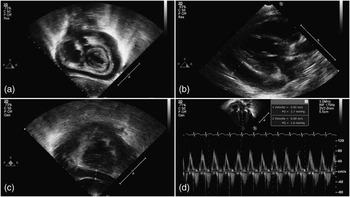
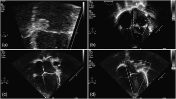
Upon arrival in the paediatric intensive care unit, the patient was febrile, tachycardic, tachypnoeic, irritable and sleepy. The blood pressure was 96/68 and pulses were normal. A pericardial friction rub was present over the precordium. The electrocardiogram was normal. Echocardiography demonstrated normal intracardiac anatomy, no vegetation, and a moderate, globally distributed pericardial effusion (Fig 1a). The effusion was echolucent, without fibrinous material in the pericardial space. There was no echocardiographic evidence of cardiac tamponade (Fig 1d) and the patient was monitored in the paediatric intensive care unit. The next day, the effusion was larger on echocardiogram (Fig 1b and c), and elective ultrasound-guided pericardiocentesis removed 55 ml of thick, yellow fluid from the pericardial space. Pericardial fluid analysis revealed: 7200 white blood cells (91% polymorphonuclear cells, 3% bands, 2% lymphocytes, 4% mononuclear cells), 4125 red blood cells, glucose <20, total protein 4.7, lactate dehydrogenase 18,471, with Gram stain for Gram-positive cocci in pairs and clusters. Blood and pericardial fluid cultures were both positive for methicillin-susceptible Staphylococcus aureus. Approximately 36 hours after pericardiocentesis, a follow-up echocardiogram demonstrated a new sessile, centrally lucent mass concerning for an abscess attached to the anterior mitral leaflet (Fig 2a). There was no associated mitral regurgitation.
(a) Subcostal coronal image at initial presentation shows a moderate, globally distributed pericardial effusion without fibrinous appearance. (b and c) Subcostal coronal image and parasternal long-axis view taken 12 hours after presentation shows enlargement of the effusion, now with a maximal dimension of 12 mm. (d) Mitral inflow velocity analysis at the time of presentation shows respiratory variation of 17%, suggesting an absence of tamponade physiology.

(a) Magnified four-chamber view obtained 48 hours after initial presentation demonstrates a sessile, centrally lucent mass at the hinge point of the anterior mitral leaflet. Note the central echolucency suggestive of an abscess. (b) A colour Doppler image 13 days after presentation demonstrates a broad-based jet of mitral regurgitation near the mass. (c and d) Follow-up images 3 months after presentation show a normal appearing mitral valve without the mass. Colour Doppler imaging shows no mitral regurgitation.

During antibiotic therapy (nafcillin 190 mg/kg intravenous, divided every 6 hours for 6 weeks and gentamicin 3 mg/kg intravenous, given every 24 hours for five doses) for infective endocarditis, serial echocardiography demonstrated progressive resolution of the mass. However, an eccentric jet of mitral regurgitation developed along the anterior mitral leaflet near the mass (Fig 2b). Around the time of discovery of the mitral regurgitation, the patient had a recurrence of fever. He was taken to the operating room where a pericardiotomy was performed and culture-negative, purulent fluid was evacuated from the posterior pericardial space. Copious irrigation with Dakin's solution was performed, and he thereafter recovered uneventfully. The patient was discharged home several days later. Late follow-up echocardiogram showed complete resolution of both the mass and the mitral regurgitation (Fig 2c and d).
Discussion
Bacterial pericarditis is a rare diagnosis.Reference Cakir, Gurkan, Balci, Eren and Dikici 1 – Reference Weir and Joffe 4 A number of case reports in the literature have helped to define the “typical” presentation and aetiology.Reference Klacsmann, Bulkley and Hutchins 2 , Reference Weir and Joffe 4 Historically, bacterial pericarditis was a disease of children and young adults, existing largely as a secondary infection with pneumonia or after trauma to the mediastinum. At present, bacterial pericarditis is more commonly found in the adult population,Reference Klacsmann, Bulkley and Hutchins 2 although it is still typically a primary infection.Reference Cakir, Gurkan, Balci, Eren and Dikici 1 – Reference Parikh, Memon, Echols, Shah, McGuire and Keeley 3 , Reference Katz, Pitlik, Porat, Biderman and Bishara 5 Formerly, the most common infecting organism was Staphylococcus pneumoniae; currently, the most common infecting organism is S. aureus.Reference Cakir, Gurkan, Balci, Eren and Dikici 1 – Reference Weir and Joffe 4 , Reference Roodpeyma and Sadeghian 6 In the past, the mortality rate for bacterial pericarditis was high because of the difficulty with diagnosis and limitations in therapy.Reference Cakir, Gurkan, Balci, Eren and Dikici 1 – Reference Weir and Joffe 4 Advances in echocardiography have made early diagnosis and treatment possible, but the mortality rate is not zero.Reference Cakir, Gurkan, Balci, Eren and Dikici 1 , Reference Parikh, Memon, Echols, Shah, McGuire and Keeley 3
Owing to the high mortality rate, once diagnosed, immediate treatment is imperative. In addition to antibiotics, pericardial drainage is beneficial to prevent constrictive pericarditis, and relieve coexisting tamponade.Reference Cakir, Gurkan, Balci, Eren and Dikici 1 , Reference Parikh, Memon, Echols, Shah, McGuire and Keeley 3 , Reference Weir and Joffe 4 , Reference Roodpeyma and Sadeghian 6 Early relief of the effusion is paramount, as most sources suggest a strong correlation with constrictive pericarditis and either delayed or absent drainage of purulent pericardial fluid.Reference Cakir, Gurkan, Balci, Eren and Dikici 1 Most disease is amenable to drainage via pericardiocentesis; only a few will require pericardiotomy.Reference Cakir, Gurkan, Balci, Eren and Dikici 1 , Reference Weir and Joffe 4 The pericardial fluid must be sent for culture and sensitivities to optimise therapy.
The spectrum of endocarditis has changed over time. What was once a disease typically caused by Streptococcus species is now a disease of Staphylococcus species. Along with changes in infectious aetiology, there has been recognition that the majority of endocarditis infections occur in individuals with normal hearts, not those with prosthetic valves or pre-existing cardiac disease.Reference Fernández Guerrero, González López, Goyenechea, Fraile and de Górgolas 7 , Reference Pasquali, He and Mohamad 8 As a result, the guidelines for endocarditis prophylaxis were changed, drastically reducing the number of patients receiving prophylaxis.Reference Wilson, Taubert and Gewitz 9 Recent population studies have shown no statistically significant change in endocarditis caused by Streptococcus species, after the reduction in the number of patients receiving prophylaxis. These studies have confirmed that Staphylococcus continues to be the most common cause of bacterial endocarditis.Reference Pasquali, He and Mohamad 8
The notion that bacterial pericarditis is almost always a secondary infection arises from the limited vascularity of the pericardium. Theoretically, it is much easier for a primary infection to become established in well-vascularised areas of the body – lung, bone – and then spread secondarily to the pericardium. Pericarditis arising from erosion of a primary endocarditis into the pericardial space can also occur,Reference Arnett and Roberts 10 usually after a valve ring abscess has formed, most commonly on the aortic valve, although it has been reported from the tricuspid and mitral valves. As such, it is very uncommon for pericarditis to be identified as the primary infection.Reference Katz, Pitlik, Porat, Biderman and Bishara 5
Mitral valve surgery is sometimes necessary after development of regurgitation due to endocarditis. According to the updated 2008 AHA guidelines, our patient did not meet criteria for mitral valve repair or replacement as he was asymptomatic and had preserved left ventricular function (Class III, level of evidence “C”). However, had he developed symptomatic, acute, severe mitral regurgitation, or had his mitral regurgitation progressively worsened to a chronic, severe level either with symptoms or with a reduced amount of left ventricular function, there would have been a clear indication for repair or replacement (Class I, level of evidence “B”).Reference Bonow, Carabello and Chatterjee 11
Our patient is unique in that he manifested an unusual presentation of pericardial effusion: bacterial pericarditis with secondary spread leading to presumed bacterial endocarditis. He presented with fever, tachypnoea and pericardial friction rub, which are the most common presenting symptoms of bacterial pericarditis. An echocardiogram on the day of presentation to the pediatric intensive care unit clearly demonstrated no evidence of intracardiac mass or thrombus of any kind. Once he had developed the abscess on the mitral valve, however, he met modified Duke criteria for the diagnosis of endocarditis.Reference Li, Sexton and Mick 12 This, in combination with an increasing pericardial effusion, was indication for drainage of the effusion, which also yielded positive cultures for S. aureus, despite having already received appropriate initial intravenous antibiotics. Constrictive pericarditis did not ensue, but fever resumed with evidence of a posterior pericardial fluid collection, which necessitated surgical pericardiotomy.
Acknowledgement
None.