Introduction
Giant prolactinoma is a rare form of prolactinoma mainly reported in the neurosurgical and endocrinological literature. It is more common in men and generally presents with symptoms of endocrine disturbance or neurological deficit. Here, we present a case of a giant prolactinoma which presented with symptoms of sinusitis and became rapidly progressive, causing multiple neurological deficits.
Case report
A normally fit and well, 30-year-old teacher first presented to the nephrologists with a six-month history of generalised abdominal pain, microscopic haematuria and proteinuria, fatigue, recurrent sinusitis, nasal discharge, and dizziness. He was noted at this time to have a mild proptosis. He was investigated for Wegener's granulomatosis but his renal biopsy was normal.
The patient was referred to the ENT clinic with ongoing symptoms of recurrent sinusitis not relieved by several courses of antibiotics. He had nasal congestion, headaches, night sweats and dizziness.
On examination, the nasal septum was significantly deviated to the left and the head of the right inferior turbinate was polypoid. There were no other abnormalities.
The patient was commenced on steroid nasal drops, and an urgent computed tomography (CT) scan of his sinuses was requested.
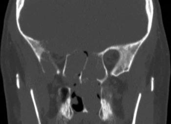
This showed a large soft tissue mass within the sphenoid sinuses, causing extensive bone erosion (Figure 1).
Coronal computed tomography scan showing a large soft tissue mass within the sphenoid sinuses, causing extensive bone erosion.

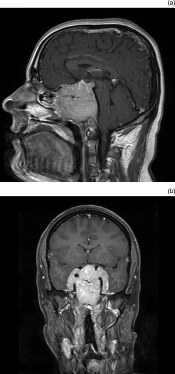
An urgent magnetic resonance imaging (MRI) scan was requested to better define the soft tissue mass. This identified an extensive skull base mass with a nasopharyngeal component (Figure 2).
(a) Sagittal and (b) coronal magnetic resonance imaging scans of the giant prolactinoma, showing an extensive skull base mass with a nasopharyngeal component.

These radiological findings suggested a nasopharyngeal carcinoma.
An examination under anaesthesia and biopsy were performed the following day, with no complications apart from bleeding during biopsy which was controlled by packing.
The histopathology report for the biopsy specimen, obtained three days after surgery, described a tumour with a nested growth pattern in a vascular stroma formed of uniform epithelial cells showing no necrosis and a low mitotic rate. The histological pattern suggested a low grade neuroendocrine tumour. This diagnosis was supported by immunohistochemically determined expression of cluster of differentiation 56 protein, synaptophysin and chromogranin, and a low Ki67 labelling rate (5 per cent). These results were initially considered consistent with a diagnosis of low grade neuroendocrine carcinoma or olfactory neuroblastoma, in the clinical context of a midline mass in the roof of the nasal cavity.
Five days post-surgery, the patient was readmitted under the care of the physicians with weakness, nausea, dizziness, headache and right-sided hearing loss. This was initially diagnosed as a middle-ear infection, for which he was treated with antibiotics. A full range of blood tests was requested, including a pituitary hormone screen.
Over a period of 48 hours, our patient developed worsening symptoms: a right-sided, House–Brackmann grade IV, lower motor neuron facial palsy; severe, bilateral tinnitus; and fasciculations of the right side of the tongue. The previously performed pituitary screening tests were reported as showing a prolactin level of over 800 000 µm/l. It was felt that the most likely diagnosis was a macroprolactinoma, and he was commenced on 0.5 mg of cabergoline, a dopamine receptor agonist.
A second, urgent CT scan showed a small area of haemorrhage in the centre of the mass, but also, more worryingly, signs of raised intracranial pressure. The patient was commenced on dexamethasone. However, his rapid neurological deterioration continued, with the onset of dysphagia and stridor, necessitating intubation and ventilation in the intensive care unit.
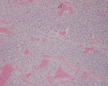
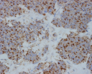
On the following day, after further examination with specific staining techniques, a giant invasive prolactinoma was confirmed histologically (Figures 3 and 4). Sadly, our patient developed a fixed, dilated left pupil as a result of brainstem compression, and underwent a right pterional craniotomy to decompress the prolactinoma. Cabergoline therapy was continued, leading to a reduction in his prolactin levels over several weeks.
Medium power photomicrograph of the tumour biopsy with haematoxylin and eosin staining. (×200)

Higher power photomicrograph of the tumour biopsy stained with an immunohistological preparation containing anti-prolactin antibodies. (×400)

Discussion
Prolactinomas are the most frequently occurring pituitary tumours, with a prevalence of 100 per million in adults. Giant prolactinomas are a rare form of prolactinoma which make up between 0.5 and 4.4 per cent of all pituitary tumours.Reference Gillam, Molitch, Lombardi and Colao 1
Giant prolactinomas are defined as prolactin-secreting adenomas greater than 4 cm in diameter and/or with more than 2 cm suprasellar extension. They are usually associated with high serum prolactin levels of 20 000 to 100 000 ng/ml.Reference Murphy, Vesely, Jordan, Flanigan and Kohler 2
Prolactinomas generally occur more frequently in women than men; however, microadenomas are more commonly found in women and macroadenomas in men. Prolactin levels tend to be higher at presentation in men regardless of whether they have a macro- or microadenoma; the size of the tumour is also generally greater in men. The presentation also varies between the genders: women often present with symptoms of sexual dysfunction, weight gain and galactorrhoea, whereas men tend to present with symptoms relating to tumour expansion.Reference Colao, Sarno, Cappabianca, Briganti, Pivonello and Somma 3
Our patient had approximately 11 months of vague symptoms prior to the onset of cranial nerve defects as a result of compression effects. He initially suffered with non-specific headache, recurrent sinusitis and rhinitis. This is a rare presentation of a pituitary tumour, being more commonly associated with skull base tumours. However, there have been previous reports of similar presentations. In 2002, Minniti et al. reported four cases of prolactinoma presenting as a skull base tumour. The symptoms in these cases included headache, nasal obstruction, progressive hypoacusia, ophthalmoplegia, dizziness, psychiatric symptoms and retro-orbital pain.Reference Minniti, Jaffrain-Rea, Santoro, Esposito, Ferrante and Delfini 4
Following biopsy of the tumour, the patient suffered an acute deterioration which eventually resulted in the need for urgent decompressive surgery. This may have been due to pituitary apoplexy, characterised by: sudden onset, severe headache; vomiting; visual impairment; ophthalmoplegia; meningism; altered level of consciousness; and endocrine dysfunction. This occurs as a result of sudden haemorrhage or infarction of the pituitary gland, usually within an adenoma.Reference Dafer and Jay 5
Diagnosis of prolactinoma involves hormonal and radiological methods. Firstly, a raised serum prolactin level must be identified. This may require repeated sampling as prolactin is a pulsatile hormone. Following confirmation of hyperprolactinaemia, CT and MRI scanning is used to confirm the presence and extent of the pituitary gland tumour. For histological diagnosis, specific immunohistochemical stains are required to confirm the presence of prolactin in tumour cells.Reference Ciccarelli and Camanni 6
In our patient, the initial histological appearance was of an olfactory neuroblastoma. However, the presence of cytokeratin expression and the absence of S100-expressing sustentacular cells argued against this diagnosis. This, together with the known overlap between the histological and immunohistochemical features of olfactory neuroblastoma and pituitary adenoma, and the demonstration of cytoplasmic expression of prolactin (using anti-prolactin antibodies), enabled the final diagnosis of a giant invasive prolactinoma.
Dopamine agonists are the primary treatment for prolactinomas. Cabergoline is known to be an effective treatment in cases of micro- and macroprolactinoma. Cho and colleagues' study found that cabergoline treatment enabled rapid and significant reduction in serum prolactin levels and tumour size in 10 male patients with giant prolactinoma.Reference Cho, Lee, Chung, Koh, Cho and Kim 7 In our patient, cabergoline therapy was commenced but he continued to deteriorate and required surgical intervention. Cabergoline therapy was continued, leading to a dramatic reduction in serum prolactin and tumour size.
Medical therapy is the mainstay of treatment for pituitary tumours. The 2005 Ninth International Pituitary Congress set out guidelines for when surgical intervention may be required, i.e. (1) for non-responders to dopamine agonist therapy; (2) for patients with intolerable side effects on medical therapy; (3) for patients with a cerebrospinal fluid fistula receiving dopamine agonists; and (4) for patients with rapidly progressing neurological deficits, as in our case.Reference Kreutzer, Buslei, Wallaschofski, Hofmann, Nimsky and Fahlbusch 8 Surgery for giant prolactinomas has been shown to be successful for debulking, but is rarely curative. Complication rates are high, with significant morbidity ranging from 10 to 64 per cent and mortality ranging from 3.3 to 31.2 per cent.Reference Gillam, Molitch, Lombardi and Colao 1
-
• Giant invasive prolactinomas are well reported in neurosurgical and endocrine literature
-
• Unusually, this case represented as a nasopharyngeal mass and skull base tumour
-
• This non-malignant neoplasm may have debilitating or fatal consequences
Our case demonstrates the aggressiveness of, and diagnostic challenge posed by, giant invasive prolactinoma. It also highlights the importance of endocrine screening in cases of invasive skull base tumour. The long-term sequelae of these tumours can be dramatic, as in our patient; therefore, their prompt diagnosis and treatment can be important determinants of the long-term outcome. Although medical therapy with dopamine agonists remains the most effective treatment, in some cases medical therapy alone will not be sufficient to control disease progression.