Introduction
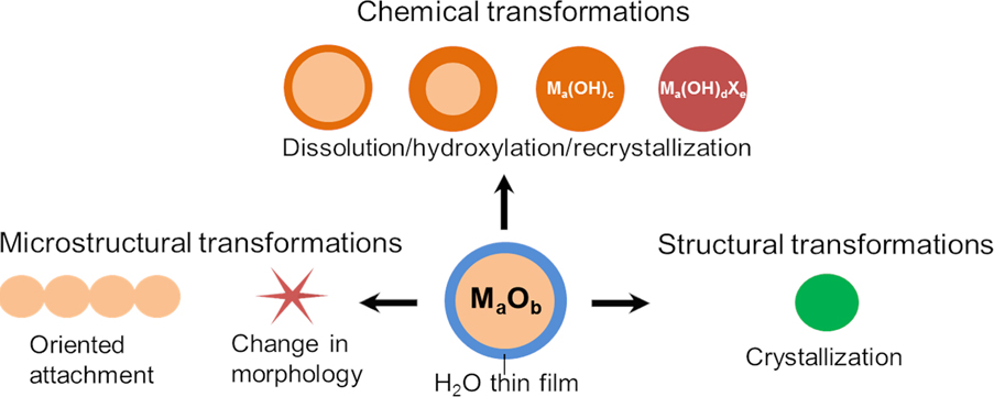
Water is ubiquitous under ambient conditions and condenses readily at metal oxide surfaces. Water adsorption, water film formation, and water-mediated reactions on metal oxide surfaces are therefore fundamentally important in environmental chemistry, in catalysis, and for processing of functional metal oxides in general [Reference Garde and Schlossman1]. During materials operation under ambient conditions, thin films of mostly undefined composition and structure cover the metal oxide nanoparticle surfaces. Therefore, the boundaries between oxide nanoparticles and the environmental phase need to be described as interfaces rather than as surfaces. Similar to bulk water, such thin water films alter the oxide surface by modifying its chemical reactivity and stability [Reference Stumm, Sigg and Sulzberger2, Reference Verdaguer, Sacha, Bluhm and Salmeron3, Reference Ewing4, Reference Ringleb, Sterrer and Freund5, Reference Bjorneholm, Hansen, Hodgson, Liu, Limmer, Michaelides, Pedevilla, Rossmeisl, Shen, Tocci, Tyrode, Walz, Werner and Bluhm6, Reference McBriarty, von Rudorff, Stubbs, Eng, Blumberger and Rosso7, Reference Mu, Zhao, Dohnálek and Gong8, Reference Sterrer, Nilius, Shaikhutdinov, Heyde, Schmidt and Freund9]. This can lead to chemical, structural, and microstructural transformations such as dissolution/precipitation, morphological changes, crystallization, high-index facet stabilization [Reference Goniakowski, Finocchi and Noguera10, Reference Noguera and Goniakowski11], or self-assembly (Fig. 1) [Reference Niederberger and Cölfen12, Reference Cölfen and Antonietti13, Reference Zhang, Shen, Liu, Kerisit, Bowden, Sushko, de Yoreo and Rosso14]. However, due to the confinement of mass transport at the oxide surface, these processes do not proceed exactly as they do in bulk liquid water.
The possible effects of thin water films on metal oxide nanostructure composition, structure, and morphology.

In this review, we will present selected examples from our group and others’ that studied water thin film–mediated transformations of well-defined metal oxide nanostructures. We focus on MgO and TiO2 nanostructures that are canonical metal oxide model systems and have been extensively studied for decades [Reference Spoto, Gribov, Ricchiardi, Damin, Scarano, Bordiga, Lamberti and Zecchina15, Reference Diebold, Li and Schmid16, Reference Erickson, Koodali and Richards17, Reference Berger, Diwald, Jupille and Thornton18]. In addition to their importance for ceramics, both metal oxides have been widely used as model systems for surface science studies and modelling. Whereas MgO is a strongly ionic metal oxide that crystallizes in the simple rock salt structure and exhibits enhanced solubility in nanoparticulate form, TiO2 is a transition metal oxide with substantial amount of covalent bonding, which, even as a nanocrystalline materials, shows almost no solubility at room temperature.
Additionally, our group has become expert in the synthesis of surfactant-free single-crystalline MgO nanocubes, which are well suited for fundamental studies due to their structural and compositional purity. They are, therefore, great substrates for such investigations and will be presented in section Vapor phase–grown MgO nanoparticles. Through this review article, we would like to introduce the reader to the rich and various ways water thin films can alter metal oxide particle structure, morphology, and composition. We will introduce water thin film formation on metal oxides (section Water thin film on metal oxides), followed by discussing its use to confine chemical reactions (section Thin water films as a two-dimensional solvent for enhanced chemical growth) and its influence on metal oxide nanostructure stability (section Residual water and transformations of metal oxide nanoparticles in high vacuum). The use of hydroxylated surfaces to drive nanostructure self-assembly in the solid state will then be discussed (section Self-assembly processes in thin water films). We will follow by presenting recent results on water thin film mediated sintering and crystallization processes at elevated temperatures (section Impact of adsorbed water on materials transformation at elevated temperatures). Finally, we will compare the effect of water vapor and liquid water on the stability of metal oxide nanostructures (section Metal oxide immersion into condensed water) and conclude with an outlook on this understudied and exciting field of research (section Concluding remarks and perspectives).
Metal oxide nanoparticles and thin water films
Vapor phase–grown MgO nanoparticles
The MgO nanoparticle cubes described in this review were synthesized by combustion reactions and ranged in size between 1 and 1000 nm. The absence of solvents during nucleation and growth prevented substantial particle aggregation and sintering. In the past, it has been demonstrated that this material represents a well-suited model system for surface science studies that aim at bridging the information gap between atomic level understanding on ordered MgO single crystal surfaces [Reference Sterrer, Berger, Diwald, Knözinger, Sushko and Shluger19, Reference Schneider, Franke, Gurrath, Röckert, Berger, Bernardi, Meyer, Steinrück, Lytken and Diwald20] and metal oxide particle powders as technologically relevant materials components. Two particle systems were used: polydisperse MgO cubes, which were collected by Mg combustion in air [Reference Spoto, Gribov, Ricchiardi, Damin, Scarano, Bordiga, Lamberti and Zecchina15, Reference Altman, Agranovski and Choi21, Reference Gajdardziska-Josifovska and Sharma22, Reference Scarano, Bertarione, Cesano, Spoto and Zecchina23, Reference Baumann, Schneider, Sternig, Thomele, Stankic, Berger, Grönbeck and Diwald24], with sizes ranging from 1 to 1000 nm, and monodisperse MgO nanocubes synthesized by chemical vapor synthesis [Reference Berger, Diwald, Jupille and Thornton18, Reference Siedl, Koller, Sternig, Thomele and Diwald25] at reduced pressures exhibiting defined morphologies with an average edge length around 4 nm. As an additional prerequisite for studies on model systems, we annealed all particle powders in high vacuum as well as pure oxygen atmosphere in order to desorb and decompose adsorbed surface contaminants. These measures were taken to eliminate potential interferences of residual adsorbates with water adsorption and subsequent surface reactions.
Despite the growing number of publications on oxide nanocrystals—predominantly on colloidal particle systems—that address the geometrical shape of crystalline polyhedra and that deal with the estimation of relative and absolute surface areas of different crystal facets, there are only rare cases of nanoparticle systems that have clean particle surfaces and well-defined particle morphologies at the same time. Vapor phase synthesis of metal oxide nanoparticles has the principal advantage to provide materials of high purity and, therefore, of better defined surface compositions than liquid phase synthesis [Reference Berger, Diwald, Jupille and Thornton18].
Water thin film on metal oxides
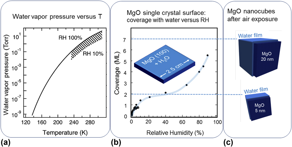
Adsorption and adsorption-induced chemical processes that occur at the metal oxide–water vapor or metal oxide–liquid water interfaces are typically accompanied by microstructural changes of nanoparticle ensembles [Reference Elser, Berger, Brandhuber, Bernardi, Diwald and Knözinger26, Reference Baumann, Elser, Auer, Bernardi, Hüsing and Diwald27]. Despite their great importance for materials science and in particular for the stability of oxide nanomaterials, the initial reactions of water vapor with solid oxide substrates (e.g., the processes of dissociative H2O chemisorption versus H2O physisorption), the thickness, and the structural properties of the thin liquid films are largely unexplored [Reference Bjorneholm, Hansen, Hodgson, Liu, Limmer, Michaelides, Pedevilla, Rossmeisl, Shen, Tocci, Tyrode, Walz, Werner and Bluhm6]. Thin water films under ambient conditions represent an intermediate case between liquid–solid and liquid–vapor interfaces that jointly determine its properties. For both natural and engineered nanomaterials, most solid surfaces in the ambient and at water partial pressures in the range between 1 and 30 Torr [Fig. 2(a)] are covered by several monolayers of water [Fig. 2(b)]. This was demonstrated for clean MgO (100) surfaces, which were investigated with FT-IR spectroscopy in atmospheres of variable relative humidity [Reference Foster, D’Agostino and Passno28]. As shown in Fig. 2(b), at a relative humidity of 20% and above, a ∼1–2 monolayer thick water layer already forms. In fact, hydroxylation and water adsorption start at a relative humidity (RH) <0.1% and significant amounts of molecular water become stabilized during the initial stages of water adsorption. Related observations underline the importance of OH–H2O interaction, which is stronger than H2O–H2O bonds [Reference Bjorneholm, Hansen, Hodgson, Liu, Limmer, Michaelides, Pedevilla, Rossmeisl, Shen, Tocci, Tyrode, Walz, Werner and Bluhm6].
The composite plot underlines that the typical water vapor pressures present in air and at room temperature (a) lead to the instantaneous coverage of almost every oxide surface with water (b). In comparison with the results of a Fourier Transform Infrared (FT-IR) study, which was performed on clean MgO (100) single crystal surfaces [Reference Foster, D’Agostino and Passno28], the significant H2O uptake of MgO nanocube powders after 20 min of air exposure is demonstrated in (c). A weight increase of 20% measured for nanocube ensembles with edge lengths between 5 and 20 nm corresponds to the formation of water multilayers at the nanocube surface. Adapted with permission from Ref. Reference Bjorneholm, Hansen, Hodgson, Liu, Limmer, Michaelides, Pedevilla, Rossmeisl, Shen, Tocci, Tyrode, Walz, Werner and Bluhm6 (Copyright 2016 American Chemical Society) and from Ref. Reference Foster, D’Agostino and Passno28 (Copyright 2005 Elsevier).

Surface science typically addresses water adsorption at the level of single molecules up to monolayer surface coverage. On two-dimensional metal oxides, surface studies reveal that the structure of interfacial water, i.e., in the first hydration layers, is distinct from bulk water [Reference Fenter and Lee29]. By means of in situ X-ray scattering techniques, it was found that the organization of hydration layers is limited to 1–2 nm, which corresponds to only a few water correlation lengths. For such hydration shell dimensions, related structures are highly dependent on characteristics of the metal oxide surface, such as local charge, spatial arrangement, and coordination of ions, and nature and abundance of additional adsorbates [Reference Zhang, Fenter, Cheng, Sturchio, Bedzyk, Předota, Bandura, Kubicki, Lvov, Cummings, Chialvo, Ridley, Bénézeth, Anovitz, Palmer, Machesky and Wesolowski30].
MgO nanocube powders with specific surface areas as large as 300 m2/g represent a model system for adsorption studies on oxide nanomaterials [Reference Schneider, Franke, Gurrath, Röckert, Berger, Bernardi, Meyer, Steinrück, Lytken and Diwald20, Reference Sternig, Koller, Siedl, Diwald and McKenna31]. Previous temperature-programmed desorption measurements and thermogravimetric analysis revealed that—depending on the exposure time—the nanoparticle powder can adsorb water between 10 and 20% of weight. H2O desorption features at T = 303 K and 573 K were observed and attributed to desorption of weakly bound surface water at T = 303 K and to the decomposition of surface Mg(OH)2 [Reference Hayun, Tran, Ushakov, Thron, van Benthem, Navrotsky and Castro32] into MgO [Reference Susman, Pham, Datye, Chinta and Rimer33] and gas phase water at T = 573 K. An estimate for the number of water molecules per Mg2+–O2− surface unit as a function of MgO nanocube size reveals that the number of water layers can be between 2 and 7 monolayers for MgO nanocubes with sizes of 5 nm and 20 nm, respectively [Fig. 2(c)].
Thin water films as a two-dimensional solvent for enhanced chemical growth
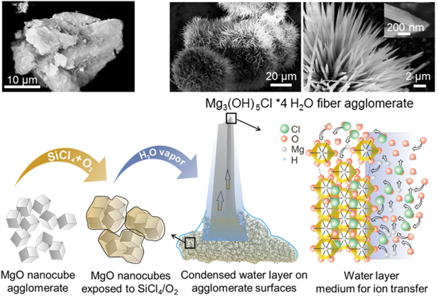
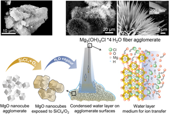
Thin water films can act as a two-dimensional solvent and give rise to the spontaneous and spatially controllable growth of hydroxide fibers in ambient air and at room temperature [Reference van Santen34, Reference Gheisi, Sternig, Redhammer and Diwald35]. Gheisi et al. recently reported the formation of Mg3(OH)5Cl·4H2O fibers on MgO nanocubes sequentially exposed to a SiCl4/O2 atmosphere at cryogenic temperatures and—in a subsequent step—to air or a water-saturated O2 atmosphere. This remarkable transformation shows that water thin films promote reactions that are confined at the metal oxide surface (Fig. 3).
Thin water films covering highly dispersed metal oxides can give rise to the spontaneous and spatially controllable growth of hydroxide fibers out of the MgO nanocube agglomerates shown in the SEM image in the left upper panel. Surface chlorinated MgO nanocubes transform upon contact with water vapor at room temperature into magnesium hydroxychloride Mg3(OH)5Cl·4H2O fibers. The formation of hydroxide fibers under ambient conditions can be applied to a variety of microstructural evolution processes that involve metastable solids and superficial water acting both as a reactant and as a reaction medium for the hydration of metal oxide particles. Figure adapted from [Reference van Santen34, Reference Gheisi, Sternig, Redhammer and Diwald35].

Residual water and transformations of metal oxide nanoparticles in high vacuum
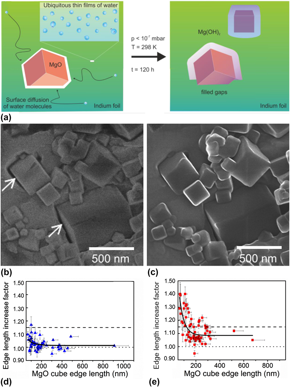
Imaging of metal oxide particles with scanning electron microscope and helium ion microscope (HIM) requires powder sample immobilization on conductive substrates. This can be achieved by pressing the sample into a soft metal that, if not cleaned and prepared prior to sample immobilization, is typically covered with water originating from the ambient atmosphere. Under such conditions, residual and surface adsorbed water molecules can act as a solvent and lead to a partial conversion of MgO into Mg(OH)2 [Reference Schwaiger, Schneider, Bourret and Diwald36]. HIM studies of MgO cubes obtained by Mg combustion (polydisperse sample with sizes ranging from 4 nm to a few micrometers) and pressed into an untreated indium foil have revealed that the samples prepared at the ambient and stored 4 days in high vacuum showed significant swelling, characteristic with a volume increase due to the formation of an Mg(OH)2 shell. On the contrary, samples prepared using an indium foil that was dried in high vacuum (10−5 mbar, 1 day) before MgO cube immobilization showed very limited swelling. Interestingly and similar to the observations made in liquid water (see section Metal oxide immersion into condensed water), it was found that the smallest MgO cubes reacted faster than the larger ones, as shown by their larger relative swelling (in relation to their initial size). The fact that the ambient water condensed at the indium foil surface was sufficient to reactively attack the MgO cubes pressed into the foil and this under high vacuum conditions is remarkable [Reference Schwaiger, Schneider, Bourret and Diwald36]. It demonstrates that spurious amounts of water can react with metal nanostructures even under high vacuum conditions and should be taken into consideration by anyone working with water sensitive metal oxide samples (Fig. 4) [Reference Baer, Engelhard, Johnson, Laskin, Lai, Mueller, Munusamy, Thevuthasan, Wang, Washton, Elder, Baisch, Karakoti, Kuchibhatla and Moon37].
(a) Proposed mechanism of superficial H2O-induced transformation of MgO cubes into MgO with Mg(OH)2 coatings. This reaction step is associated with characteristic volume expansion effects that are consistent with the observations described above. (b and c) High-magnification HIM images of MgO cubes embedded in an indium foil after 1 h (b), and 4 days in the HIM chamber at a chamber pressure P < 3 × 10⁻⁷ mbar (c). Significant volume increase, rounding of the edges, and partial fusion of the particles were observed (see arrows in micrographs). (d and e) Curves showing the edge length increase factor of MgO cubes (L 2/L 1) as a function of the edge length L 1, where L 1 and L 2 are the edge lengths before and after 4 days storage in the vacuum chamber of the HIM, respectively. (d) The indium foil was dried under vacuum before being used as a substrate. (e) The indium foil was used without any further treatment. The dashed line corresponds to a full transformation of MgO into Mg(OH)2 (i.e., L 2/L 1 = 1.15). The dotted line corresponds to zero expansion (L 2/L 1 = 1). Adapted from Ref. Reference Schwaiger, Schneider, Bourret and Diwald36.

Self-assembly processes in thin water films
Water-mediated forces play an important role in nanoparticle synthesis and structure formation at the mesoscale [Reference Shen, Chun, Rosso and Mundy38]. As an example, oriented attachment of nanoparticles represents a key process in the solution-phase growth of hierarchically organized nanostructures. The underlying self-assembly in aqueous dispersions results from the subtle balance between colloidal (i.e., hydration, electrostatic, and dispersion forces) and hydrodynamic forces [Reference Shen, Chun, Rosso and Mundy38, Reference Welch, Woehl, Park, Faller, Evans and Browning39]. Experimentally, oriented attachment of particles in bulk water has been reported for a variety of metal oxides such as TiO2 [Reference Niederberger and Cölfen12, Reference Cölfen and Antonietti13], ZnO [Reference Li, Nielsen, Lee, Frandsen, Banfield and De Yoreo40], iron oxide, or FeOOH [Reference Soltis and Penn41]. Complementary molecular dynamics simulations suggest that oriented attachment preferentially involves surfaces that have a higher propensity to hydroxylate and to dissociate water [Reference Raju, van Duin and Fichthorn42]. Therefore, hydroxylation and hydrogen bonding seem to govern particle alignment effects that are followed by coalescence.
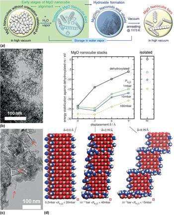
Our group recently reported the use of water vapor to induce the self-organization of MgO nanocubes into regularly staggered one-dimensional structures (Fig. 5) [Reference Thomele, Bourret, Bernardi, Bockstedte and Diwald43]. Exposure to water vapor (partial pressure: 30 mbar) and subsequent vacuum annealing at 1173 K results in the reorganization of the MgO nanocubes into one-dimensional structures, that is, MgO nanobars. This transformation evolves via an initial alignment of the MgO cubes [Fig. 5(a)], the formation of intermediate elongated Mg(OH)2 structures, and their reconversion into staggered MgO cube structures by thermal annealing [Fig. 5(c)]. The particles spontaneously orient themselves to form one-dimensional arrays, which, instead of forming perfect stacked cube assemblies, such as observed for metal nanocubes in solution [Reference Gao, Arya and Tao44], form staggered MgO nanocube assemblies, where the cubes are offset from each other. Ab initio Density Functional Theory (DFT) modelling identifies surface energy changes [Fig. 5(d)] associated with the cube surface hydration and hydroxylation to promote the uncommon staggered assembly of the cubes [Reference Thomele, Bourret, Bernardi, Bockstedte and Diwald43]. This first observation of metal oxide nanoparticle self-organization occurring outside a bulk solution may pave novel routes for inducing texture in ceramics and represents a great test bed for new surface science concepts.
(a) Illustration of water adsorption–induced arrangement of MgO nanocubes into nanobars. (b and c) TEM micrographs of the MgO nanocubes before (b) and after hydroxylation and and dehydroxylation (c), where the arrows indicate the experimentally observed MgO nanocube bars. (d) Stabilization energy of the hydroxylated stacks relative to the dehydroxylated nanocube versus displacement δ along the surface diagonal for different water pressures at T = 300 K. Bottom: Geometries of the hydroxylated straight and displaced stacks—oxygen ions are red spheres and magnesium ions are light red spheres. Oxygen ions bound in OH groups or in water are blue spheres and hydrogen atoms are white spheres. The hydroxylated nanocube stacks are stable within the range of water vapor partial pressure indicated below the geometries. Adapted from Ref. Reference Thomele, Bourret, Bernardi, Bockstedte and Diwald43.

Impact of adsorbed water on materials transformation at elevated temperatures
Thin water films for sintering
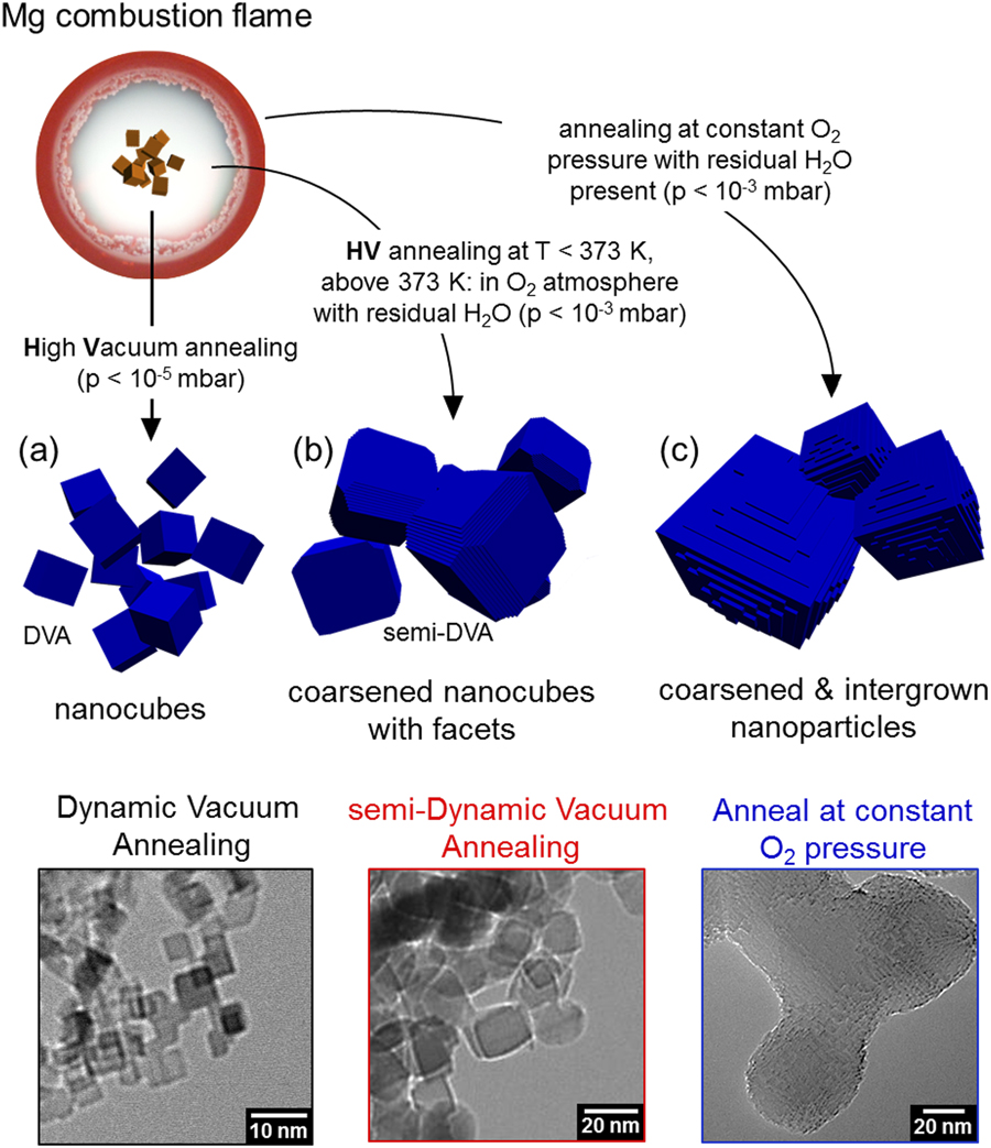
Thin water films can also influence sintering processes where a loose metal oxide nanoparticle powder is converted into a dense nanocrystalline material. Sintering is crucial in ceramics processing [Reference Castro, Gouvêa and Wakai45, Reference Gheisi, Niedermaier, Tippelt, Lottermoser, Bernardi and Diwald46, Reference Bordia, Kang and Olevsky47] and can be used to consolidate metal oxide nanoparticle systems with enhanced physicochemical properties [Reference Lu48, Reference Lu49]. It is a surface elimination process that starts with an assembly of particles in close contact. The initial interfaces or interfacial spots range from point of contact to highly deformed interfaces. With sintering, the contacts grow in size and there is extensive loss of surface area in the initial stage [Reference Castro, Gouvêa and Wakai45, Reference Phokha, Klinkaewnarong, Hunpratub, Boonserm, Swatsitang and Maensiri50]. The determining role of interfacial energies with regard to the densification of consolidated particle systems is well accepted [Reference Phokha, Klinkaewnarong, Hunpratub, Boonserm, Swatsitang and Maensiri50, Reference Folly, Soriano, Sinnecker and Novak51]. Since a new type of interface is being created during neck formation, sintering has an energy cost that is proportional to the grain boundary energy [Reference Hayun, Tran, Ushakov, Thron, van Benthem, Navrotsky and Castro32, Reference Castro, Gouvêa and Wakai45, Reference Castro, Tôrres, Pereira and Gouvêa52]. It was recently found that water adsorbed from ambient on MgO crystalline nanocubes (4 nm) can significantly affect their morphology during vacuum annealing depending on the reactor pressure (Fig. 6). While dynamic vacuum annealing at 1173 K (continuous pumping at pressure <10−5 mbar during annealing) does not affect the particle size and shape [Fig. 6(a)], semi-dynamic annealing [Fig. 6(b)], where dynamic pumping is replaced with a constant static O2 pressure, at T = 373 K leads to larger cubic particles (size ranging from 4 nm to ∼15 nm) with rounded edge features. The latter phenomenon is due to the increased abundance of step edges that are quite different from the clear-cut sharp edges specific to the parent 4 nm MgO cubes typically obtained from annealing in dynamic vacuum. Annealing at a constant O2 pressure and without continuous removal of water molecules leads to a significant change in morphology of the particles that become facetted and show significant particle fusion [Fig. 6(c)]. These experiments show that adsorbed water can lead to significant coarsening of the particles. The changes are attributed to enhanced mass transport in the thin water film that acts as a two-dimensional solvent for Mg2+ hydration and transport, promoting particle sintering and coarsening at high temperatures.
Schematic representation of how different annealing protocols affect the growth of MgO nanocubes synthesized in the Mg combustion flame. Adapted with permission from Ref. Reference Thomele, Gheisi, Niedermaier, Elsässer, Bernardi, Grönbeck and Diwald53 (Copyright 2018 WILEY VCH). DVA stands for dynamic vacuum annealing and indicates that annealing was performed under dynamic pumping with a base pressure of p< 10−5 mbar.

Instead of using high temperatures and residual water to sinter hydroxylated metal oxide particles, thin water films can also be used to promote the low-temperature sintering of TiO2 nanoparticles and prepare conducting nanocrystalline TiO2 films. Such films are of great importance due to their uses in many technological applications such as solar cells, photoelectrodes, photocatalysts, and gas sensors [Reference Berger, Monllor-Satoca, Jankulovska, Lana-Villarreal and Gómez54, Reference Sun, Haidry, Fatima, Li and Yao55]. They are usually prepared via high-temperature sintering, typically performed at 450 °C, and are thus not compatible with the use of heat-sensitive substrates such as flexible polymeric films. Annealing a mixture of TiO2 nanoparticles with Ti(IV) precursors such as titanium tetraisopropoxide [Ti(OiPr)4] in water vapor at 100 °C was found to be an efficient route for the preparation of conducting crystalline porous TiO2 with enhanced photoelectrochemical properties (Fig. 7). While annealing ethanolic solution of Ti(OiPr)4 under dry conditions at 100 °C yields an amorphous solid, annealing under water vapor at 100 °C yields rutile [Fig. 7(b)]. By using different titanium precursors, it is possible to obtain either rutile or anatase. Such a report shows the beneficial influence of water thin films in the consolidation and preparation of functional ceramics and represents an alternative to high-temperature annealing that is usually necessary for these materials.
(a and b) SEM photographs of the TiO2 films produced by mixing P25 particles and Ti(OiPr)4 before (a) and after annealing in water vapor at 100 °C (b). The undefined matrix seen in (a) is the amorphous Ti(OiPr)4 film that embeds the TiO2 particles within. After sintering/crystallization, one can clearly see the TiO2 crystalline particles. (c) X-ray diffraction (XRD) patterns of the films prepared by heating a solution of at 100 °C for 12 h in a dry oven (1) or by treating the following chemicals in water vapor at 100 °C for 12 h: Ti(OiPr)4 (2), TiOSO4 (3), and TiCl4 (4). Adapted with permission from Ref. Reference Zhang, Yoshida and Minoura56 (Copyright 2003 WILEY VCH).

Water-assisted crystallization
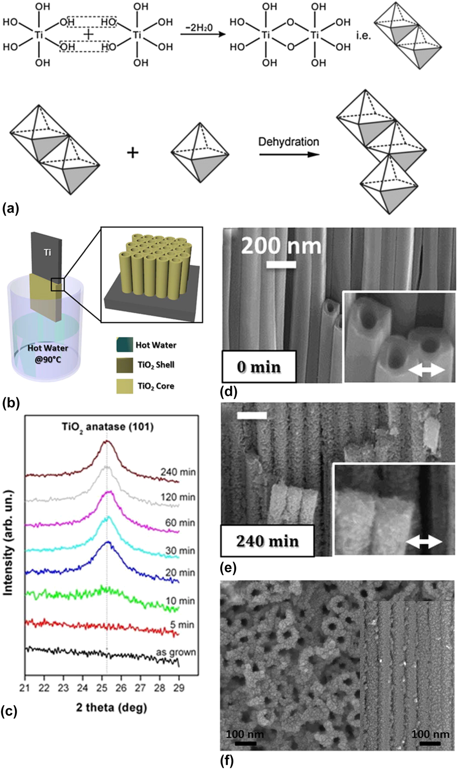
In addition to promoting sintering, water thin films can also be used to crystallize amorphous metal oxide substrates via dissolution/precipitation processes. For example, some groups have reported on the use of water vapor to crystallize anodized amorphous TiO2 nanotubes into anatase at moderate temperatures (i.e., lower than 100 °C) [Reference Lamberti, Chiodoni, Shahzad, Bianco, Quaglio and Pirri57]. The anodic oxidation of Ti foil into TiO2 nanotubes has led to a vast field of research [Reference Roy, Berger and Schmuki58]. Unfortunately, the as-produced TiO2 structures after anodization are amorphous and need an additional annealing step in order to be used as functional TiO2 photocatalysts, typically done at temperatures around 450 °C. Instead of using thermal energy only to crystallize TiO2, Lamberti et al. [Reference Lamberti, Chiodoni, Shahzad, Bianco, Quaglio and Pirri57] recently reported that incubation of amorphous anodized TiO2 nanotubes for 30 min in water vapor at 90 °C converts amorphous TiO2 into anatase (Fig. 8). Although at present the mechanism is unclear, a dissolution precipitation reaction is expected to occur due to the changes in the TiO2 structure morphology that was observed. It is likely that a thin water film hydroxylates the TiO2 surface, similarly to what occurs at room temperature on MgO surfaces. Dissolution of the Ti(III/IV) species as [Ti(OH)6]2− in the water film then leads to a supersaturated solution [Eq. (1)], eventually yielding crystallization of anatase via a sequential dehydration step [Eq. (2)] [Reference Huo, Wang, Zhang, Cao and Chu59].
 $${\rm{Ti}}{{\rm{O}}_x} + 4{{\rm{H}}_{\rm{2}}}{\rm{O}} + \left( {1 - {x \over 2}} \right){{\rm{O}}_2} \to {\rm{Ti}}\left( {{\rm{OH}}} \right)_6^{2 - } + 2{{\rm{H}}^ + }\left( {1 \lt x \le 2} \right)\quad ,$$
 $${\rm{Ti}}\left( {{\rm{OH}}} \right)_6^{2 - } + 2{{\rm{H}}^ + } \to {\rm{Ti}}{{\rm{O}}_2} + 4{{\rm{H}}_{\rm{2}}}{\rm{O}}\quad .$$
(a) Proposed crystallization mechanism from hydroxylated TiO2. From Ref. Reference Chen and Zhou62. (b) Scheme of the experimental setup used for the crystallization of the amorphous TiO2 nanotubes in water vapor. (c) X-ray diffraction (XRD) data showing the evolution of the main anatase X-ray diffraction peak [(101) crystal face] during the water vapor exposure. (d and e) SEM images of the TiO2 tubes before (d) and after 240 min of water vapor annealing at 90 °C (e). (b–e) Adapted with permission from Ref. Reference Lamberti, Chiodoni, Shahzad, Bianco, Quaglio and Pirri57. (f) SEM image of the TiO2 tubes after 240 min annealing at 90 °C in liquid water. Adapted with permission from Ref. Reference Krengvirat, Sreekantan, Mohd Noor, Negishi, Kawamura, Muto and Matsuda60 (Copyright 2013 Elsevier).

A significant change in the amorphous TiO2 tubular morphology was observed during water vapor annealing, which was converted into solid anatase wires [Reference Lamberti, Chiodoni, Shahzad, Bianco, Quaglio and Pirri57]. This is quite different from what occurs during annealing in liquid water at 90 °C, where the smooth amorphous TiO2 tube surface is instead replaced with a rough polycrystalline surface composed of granular anatase nanoparticles [Fig. 8(f)] [Reference Krengvirat, Sreekantan, Mohd Noor, Negishi, Kawamura, Muto and Matsuda60]. As expected, the photocatalytic activity of the water vapor–crystallized TiO2 substrate was much higher than that of the as-synthesized amorphous TiO2 nanotubes, as shown by the higher decomposition rate of methylene blue under UV illumination of the crystallized TiO2 substrate [Reference Lamberti, Chiodoni, Shahzad, Bianco, Quaglio and Pirri57]. It is interesting to note that the substrates crystallized in water vapor were also slightly more photoactive than the TiO2 tubes annealed at 450 °C under dry conditions. This is also consistent with a previous work showing that annealing similar TiO2 nanotubes under water vapor in an autoclave at 180 °C yielded a much higher photo-electrochemical activity than the same substrate annealed at 180 °C and atmospheric pressure (10 times increase in the photocurrent under similar conditions) [Reference Liu, Liu, Zhang, Zhai and Jiang61].
Metal oxide immersion into condensed water
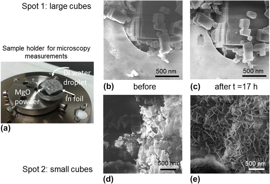
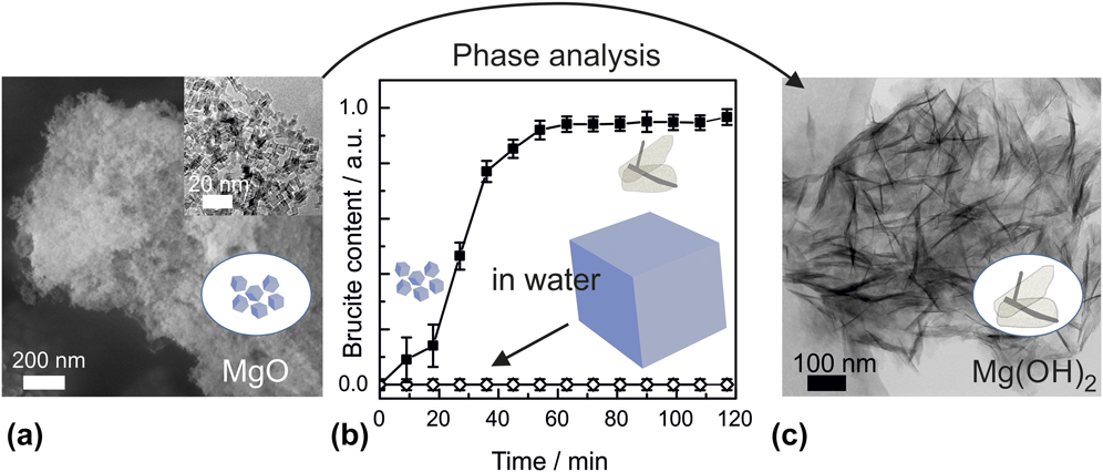
Bulk and thin water films can induce strong changes in the composition, structure, and microstructure of both isolated and ensembles of metal oxide nanostructures. While thin water films confine these transformations by limiting mass transport and favoring precipitation at the oxide surface, liquid water usually promotes dissolution/precipitation into bulk water. For example, while MgO particles tend to form core shell structures in the presence of thin water films under high vacuum conditions due to partial hydroxylation, they tend to partially or even completely dissolve in liquid water. Figure 9 shows HIM images of MgO micro- and nanocubes pressed onto an In foil and imaged at two different locations before and after being in contact with a water droplet for 17 h. While large microcubes show an increase in particle size due to partial hydroxylation (spot 1), the smallest MgO cubes (spot 2) completely dissolve and precipitate as brucite nanosheets [Mg(OH)2]. Due to the mass transport limitation imposed by the limited thickness of water thin films, such a situation does not occur when the same MgO cubes react with environmental adsorbed water (Fig. 4).
Effect of liquid water on the microstructure of MgO cube and nanocube powders. The HIM images of MgO cubes were taken from one and the same sample (a) on two different areas labelled spot 1 (b, c) and spot 2 (d, e), before (b, d) and after (c, e) exposure to a droplet of liquid water. Clearly, different types of nanostructures can result from the interaction of MgO particle surfaces with H2O depending on their size and distribution at the sample surface. Comparison of (b) and (c) points to the volume expansion during hydration and hydroxylation, while (d) and (e) reveal that the substantially smaller sized MgO cubes (d) have undergone a dissolution and Mg(OH)2 precipitation mechanism (e), as shown by the formation of network of brucite sheets after exposure to liquid water. Adapted from Ref. Reference Schwaiger, Schneider, Bourret and Diwald36.

It is interesting to note that the dissolution behavior in liquid water is significantly affected by the structure size: MgO microcubes were found to dissolve much slower than MgO nanocubes (4 nm edge length). It was proposed that the Mg(OH)2 passivation layer grown at the MgO nanocube surface [Reference Hayun, Tran, Ushakov, Thron, van Benthem, Navrotsky and Castro32] due to the presence of water molecules is not thick enough to influence their dissolution rate, leading to fast and complete dissolution. The situation changes for MgO microcubes whose dimensions allow for the growth of a thick brucite passivating layer [Reference Ringleb, Sterrer and Freund5] that can significantly slow down the dissolution, explaining the different dissolution rate observed for micro- and nanocubes (Fig. 10). Analysis of the dissolution products as a function of time shows that the dissolution rate constant is one order of magnitude higher in the case of the nanocubes [with k Diss = 3.6 × 10⁻⁶ mol/(m2 s)] and is not due to an increase in surface area. Such observations demonstrate that the stability of metal oxide nanostructures in liquid water can be strongly affected by their sizes [Reference Baumann, Schneider, Sternig, Thomele, Stankic, Berger, Grönbeck and Diwald24].
(a and c) Electron microscopy images of native MgO nanocubes (a), and after complete transformation into Mg(OH)2 nanosheets (c). (b) Size-dependent dissolution of MgO nanocubes (top curve) and MgO microcubes (bottom curve) in liquid water. Adapted with permission from Ref. Reference Altman, Agranovski and Choi21 (Copyright (2015) American Chemical Society).

These experiments illustrate the difference in complexity between water thin films and bulk water: water thin films on metal oxides are quite well defined due to the limited number of water molecule layers that can be condensed at the substrate surface, while metal oxides immersed in liquid water are much more complex systems. We believe that water thin films on metal oxides are a great test bed to understand and study the interaction between molecular water and metal oxide nanostructures.
Concluding remarks and perspectives
Water adsorption on oxide nanomaterials plays an important but so far underestimated role in materials chemistry and related characterization approaches. For materials synthesis under nonaqueous conditions, the unintended presence of spurious amounts of water may lead to surfactant effects that influence morphology evolution via the stabilization of specific crystal faces. An important example is the stabilization of polar (111) surfaces [Reference Noguera and Goniakowski11, Reference Hu, Zhu, Chen, Kubel and Richards63, Reference Cadigan, Corpuz, Lin, Caskey, Finch, Wang and Richards64] where surface hydroxylation compensates for surface dipole formation and therefore eliminates the energetic reasons for the instability of corresponding faces. This provides an important handle to tune both the adsorption properties and the catalytic activity of metal oxide surfaces using surface hydroxyls as ubiquitous but—at the same time—structure-directing ligands [Reference Cadigan, Corpuz, Lin, Caskey, Finch, Wang and Richards64].
Moreover, the fact that high surface area materials can adsorb substantial amounts of water from the ambient atmosphere has a key influence on particle growth and structural reorganization during subsequent thermal processing and sintering [Reference Castro, Gouvêa and Wakai45, Reference Lu65]. Adsorption-induced changes in the hierarchy of surface energies [Reference Thomele, Gheisi, Niedermaier, Elsässer, Bernardi, Grönbeck and Diwald53, Reference Geysermans, Finocchi, Goniakowski, Hacquart and Jupille66, Reference Sternig, Klacar, Bernardi, Stöger-Pollach, Grönbeck and Diwald67] lead to substantially different particle habits as compared with oxide nanoparticles with bare and dehydroxylated surfaces that are processed under dry vacuum conditions [Reference Thomele, Gheisi, Niedermaier, Elsässer, Bernardi, Grönbeck and Diwald53]. Apart from the hydroxylation-induced energetic stabilization of specific surface planes that determine the evolution of different crystal shapes [Reference Barnard68, Reference Guo and Barnard69], thin water films provide a so far unnoticed solvent and reaction medium that converts metastable nanomaterials into new structures at room temperature and at different length scales [Reference Gheisi, Sternig, Rangus, Redhammer, Hartmann and Diwald70]. This is reminiscent of mineral-based binders where hydration reactions at ambient conditions provide materials of sufficient strength and stability to be used in construction applications.
In this short review, we highlighted water-induced chemical and/or structural changes that are mediated by water vapor and resulting thin films of adsorbed water on MgO and TiO2 nanostructures. Both metal oxides are canonical model compounds. At the same time, their chemical bondings show characteristic differences with regard to their degree of covalency/ionicity. This determines their interaction with water and their stability in aqueous environments. Being pH dependent, the solubility values of metal oxides range from moderate (>80 mg/L for MgO at pH = 7) to practically insoluble (0.15 mg/L for TiO2, at pH = 7) [Reference Rogers, Franklin, Apte and Batley71]. Additionally, variations in particle size, crystal modification, resulting surface terminations and particle shape, pH-dependent surface charges [Reference Söhnel72], and—last but not least—nature and abundance of reactive surface defects determine the overall particle stability in aqueous environments. Considering the variations in surface reactivity between metal oxide nanocrystals of different composition and synthetic origin, there may exist different mechanistic pathways for water adsorption–induced particle organization and aggregation. However, one can expect the resulting particle-based architectures to have a more characteristic structure at the mesoscale when the individual nanocrystals are facetted and exhibit anisotropic crystal habits [Reference Shen, Chun, Rosso and Mundy38, Reference Raju, van Duin and Fichthorn42, Reference Thomele, Bourret, Bernardi, Bockstedte and Diwald43]. Further experimental and theoretical efforts are needed to explore this exciting playground of mesostructure formation in gaseous or thin liquid film environments that are different from bulk water [Reference Zhang, Shen, Liu, Kerisit, Bowden, Sushko, de Yoreo and Rosso14, Reference Shen, Chun, Rosso and Mundy38].
The investigation of metal oxide particle surfaces at ambient relative humidity has continuously benefited from the development of experimental techniques in the field of surface science [Reference Ringleb, Sterrer and Freund5, Reference Bjorneholm, Hansen, Hodgson, Liu, Limmer, Michaelides, Pedevilla, Rossmeisl, Shen, Tocci, Tyrode, Walz, Werner and Bluhm6, Reference Baer, Engelhard, Johnson, Laskin, Lai, Mueller, Munusamy, Thevuthasan, Wang, Washton, Elder, Baisch, Karakoti, Kuchibhatla and Moon37, Reference Kaden, Pomp, Sterrer and Freund73, Reference Brown, Redondo, Sterrer, Winter, Pacchioni, Abbas and van Bokhoven74], where the interest in solid–liquid interfaces has grown over the last decades. Moreover, the increasing availability of environmental electron microscopy techniques [Reference Gajdardziska-Josifovska and Sharma22, Reference Tao and Crozier75, Reference Tao and Salmeron76] will provide access to complementary information not only to study purely water-induced transformation effects of metal oxide nanomaterials but also to identify surface adsorbed water as a source of experimental artifacts [Reference Schwaiger, Schneider, Bourret and Diwald36, Reference Baer, Engelhard, Johnson, Laskin, Lai, Mueller, Munusamy, Thevuthasan, Wang, Washton, Elder, Baisch, Karakoti, Kuchibhatla and Moon37].
Acknowledgments
Financial support of the Austrian Science Fund FWF for project P-28797 “Intergranular regions in nanocrystalline ceramics” is gratefully acknowledged. Partial support comes from the University of Salzburg within the Allergy Cancer BioNano (ACBN) Research initiative.