Introduction
Migration of dental implants into the maxillary sinus is an uncommon but recognised complication of dental practice.Reference Galindo, Sanchez-Fernandez, Avila, Cutando and Fernandez1–Reference El Charkawi, El Askary and Ragab4 Such patients may present to ENT surgeons, either with secondary sinonasal symptoms, or as a referral from dental colleagues for consideration of endoscopic intervention. Left untreated, these foreign bodies may give rise to serious complications.
We report two such patients, one of whom experienced symptoms due to the migrated implant. Both patients were subsequently managed by endoscopic sinus surgery to remove the implant. To our knowledge, this paper represents the first report on the benefits of using image guidance in such cases.
Case reports
Patient one
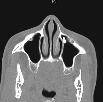
A 53-year-old woman was referred by her dentist after undergoing osseointegrated dental implantation in the left upper first molar (left upper 6). At the time of this procedure, the implant had been displaced. It had been noted at the time of surgery that there was only a small amount of bone, and that the maxillary sinus floor was in close proximity. An Orthopantogram (OPG) X-ray had revealed the implant in the maxillary sinus, lying in a horizontal plane. On computed tomography (CT) (Figure 1), the implant had been identified in the anterior-inferior wall of the left antrum. The patient had been commenced on antibiotics and analgesia.
Axial computed tomography scan of patient one, demonstrating the implant in the left maxillary sinus.

The patient presented to us symptom-free, two days after her dental procedure, and remained so until the removal procedure a month later.
A BrainLab protocol (BrainLab AG; Heimstetten, Germany) was used to create CT images for guidance.
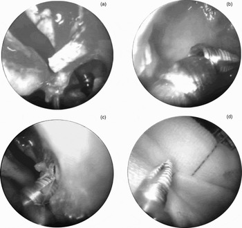
Endoscopic removal involved nasal preparation with Moffett's solution followed by left uncinectomy and middle meatal antrostomy (Figure 2). The titanium implant was removed from the maxillary antrum using suction, and adrenaline-soaked packs were applied to the area.
Intra-operative, endoscopic images for patient one, showing: (a) visualisation of the implant in the maxillary sinus; (b) the implant captured by suction; (c) removal of the implant through the middle meatal antrostomy; and (d) the implant.

Post-operative nasal douching was prescribed. No long-term follow up was required.
Patient two
A 52-year-old woman presented to the ENT clinic five months after a dental procedure to place an osseointegrated dental implant in the right upper second molar (right upper 7). She had subsequently developed sinogenic symptoms. The implant had displaced into the sinus at the time of her dental procedure.
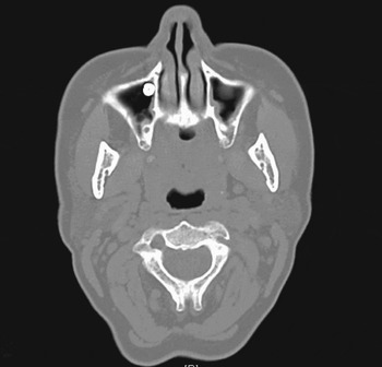
When the patient presented to the ENT clinic, she was experiencing occasional obstruction in the right nostril with crusting. An OPG X-ray confirmed a high and posterior position of the implant in the right maxillary sinus, with later movement to a medial position. A CT scan (Figure 3) confirmed that the implant was in the anterior right maxillary antrum.
Axial computed tomography scan of patient two, demonstrating the implant in the anterior wall of the right maxillary antrum.

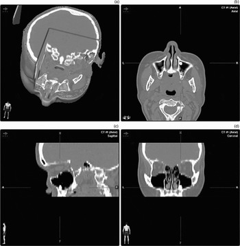
Endoscopic removal of the implant was ultimately necessary. This procedure was image-guided, after the patient had undergone the BrainLab protocol to generate images (Figure 4).
BrainLab protocol scans: (a) three-dimensional navigational reconstruction for patient two; (b) axial computed tomography (CT) scan; (c) sagittal CT scan; and (d) coronal CT scan. A = anterior; P = posterior; R = right; L = left; H = head; F = feet

The procedure involved nasal preparation followed by uncinectomy and right middle meatal antrostomy. When the implant could not be visualised on the floor of the sinus, a right inferior meatal antrostomy was performed. The implant was visualised and removed, the mucosal flap was replaced to close the inferior meatal antrostomy, and adrenaline-soaked packs were applied.
The patient was discharged after an uncomplicated post-operative course, with a follow-up appointment scheduled two months hence.
Both patients were successfully treated with the described surgical approach, resulting in complete resolution of symptoms.
Discussion
The aim of dental implants is to provide a foundation for replacement teeth that appear, feel and function like natural teeth. The implants themselves are small titanium posts inserted surgically into the mandible or maxilla. The titanium implant integrates with the bone through the process of osseointegration. Approximately six months later, artificial teeth or crowns are attached to the implants. Unfavourable factors include lack of bone, low bone density, alveolar ridge resorption and short maxillary ridge distance.Reference Galindo, Sanchez-Fernandez, Avila, Cutando and Fernandez1 These conditions occur mainly in the posterior maxilla. As there is a risk of infectious complications, early removal of migrated implants is indicated.
Dental implant migration
The pathophysiology of dental implant migration is not fully understood.
There are several possible reasons for early migration. Low bone density in an edentulous maxilla may lead to inadequate implant anchorage and lack of stability. Poor surgical technique, previous alveolar infection and bone destruction may all contribute to the process.
Late migration of dental implants into the maxillary sinus poses a more challenging question. Three theories have been proposed to explain such migration. The first is that changes in intra-sinus and nasal pressures may produce a negative pressure and subsequently a suction effect. The second theory is that migration results from an autoimmune reaction causing peri-implant bone destruction and compromise of osseointegration. A final possible explanation is that early prosthetic loads can produce resorption and may then interfere with long-term osseointegration.Reference Galindo, Sanchez-Fernandez, Avila, Cutando and Fernandez1
Further dental implant migration from the maxillary sinus into the ethmoid or sphenoid sinus has been reported; therefore, timely removal is important even in asymptomatic patients.Reference Felisati, Lozza, Chiapasco and Borloni5 There is also the theoretical risk of further movement of the implant from the sinus into the nasopharynx, compromising the airway.
Surgical techniques
Established techniques for the removal of migrated dental implants in the maxillary sinus include suction via the socket of the extracted tooth and open surgery via the canine fossa. The main benefit of transnasal endoscopic surgery is the minimally invasive aspect of the technique, allowing better visualisation and access to the sinus with lower post-operative morbidity. This technique has not often been described as the sole approach, as the Caldwell-Luc procedure has hitherto been the primary treatment.Reference Lubbe, Aniruth, Peck and Liebenberg6 However, comparison of endoscopic sinus surgery and the Caldwell-Luc operation undertaken for chronic sinusitis has shown that the former results in a significantly reduced operation time and earlier discharge from hospital.Reference Ideka, Hirano, Oshima, Shimomura, Suzuki and Sunose7, Reference Varol, Turker, Goker and Basa8
• Removal of dental implants which have migrated into the maxillary sinus previously involved invasive surgical techniques, including the Caldwell-Luc procedure and retrieval through the implant site
• An image-guided, transnasal endoscopic approach allows: better visualisation of and access to the migrated implant; minimal invasiveness; reduced operation time; less destruction of nasal and sinus tissue; and earlier discharge from hospital
The transnasal endoscopic approach may be more difficult if the implant is in a more anterior and medial position in the maxillary sinus, and if the technique is unfamiliar to the operator. The former problem was illustrated by a case reported by El Charkawi et al., in which an initial endoscopic approach was abandoned in favour of a Caldwell-Luc procedure as the implant was not in a favourable position for transnasal removal.Reference El Charkawi, El Askary and Ragab4 However, image guidance facilitates precise, real-time localisation of the implant, as was demonstrated in our patients (whose migrated implants were retrieved from difficult anatomical positions). Furthermore, given the morbidity of the Caldwell-Luc procedure and the fact that future implant retrieval attempts are compromised, primary consideration of a transnasal endoscopic approach is warranted. Due to the dynamic nature of foreign body position within the sinuses, the transnasal approach may lead to a higher retrieval rate with fewer intra- and post-operative complications, compared with other, previously used, invasive surgical approaches.
We report these cases in order to raise awareness of this pathology as a cause of maxillary sinusitis and oroantral fistulae, and to highlight ENT surgeons' potentially increasing role in the management of this dental complication.