INTRODUCTION
Trypanosomes of the Trypanosoma brucei group are the aetiological agents of human African trypanosomiasis, a parasitic disease that affects over 36 countries in subSaharan Africa (Barrett et al. Reference Barrett, Burchmore, Stich, Lazzari, Frasch, Cazzulo and Krishna2003). The T. brucei life-cycle is complex, and the parasite adapts to life in both its insect host (tsetse fly) and in the mammalian hosts. These distinct environments require the parasite to remodel its metabolism to thrive within each. In the D-glucose-rich environment of the mammalian bloodstream the parasite relies solely on D-glucose to produce energy. The first 7 steps of glycolysis occur sequestered in a peroxisome-like organelle, the glycosome (Opperdoes and Borst, Reference Opperdoes and Borst1977; Michels et al. Reference Michels, Bringaud, Herman and Hannaert2006).
In procyclic-form trypanosomes, the mitochondrion has traditionally been held to produce the major proportion of the cell's ATP (Bochud-Allemann and Schneider, Reference Bochud-Allemann and Schneider2002). This is largely due to the use of alternative carbon sources such as L-proline as the input for mitochondrial metabolism (Besteiro et al. Reference Besteiro, Barrett, Riviere and Bringaud2005). ATP can be generated by oxidative phosphorylation (electron transport chain), or by substrate-level phosphorylation, with an unusual reaction catalysed by succinyl-CoA synthase coupled to acetate formation being key (van Hellemond et al. Reference van Hellemond, Opperdoes and Tielens1998; Bochud-Allemann and Schneider, Reference Bochud-Allemann and Schneider2002; Riviere et al. Reference Riviere, van Weelden, Glass, Vegh, Coustou, Biran, van Hellemond, Bringaud, Tielens and Boshart2004). Recently, the exact contribution of the Kreb's cycle to energy generation has been looked at in detail (van Weelden et al. Reference van Weelden, Fast, Vogt, van der, Saas, van Hellemond, Tielens and Boshart2003, Reference van Weelden, van Hellemond, Opperdoes and Tielens2005), and it appears that in spite of most Kreb's cycle enzymes being present, the flux of substrates through these enzymes does not occur in the classical manner. However, these studies – as with many related to procyclic metabolism – were performed on trypanosomes grown in D-glucose-rich medium. D-glucose apparently represses L-proline metabolism in procyclic trypanosomes (Evans and Brown, Reference Evans and Brown1972; Lamour et al. Reference Lamour, Riviere, Coustou, Coombs, Barrett and Bringaud2005). In these conditions the parasite appears capable of fulfilling energy needs without using the Kreb's cycle and by producing ATP primarily by substrate level phosphorylation (oxidative phosphorylation is not essential).
In the mammalian host, trypanosomes divide as long slender bloodstream forms, covered by a dense coat of variant surface glycoproteins (VSG) (Cross, Reference Cross1975; Roditi, Reference Roditi1996). Short stumpy bloodstream forms are cell-cycle arrested and pre-adapted to life within the tsetse fly (Roditi et al. Reference Roditi, Schwarz, Pearson, Beecroft, Liu, Richardson, Buhring, Pleiss, Bulow, Williams and Overath1989). When ingested by the fly in the bloodmeal, the stumpy forms differentiate into procyclic-form trypanosomes that are able to colonize the midgut of the fly (Roditi et al. Reference Roditi, Schwarz, Pearson, Beecroft, Liu, Richardson, Buhring, Pleiss, Bulow, Williams and Overath1989). Parasite differentiation is accompanied by the replacement of the VSG coat by an invariant coat of EP and GPEET procyclins (Roditi et al. Reference Roditi, Schwarz, Pearson, Beecroft, Liu, Richardson, Buhring, Pleiss, Bulow, Williams and Overath1989, Reference Roditi, Furger, Ruepp, Schurch and Butikofer1998). EP expression continues throughout the fly stage of the procyclic form whereas GPEET expression is downregulated in late procyclics (Vassella et al. Reference Vassella, Den Abbeele, Butikofer, Renggli, Furger, Brun and Roditi2000; Acosta-Serrano et al. Reference Acosta-Serrano, Vassella, Liniger, Kunz, Brun, Roditi and Englund2001). However, in culture, GPEET expression can be modulated by glycerol or D-glucose (Vassella et al. Reference Vassella, Den Abbeele, Butikofer, Renggli, Furger, Brun and Roditi2000). RNAi silencing of the enzyme catalysing the first step of glycolysis, hexokinase, caused a dramatic switch from EP-procyclin to GPEET-procyclin expression (Morris et al. Reference Morris, Wang, Drew and Englund2002); suggesting that removal of D-glucose enhances GPEET-procyclin expression. Regulation of expression of EP- and GPEET-procyclin isoforms appears to be controlled by mitochondrial enzymes of the acetate:succinate CoA-transferase (ASCT) cycle (Vassella et al. Reference Vassella, Probst, Schneider, Studer, Renggli and Roditi2004). Silencing of the hexose transporter and the phosphofructokinase enzyme also upregulated GPEET-procyclin expression, indicating that a reduction in glycolytic flux correlates with the EP to GPEET switch.
In addition to these profound differences in glucose metabolism between bloodstream form and procyclic form trypanosomes, uptake of this carbohydrate also differs in bloodstream-form and procyclic-form Trypanosoma brucei brucei. Different hexose transporter isoforms make the major contribution to uptake in the two life-cycle stages. Trypanosome Hexose Transporter 1 (THT1) is expressed in bloodstream forms at a level 40 times that of THT2, while procyclic form trypanosomes express only the THT2 isoform (Bringaud and Baltz, Reference Bringaud and Baltz1993; Barrett et al. Reference Barrett, Tetaud, Seyfang, Bringaud and Baltz1995). The C2 substituents of D-glucose, D-glucosamine (GlcN) and N-acetyl D-glucosamine (GlcNAc) have previously been shown to interact with the hexose transporter of bloodstream-form trypanosomes (Eisenthal et al. Reference Eisenthal, Game and Holman1989). While D-glucose and GlcN uptake has been demonstrated, there was no apparent uptake of GlcNAc in bloodstream forms (Azema et al. Reference Azema, Bringaud, Blonski and Perie2000). However, interaction of GlcNAc with the hexose transporter was indicated by the inhibition of uptake of 2-DOG and GlcN by GlcNAc. The ability of procyclic-form trypanosomes to accumulate GlcNAc has not been studied.
Establishment of trypanosome infection is as complicated in the fly as it is in the mammalian system. The parasite must establish itself as a dividing procyclic population before development can progress. Natural infection rates are low, a fact attributed to many environmental and intrinsic fly factors (Aksoy, Reference Aksoy2003). Like other insects, the tsetse fly has an effective innate immune system and several components, including midgut lectins, have been implicated in trypanosome killing (Mihok et al. Reference Mihok, Otieno, Darji and Munyinyi1992; Welburn and Maudlin, Reference Welburn and Maudlin1999; Aksoy, Reference Aksoy2003; Abubakar et al. Reference Abubakar, Bulimo, Mulaa and Osir2006; Hu and Aksoy, Reference Hu and Aksoy2006). The role of lectins in preventing trypanosome infection was inferred from enhanced infection rates after feeding the lectin-inhibitory amino sugars GlcN and GlcNAc. These sugars block lectin-mediated trypanosome killing as well as inhibiting trypsin activity (Osir et al. Reference Osir, Imbuga and Onyango1993; Abubakar et al. Reference Abubakar, Bulimo, Mulaa and Osir2006), and GlcN may also have anti-oxidant properties that aid in establishing infection (Macleod et al. Reference Macleod, Maudlin, Darby and Welburn2007).
In addition, GlcNAc was found to stimulate trypanosome growth in vivo in the fly gut. GlcNAc also stimulates in vitro growth in the absence of fly derived factors (Peacock et al. Reference Peacock, Ferris, Bailey and Gibson2006). Here we have investigated the underlying basis for this effect. Given that bloodstream-form trypanosomes do not show any apparent uptake of GlcNAc (Azema et al. Reference Azema, Claustre, Alric, Blonski, Willson, Perie, Baltz, Tetaud, Bringaud, Cottem, Opperdoes and Barrett2004), we aimed to determine whether procyclic forms could accumulate GlcNAc, or whether its effects might be due to other mechanisms, for example, either as a simple direct growth factor or alternatively, through inhibition of D-glucose uptake leading to an induced metabolic shift that enhances growth.
MATERIALS AND METHODS
Growth and maintenance of trypanosomes
Procyclic-form trypanosomes were cultivated at 25–28°C in 4 different media supplemented with 5 μg/ml hemin and 10% heat-inactivated foetal calf serum: Cunningham's medium (CM) (Cunningham, Reference Cunningham1977), ‘no sugar Cunningham's medium’ (NSCM) made from CM by omitting all 3 sugars (D-glucose, D-fructose and sucrose), SDM80 (first employed by Lamour et al. Reference Lamour, Riviere, Coustou, Coombs, Barrett and Bringaud2005) and SDM79 (Brun and Schonenberger, Reference Brun and Schonenberger1979). For the glucose-depleted SDM80 medium, SDM80 was supplemented with 5 μg/ml hemin, 9% (v/v) heat-inactivated foetal calf serum dialysed by ultrafiltration against 0·15 m NaCl and 1% (v/v) heat-inactivated foetal calf serum. Development of the culture was monitored by microscopy and cell numbers were determined using a Neubauer haemocytometer (counting chamber). The strains of Trypanosoma brucei used were TREU 927, EATRO1125, J10 and KETRI 1738. All procyclic-form trypanosomes used were from established cultures.
Transport assays
Procyclics of TREU 927 were harvested during the mid-log phase of growth, by centrifugation at 2500 rpm in a bench-top centrifuge for 10 min and washed 3 times with PBS buffer (137 mm NaCl, 2·7 mm KCl, 10 mm phosphate buffer solution, pH 7·4) at 4°C. Parasites were resuspended in PBS at the density of 2×10⁸ parasites/ml and kept on ice, then brought to room temperature when the experiment was about to proceed.
Uptake of radio-isotope labelled sugars was determined using a rapid stop/oil-spin protocol, as previously described (Carter and Fairlamb, Reference Carter and Fairlamb1993; Barrett et al. Reference Barrett, Tetaud, Seyfang, Bringaud and Baltz1995; Azema et al. Reference Azema, Bringaud, Blonski and Perie2000). Transport was initiated with 100 μl of cells being mixed with 100 μl of assay buffer, containing isotope-labelled sugar and uptake was left to proceed over the time-points specified in the Results section. [6-14C] 2-deoxy-D-glucose (40 Ci/mmol), [1-14C] GlcN (57·9 Ci/mmol), [3H] GlcNAc (12 Ci/mmol) and [3H] L-proline (85 Ci/mmol) were from Amersham Bioscience. All were included at specific activities stated in the text or figure legend.
To separate trypanosomes from isotope label, cells in uptake solution were layered over a 100 μl cushion of oil (1-Bromodo-decane, density: 1·066 gcm−3). Upon centrifugation, trypanosomes pass through the oil. The tube was then flash frozen in liquid nitrogen, the pellet separated from the remnants of the tube using a tube cutter, lysed in 200 μl of SDS (2%) and mixed with 3 ml of scintillation fluid (Ecoscint A, National Diagnostics). Incorporated radioactivity was counted after 24 h (to avoid luminescence) using a liquid scintillation counter (Perkin Elmer, liquid scintillation and luminescence counter, 1450 Microbeta). All experiments (at each time-point for each substrate) were carried out, at least in duplicate, and 3 times independently. The results presented show the average of the 3 replicates.
Non-transported isotope-labelled compounds, associated with cells or in interstitial spaces, were measured by performing control uptake determinations using cells kept on ice and centrifuged as quickly as possible after adding cells.
Tsetse fly infection
Experimental tsetse flies were from the Bristol Laboratory colony of Glossina morsitans morsitans. Male flies were used for experiments and were kept at 25°C and 70% relative humidity, and fed on sterile defibrinated horse blood via a silicone membrane. The infected bloodmeal consisted of washed horse red blood cells (RBC) resuspended in Hanks Balanced Salt Solution (HBSS) mixed with procyclic trypanosomes (strains TREU 927, J10) from in vitro culture that had been grown in CM or NSCM for a minimum of 5 days. Before use, the trypanosomes were washed 3 times in NSCM, resuspended in HBSS and added to the washed horse RBC at a final concentration of approximately 106 trypanosomes/ml. GlcNAc was added to the infective feed at a final concentration of 60 mm using a filter-sterilized 1 m stock solution (Peacock et al. Reference Peacock, Ferris, Bailey and Gibson2006); an equal volume of PBS was added to control bloodmeals. Flies were dissected 1–2 weeks later and midguts examined for infection by microscopy. The chi-squared test was used for comparison of infection rates.
Determination of oligomycin sensitivity
The EATRO1125 procyclic cells (2×10⁶ cells/ml) were incubated in SDM80 either containing or lacking 6 mm D-glucose, in the presence of decreasing quantities of oligomycin (from 50 μg/ml to 0·01 ng/ml). The assay was performed in 96-well microtitre plates with 100 μl of cell suspension per well and the cell viability was determined optically. The oligomycin concentration required to kill all of the cells (lethal dose 100: LD100) was determined 72 h after drug addition.
The Alamar Blue assay (Raz et al. Reference Raz, Iten, Grether-Buhler, Kaminsky and Brun1997) was employed to test the effect of oligomycin with strain TREU 927. Briefly, 106 cells/ml were incubated in the presence of 0·1 ng/ml oligomycin, 5 mm L-proline, and doubling dilutions of D-glucose (10 mm starting), with or without 60 mm GlcNAc for 72 h in 96-well flat-bottomed microtitre plates. Alamar Blue reagent (Resazurin), stored and protected from light at 4°C, was added after incubation for 48 h (at 10% – 20 μl per well). Measurement of fluorescence was carried out in a Millipore Cytofluor plate reader at 530 nm excitation wavelength and 590 nm emission wavelength. All results represent an average of 3 individual experiments.
Determination of D-glucose and L-proline consumption
To determine the rate of D-glucose and L-proline consumption, cells (inoculated at 1–1·5×107 cells/ml) were grown in 10 ml of SDM80 medium (Lamour et al. Reference Lamour, Riviere, Coustou, Coombs, Barrett and Bringaud2005) supplemented with 1 mm L-glutamine, 10 mm L-proline, 4 mm pyruvate, 3 mm L-threonine, 0–6 mm D-glucose, 9% (v/v) heat-inactivated FCS dialysed by ultrafiltration against 0·15 m NaCl and 1% (v/v) heat-inactivated FCS. Aliquots of the growth medium (500 μl) were collected 0, 6, 9, 10, 23 and 24 h after incubation at 27°C. The quantity of D-glucose present in the medium was determined using the ‘glucose GOD-PAP kit’ (Biolabo SA). L-proline concentration was determined with a colorimetric assay as previously described (Bates et al. Reference Bates, Waldren and Teare1973) after deproteinization of the samples by perchloric acid treatment. At the beginning of the experiment, at 9 h and at 24 h, the cell density and the cellular protein concentration were determined to estimate the amount of carbon source consumed per mg of cellular proteins.
RESULTS
GlcNAc stimulates growth of procyclic T. brucei in vitro
GlcNAc has previously been shown to stimulate growth of procyclic-form trypanosomes (T. b. gambiense strain TH2 and T. b. brucei strain J10) in vitro using Cunningham's medium (CM) (Peacock et al. Reference Peacock, Ferris, Bailey and Gibson2006). Here we confirmed strain J10's enhanced growth with GlcNAc (Fig. 1A), and also tested to see if a similar growth profile was seen in cells grown in medium deficient in sugars (NSCM). Comparison of growth of strain J10 in CM, with and without sugars and GlcNAc revealed that the addition of GlcNAc to CM significantly increased the number of trypanosomes (Fig. 1A). Growth in NSCM was also greater than in CM and addition of GlcNAc did not further enhance growth in NSCM.
(A) Growth of procyclic-form trypanosomes (strain J10) in CM (open circles), CM supplemented with 60 mm GlcNAc (closed circles), NSCM (open squares), and NSCM supplemented with 60 mm GlcNAc (closed squares). (B) Cell counts of procyclic-form trypanosomes (strain TREU 927), at a starting density of 5×10⁵ cells per ml, grown in CM (open circles) and CM supplemented with 60 mm GlcNAc (closed circles). (A) and (B) represent data from 3 similar repeats, error bars are±S.E.M. (C) Procyclic-form trypanosomes (strain TREU 927) grown in SDM80 (D-glucose-L-proline-depleted medium) (open circles) supplemented independently with 60 mm GlcNAc (closed circles), 10 mm L-proline (open squares), 10 mm D-glucose (closed squares) or 10 mm L-proline and 10 mm D-glucose (open triangles).

We tested whether a similar effect was seen using the T. brucei strain TREU 927, which was used for the genome sequencing project (Berriman et al. Reference Berriman, Ghedin, Hertz-Fowler, Blandin, Renauld, Bartholomeu, Lennard, Caler, Hamlin, Haas, Bohme, Hannick, Aslett, Shallom, Marcello, Hou, Wickstead, Alsmark, Arrowsmith, Atkin, Barron, Bringaud, Brooks, Carrington, Cherevach, Chillingworth, Churcher, Clark, Corton, Cronin, Davies, Doggett, Djikeng, Feldblyum, Field, Fraser, Goodhead, Hance, Harper, Harris, Hauser, Hostetler, Ivens, Jagels, Johnson, Johnson, Jones, Kerhornou, Koo, Larke, Landfear, Larkin, Leech, Line, Lord, MacLeod, Mooney, Moule, Martin, Morgan, Mungall, Norbertczak, Ormond, Pai, Peacock, Peterson, Quail, Rabbinowitsch, Rajandream, Reitter, Salzberg, Sanders, Schobel, Sharp, Simmonds, Simpson, Tallon, Turner, Tait, Tivey, Van Aken, Walker, Wanless, Wang, White, White, Whitehead, Woodward, Wortman, Adams, Embley, Gull, Ullu, Barry, Fairlamb, Opperdoes, Barrell, Donelson, Hall, Fraser, Melville and El Sayed2005). GlcNAc at 60 mm enhanced survival and increased final density, with an increase in growth rate clear by 4 days (Fig. 1B), suggesting that the effect of GlcNAc is common to different strains of T. brucei procyclics. Further tests with T. brucei strains J10 and KETRI 1738 (data not shown) suggested that the effect on growth rate was density dependent, being more pronounced at starting densities <106 ml−1. To test whether GlcNAc itself was capable of supporting growth, we tested the ability of trypanosomes to grow in medium containing this amino sugar but no D-glucose or L-proline. While either L-proline or D-glucose supported growth, GlcNAc failed to do so, ruling out a role for GlcNAc as an energy source for these parasites (Fig. 1C).
It has previously been reported that the presence of D-glucose inhibits L-proline catabolism in several T. brucei strains. L-proline transport and the enzyme proline dehydrogenase were both shown to be down regulated in the presence of D-glucose (Lamour et al. Reference Lamour, Riviere, Coustou, Coombs, Barrett and Bringaud2005). Here we show that supplementation of SDM80 with both D-glucose and L-proline gave a similar growth curve to supplementation with D-glucose alone (Fig. 1C), suggesting that D-glucose is the preferred carbon source for these parasites.
GlcNAc is not internalized by procyclic-form trypanosomes
Interaction of GlcNAc with the hexose transporter without uptake has previously been reported in bloodstream forms of the parasite (Azema et al. Reference Azema, Claustre, Alric, Blonski, Willson, Perie, Baltz, Tetaud, Bringaud, Cottem, Opperdoes and Barrett2004) and we investigated whether this was also the case in procyclic trypanosomes. The ability of the procyclic hexose transporter to recognize GlcNAc and GlcN was assessed by determining the ability of each sugar to inhibit uptake of isotope-labelled 2-DOG (2-DOG has previously been shown to be a suitable probe to study D-glucose uptake in procyclic form trypanosomes) (Barrett et al. Reference Barrett, Tetaud, Seyfang, Bringaud and Baltz1995).
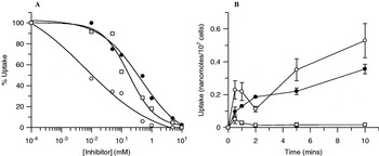
IC50 values (Fig. 2A) were calculated for each inhibitor (2-DOG=16·5±9·3 μm, GlcNAc=176·18±65·28 μm, GlcN=443·96±18·39 μm). This suggests that GlcNAc and GlcN do exhibit interaction with the procyclic hexose transporter, although they show a relatively low affinity (10- and 30-fold lower than 2-DOG, respectively). However, interaction with the transporter does not necessarily mean internalization of the analogue, and accumulation of the analogues was tested using uptake assays of isotope-labelled sugars (Fig. 2B). As previously shown with the bloodstream-form hexose transporter, there was no internalization of GlcNAc into procyclics. In comparison, the D-glucose analogues 2-DOG and GlcN were both recognized and internalized by the parasite. Thus GlcNAc neither acts as a carbon source nor enters trypanosomes, although it does interact with the hexose transporter and thereby inhibits D-glucose uptake.
(A) Inhibition of uptake of 10 μm [6-14C] 2-DOG with cold GlcNAc (open squares), GlcN (closed circles) and 2-DOG (open circles) over a range of concentrations (10 μm, 50 μm, 100 μm, 500 μm, 1 mm, 5 mm, 10 mm). IC50 values were calculated for each inhibitor. (B) Uptake of 1 mm[6-14C] -2-DOG (open circles), [1-14C] GlcN (closed circles) and [3H] GlcNAc (open squares) (n=3, error bars are±S.E.M.).

GlcNAc causes a switch to L-proline metabolism
The rate of L-proline consumption was also measured as a function of D-glucose concentration in the EATRO1125 procyclic trypanosome (Fig. 3A). L-proline consumption as a function of D-glucose concentration shows a dose-dependent response to D-glucose, indicating a gradual replacement of D-glucose metabolism by L-proline metabolism – rather than an ‘on-off’ switch stimulated by D-glucose reaching a critical limiting concentration.
L-proline consumption (A) and oligomycin sensitivity (B) in the presence of increasing amounts of D-glucose in the EATRO1125 procyclic-form trypanosome strain.

Oligomycin is the most sensitive known inhibitor of the F0/F1 ATPase enzyme, which is critical in ATP generation using the mitochondrial respiratory chain, but not when cells produce ATP by substrate level phosphorylation. It has been reported that procyclic-form trypanosomes growing in a D-glucose-rich medium are over 1000 times less sensitive to oligomycin than parasites growing in D-glucose-depleted medium (Coustou et al. Reference Coustou, Besteiro, Biran, Diolez, Bouchaud, Voisin, Michels, Canioni, Baltz and Bringaud2003; Lamour et al. Reference Lamour, Riviere, Coustou, Coombs, Barrett and Bringaud2005).
Oligomycin sensitivity, as a metabolic determinant, was tested in the EATRO1125 procyclic-form trypanosome strains. Usage of L-proline is upregulated as a direct result of D-glucose reduction (Fig. 3A), and consequently, oligomycin sensitivity increases as a direct consequence of the switch to oxidative phosphorylation (Fig. 3B). It is evident that there is a tightly regulated link between different modes of metabolism as a function of relative D-glucose availability, and we wanted to test if this link could be modulated by the presence of GlcNAc. Since GlcNAc inhibits D-glucose uptake without itself being internalized by procyclic-form trypanosomes, its effect on trypanosome growth may be related to its capacity to diminish D-glucose uptake and to induce a metabolic shift.
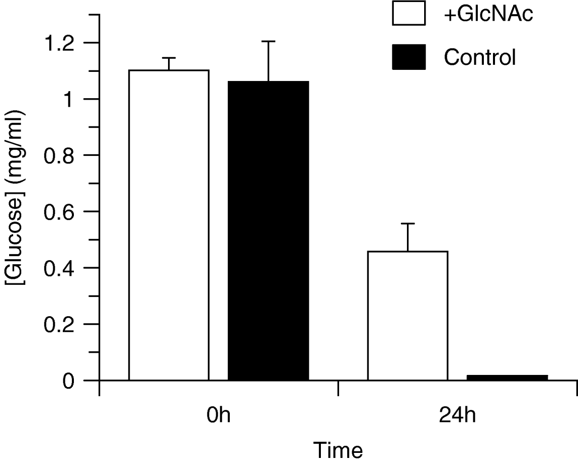
We analysed the D-glucose present in SDM79 medium after 24 h during log phase growth of trypanosomes. With GlcNAc present in the medium, D-glucose is consumed at a lower rate by the parasites (Fig. 4). The quantity of L-proline consumed by the procyclic trypanosomes has previously been shown to be decreased when D-glucose is available as the principal carbon source (Lamour et al. Reference Lamour, Riviere, Coustou, Coombs, Barrett and Bringaud2005). Procyclic-form trypanosomes were grown in CM (containing 4 mm D-glucose and 60 mm L-proline) plus or minus GlcNAc (60 mm), SDM80 supplemented with D-glucose (10 mm), or L-proline (10 mm) and then assayed for L-proline uptake.
Residual D-glucose concentration in culture medium during log phase growth of procyclic-form trypanosomes (strain TREU 927) in the presence and absence of GlcNAc (60 mm) (n=3, error bars are±S.E.M.).

Pre-adaptation to D-glucose-deficient conditions (in SDM80-based medium) increased L-proline uptake in procyclic-form trypanosomes, to a rate twice that in cells grown in high D-glucose conditions (Fig. 5). Cells grown in CM showed an even further reduction in L-proline uptake. However, addition of GlcNAc to the medium induced L-proline uptake to levels comparable to cells grown in high L-proline medium. It thus appears that the presence of GlcNAc induced increased L-proline consumption, probably as a consequence of its capacity to diminish uptake of D-glucose.
L-proline uptake. Procyclic-form trypanosomes pre-adapted to different medium concentrations and then assayed for L-proline uptake. Uptake of [3H] L-proline into procyclic (strain TREU 927) trypanosomes grown in Cunningham's medium (CM containing 4 mm D-glucose and 60 mm L-proline) (white square), SDM80 supplemented with 10 mm D-glucose (black circles), SDM80 supplemented with 10 mm L-proline (white circles) and CM supplemented with 60 mm GlcNAc (black squares). Uptake was measured at a single 30 s time-point over a range of concentrations (up to 200 μm) (n=3, error bars=±S.E.M.).

A modified Alamar Blue method was employed, where procyclic trypanosomes were grown in the presence of oligomycin (0·1 ng/ml) and L-proline (5 mm) and the D-glucose concentration was titred in doubling dilutions (5 mm starting concentration). The D-glucose IC50 value for oligomycin sensitivity significantly increases by more than 10-fold from 48·6 μm to 674·5 μm in the presence of GlcNAc (data not shown). The metabolic switch appears to be a graded response, with oligomycin sensitivity increasing as D-glucose levels decrease, indicating a tightly regulated balance between the two. From this it is evident that, the same link between D-glucose availability, L-proline consumption and oligomycin sensitivity is observed.
GlcNAc enhancement of tsetse colonization is not due to a metabolic shift
GlcNAc has also been shown to promote colonization of tsetse flies by procyclic T. brucei (Peacock et al. Reference Peacock, Ferris, Bailey and Gibson2006). We tested whether this effect was related to the capacity of GlcNAc to diminish glucose uptake and induce a metabolic shift to L-proline utilization, thereby pre-adapting trypanosomes to low glucose/high proline availability in the fly midgut. Flies were infected with procyclic trypanosomes pre-adapted to growth in CM or NSCM medium. The highest infection rates occurred when 60 mm GlcNAc was added to the infective feed, regardless of whether the procyclic trypanosomes had been grown in the presence or absence of D-glucose (Fig. 6). There was no significant difference in infection rates between flies that had been fed trypanosomes grown in CM or NSCM. Thus, trypanosomes that enter flies already using L-proline as their primary energy source have no advantage, and the effect of GlcNAc on tsetse midgut infection rates is probably due to effects of GlcNAc other than its capacity to induce a metabolic shift. Inhibition of trypanocidal lectins has previously been proposed as a mechanism of GlcNAc action (Welburn and Maudlin, Reference Welburn and Maudlin1999).
Fly infectivity. Effect of GlcNAc and NSCM on the frequency of midgut Trypanosoma brucei strains TREU 927 (A) and J10 (B) infections. The difference between CM and CM plus GlcNAc; and NSCM and NSCM plus GlcNAc is significant at P⩽0·001 for TREU 927 and P⩽0·05 for J10. The numbers of flies dissected are given as n in the figure.

DISCUSSION
It has been long established that GlcNAc enhances colonization of tsetse by trypanosomes, which has been causally linked to GlcNAc inhibition of midgut trypanocidal lectins (Mihok et al. Reference Mihok, Otieno, Darji and Munyinyi1992; Osir et al. Reference Osir, Imbuga and Onyango1993; Welburn and Maudlin, Reference Welburn and Maudlin1999; Abubakar et al. Reference Abubakar, Bulimo, Mulaa and Osir2006). The demonstration that GlcNAc also stimulates growth in vitro, in the absence of any fly-derived factors, was surprising (Peacock et al. Reference Peacock, Ferris, Bailey and Gibson2006). We show here that the stimulation of growth is not as a direct result of GlcNAc utilization as a carbon source, because GlcNAc is not internalized by the parasite and cannot replace D-glucose or L-proline as a carbon source for energy. However, there is interaction with the hexose transporter, demonstrated by GlcNAc inhibition of uptake of 2-DOG.
We therefore propose that GlcNAc, through blocking of the procyclic hexose transporter, promotes a metabolic shift in procyclic-form trypanosomes leading to an increased consumption of L-proline, as previously noted when procyclic-form trypanosomes were grown in glucose-depleted media (Lamour et al. Reference Lamour, Riviere, Coustou, Coombs, Barrett and Bringaud2005). L-proline metabolism in procyclic-form trypanosomes is more efficient than D-glucose metabolism. When D-glucose and L-proline are both present in the growth medium, however, the procyclic-form trypanosome will preferentially use D-glucose as the main carbon source (Cross et al. Reference Cross, Klein and Linstead1975; Bringaud et al. Reference Bringaud, Riviere and Coustou2006).
It appears, therefore, that the in vitro growth stimulatory effect of GlcNAc relates to the switch from D-glucose to L-proline catabolism, leading to an increase in L-proline consumption. This is correlated to increased oligomycin sensitivity. The switch is a graded response, rather than a binary distinction, since L-proline consumption is gradually reduced as D-glucose levels rise, and there is a corresponding gradual decrease in susceptibility to oligomycin. The repressive effect of D-glucose on L-proline metabolism seems to be related to the overall metabolic status of the cell rather than any allosteric action of D-glucose on its own, since cells adapted to growth without D-glucose retain high levels of L-proline uptake rates for many hours even when D-glucose is added (data not shown).
In spite of the metabolic shift that accompanies growth in L-proline, procyclics using L-proline as the main carbon source show no infectivity advantage to tsetse flies over control procyclics grown in normal D-glucose-rich medium. This suggests that the in vivo effects of GlcNAc are not a result of any metabolic advantage of the trypanosomes, but rather are a consequence of reduction in fly anti-microbial defences such as inhibition of lectins (Welburn and Maudlin, Reference Welburn and Maudlin1999).
In addition to its impact on L-proline catabolism, D-glucose exerts an influence on other aspects of procyclic trypanosome biology, including procyclin expression as well as mitochondrial energy generation (Butikofer et al. Reference Butikofer, Ruepp, Boschung and Roditi1997; Vassella et al. Reference Vassella, Acosta-Serrano, Studer, Lee, Englund and Roditi2001; Morris et al. Reference Morris, Wang, Drew and Englund2002). The reason for the negative regulation exerted by D-glucose on L-proline metabolism is far from clear, especially in the light of the fact that L-proline provides a markedly better growth stimulus than D-glucose. A possible explanation could be linked to the developmental cues associated with reduced D-glucose availability. When bloodstream-form trypanosomes are taken up by the tsetse vector, glucose availability presumably drops rapidly during digestion of the bloodmeal in the tsetse midgut. It is possible that D-glucose availability is one marker involved in life-cycle progression of the parasite. It will therefore be of interest to determine whether the response of procyclics to D-glucose with regard to L-proline catabolism is linked to the response in expression of particular procyclin surface membrane protein isoforms or is related to processes leading to developmental differentiation of the parasite.
C. Ebikeme was funded by the Medical Research Council. F. Bringaud, V. Coustou and L. Rivière were supported by the CNRS, the Conseil Régional d'Aquitaine and the Ministère de l'Education Nationale de la Recherche et de la Technologie (Action Microbiologie). Work in Bristol was supported by The Wellcome Trust. We thank Mrs Vanessa Ferris (Bristol) for assistance with tsetse and trypanosome culture. The British Council and Ministère des Affaires Etrangères and the Ministry of Research are gratefully acknowleged for funding collaborative links between M. P. B and F. B. through their Alliance Programme.