Introduction
Cystic echinococcosis (CE) is a disease with a history dating back to ancient times, and yet it is still an important zoonosis with socio-economic impact in modern society (Eckert & Thompson, Reference Eckert and Thompson2017). The causative agent of the disease is Echinococcus granulosus, a small tapeworm that typically lives in the intestine of carnivores, including dogs, wolves and foxes, which are the known definitive hosts of the parasite. These animals become infected by eating offal of the intermediate hosts harbouring the parasite metacestode. Herbivores are the main intermediate hosts, yet rodents can be also infected. The intermediate hosts become infected by ingesting parasite eggs, which are shed into the environment by faeces excreted by infected definitive hosts (Pakala et al., Reference Pakala, Molina and Wu2016; Brehm & Koziol, Reference Brehm and Koziol2017).
Echinococcus granulosus can accidentally infect humans who then technically become intermediate hosts. Humans mostly become infected either by direct contact with an infected dog or by ingesting contaminated food, water or soil carrying the parasite eggs (Pakala et al., Reference Pakala, Molina and Wu2016). Human infection is asymptomatic at the beginning of infection. At this stage, oncospheres released from ingested eggs penetrate the human intestinal wall and, via the portal venous system, they may access the liver, lungs and various other organs, where they may develop into hydatid cysts (Lewall, Reference Lewall1998; Siracusano et al., Reference Siracusano, Delunardo, Teggi and Ortona2012; Brehm & Koziol, Reference Brehm and Koziol2017).
Echinococcus granulosus exhibits fairly wide genetic diversity. The current taxonomy of E. granulosus involves different species and strains (denoted as G1–G10), which have been classified using mitochondrial DNA sequences. These include the most common E. granulosus G1 (sheep strain), E. granulosus G2 (Tasmanian sheep strain), E. granulosus G3 (buffalo strain), E. equinus G4 (horse strain), E. ortleppi G5 (cattle strain), E. intermedius G6 (camel strain), E. intermedius G7 (pig strain), E. canadensis G8 and G10 (cervid strains) and E. felidis (Thompson, Reference Thompson2017).
Echinococcus granulosus G1 strain is responsible for most cases of human CE and is distributed widely throughout the globe (Siracusano et al., Reference Siracusano, Delunardo, Teggi and Ortona2012; Carmena & Cardona, Reference Carmena and Cardona2013). The Tasmanian sheep strain (G2) is geographically distributed in Tasmania and Argentina, although there are recent reports of its existence in some European countries. The buffalo strain (G3) is distributed in Asia and in some European countries (Siracusano et al., Reference Siracusano, Delunardo, Teggi and Ortona2012). All of the three strains mentioned above have wide geographical distributions, demonstrate no intermediate host specificity and occur sympatrically (Thompson, Reference Thompson2008). Echinococcus ortleppi (G5) is distributed in Nepal, Switzerland, Brazil, India, Nepal and The Netherlands. Echinococcus intermedius (G6) has been reported from Kenya, Somalia, Sudan, China, Iran, Mauritania, Argentina and Nepal. Echinococcus intermedius (G7) or pig strain is found in Poland, Slovakia, Ukraine, Argentina and Spain. Echinococcus canadensis strains (G8 and G9) are reported from the United States and Poland, respectively (McManus & Thompson, Reference McManus and Thompson2003). Considerable intra-strain genetic variation has been observed in E. granulosus isolates from Iran (Fadakar et al., Reference Fadakar, Tabatabaei, Borji and Naghibi2015; Sharbatkhori et al., Reference Sharbatkhori, Tanzifi, Rostami, Rostami and Fasihi Harandi2016; Spotin et al., Reference Spotin, Mahami-Oskouei, Harandi, Baratchian, Bordbar, Ahmadpour and Ebrahimi2017), as from the other parts of the globe (Thompson, Reference Thompson2008).
In Iran, E. granulosus G1, G3 (Sharafi et al., Reference Sharafi, Rostami-Nejad, Moazeni, Yousefi, Saneie, Hosseini-Safa and Yousofi-Darani2014), G2 (Rostami et al., Reference Rostami, Shariat Torbaghan, Dabiri, Babaei, Ali Mohammadi, Sharbatkhori and Fasihi Harandi2015) and E. intermedius G6 (Nikmanesh et al., Reference Nikmanesh, Mirhendi, Ghalavand, Alebouyeh, Sharbatkhori, Kia, Mohebali, Eghbali and Rokni2014) have been isolated from human cases. Moreover, E. granulosus G1, G2, G3, E. ortleppi G5, E. intermedius G6 (Sharafi et al., Reference Sharafi, Rostami-Nejad, Moazeni, Yousefi, Saneie, Hosseini-Safa and Yousofi-Darani2014) and G7 (Fadakar et al., Reference Fadakar, Tabatabaei, Borji and Naghibi2015) have been reported in animals. Thus, the present study aimed to identify the genetic variation of E. granulosus isolates from clinical samples of human CE, based on polymerase chain reaction (PCR) sequencing of two mitochondrial housekeeping genes, cytochrome c oxidase I (cox1) and NADH dehydrogenase subunit I (nad1).
Materials and methods
Study area
Isfahan is one of the central provinces of Iran and is situated between the central Zagros mountain range and the Grand Desert. It is situated between 30°42′ and 34°30′N latitude and 49°36′, 55°32′E longitude, reaches an altitude of 1590 m above sea level and has an arid climate.
Sample collection
In the present cross-sectional study, 50 surgically removed hydatid cysts from patients in Al-Zahra Hospital, Isfahan, Iran, were collected between April 2015 and March 2017. In terms of organ sourcing of the samples, 44 were isolated from the liver, five from the lungs and one from the kidney. The hydatid cyst samples were brought to the laboratory and protoscoleces were isolated by centrifugation of hydatid fluid and preserved in 70% ethanol at −20°C. A specimen from the cyst walls was also preserved.
DNA extraction
The collected samples were washed twice with phosphate-buffered saline (PBS) in order to remove ethanol. DNA was extracted from the tissue using a commercial genomic DNA extraction kit (PrimePrep™, Genet Bio, Korea). The procedure was performed according to the manufacturer's instructions. Extracted DNAs were kept frozen at −20°C for future application.
Polymerase chain reaction (PCR)
The PCR reaction was performed using the two previously described primers, JB3 forward (5′-TTTTTTGGGCATCCTGAGGTTTAT-3′), JB4.5 reverse (5′-TAAAGAAAGAACATAATGAAAATG-3′) for cox1 (Bowles et al., Reference Bowles, Blair and McManus1992) and MS1 forward (5′-CGTAGGTATGTTGGTTTGTTTGGT-3′) and MS2 reverse (5′-CCATAATCAAATGGCGTACGAT-3′) for nad1 (Sharbatkhori et al., Reference Sharbatkhori, Mirhendi, Jex, Pangasa, Campbell, Kia, Eshraghian, Harandi and Gasser2009). PCR amplification was performed in a 50-μl reaction volume containing 25 μl Taq DNA polymerase, 2× Master Mix (Ampliqon, Denmark; 3 mm MgCl2, 0.4 mm deoxyribonucleosides (dNTPs), 0.2 units/μl Taq DNA polymerase), 19 μl nuclease-free water (Merck Millipore, Darmstadt, Germany), 3 μl template DNA, 1.5 μl reverse primer and 1.5 μl forward primer (10 pmol). The amplification conditions for both genes were as follows: initial denaturation at 95°C for 5 min; followed by 35 cycles of 94°C for 45 s (denaturation), 55°C for 30 s (annealing) and 72°C for 30 s; with a final extension step at 72°C for 5 min. Gel electrophoresis of PCR products was performed on 1.5% agarose gels containing GelRed™ (Biotium Inc., Fremont, California, USA) as a nucleic acid stain. Results were studied using a transilluminator device.
Sequencing and phylogenetic analysis
The amplicons were purified (Wizard SV Clean-up System; Promega, Madison, Wisconsin, USA) and sequenced directly by Sanger dideoxy-sequencing technology in both directions using a capillary 3730xl DNA analyser (Applied Biosystems, Foster City, California, USA). Ambiguous sites were edited using the standard International Union of Pure and Applied Chemistry (IUPAC) codes for combinations of two or more bases. Sequence contigs of the samples were aligned and edited visually in consensus positions, and compared with sequences from GenBank using the software Sequencher v.4.1.4 (Gene Codes Corporation, Ann Arbor, Michigan, USA) (Spotin et al., Reference Spotin, Mahami-Oskouei, Harandi, Baratchian, Bordbar, Ahmadpour and Ebrahimi2017). The sequences were aligned using ClustalW and translated into protein sequences using Mega v.7 (Pennsylvania State University, USA). Codons were aligned using multiple sequence alignment with hierarchical clustering (http://multalin.toulouse.inra.fr/). Diversity indices (haplotype diversity, nucleotide diversity, Tajima's D and Fu's F values) and the number of segregating sites were estimated by DnaSP software v.5.10 (Rozas et al., Reference Rozas, Sanchez-DelBarrio, Messeguer and Rozas2003). A network of mitochondrial haplotypes was drawn based on the DNA sequences reflecting the nad1 and cox1 genes by the PopART software (http://popart.otago.ac.nz) using statistical parsimony. A phylogenetic tree was built by Maximum Likelihood using the Tamura–Nei model (MEGA v.7) (Tamura & Nei, Reference Tamura and Nei1993).
Results
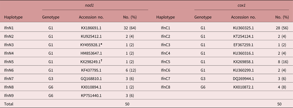
A total of 50 hydatid cyst isolates were collected. DNA sequences representing the nad1 (398 bp) and cox1 (444 bp) genes were amplified successfully from all 50 isolates. The sequence alignment was trimmed manually, and 346 bp of the nad1 and 386 bp of the cox1 genes, respectively, were analysed. Based on GenBank reference sequences, 43 (86%), 3 (6%) and 4 (8%) isolates were identified as E. granulosus (G1), E. granulosus (G3) and E. intermedius (G6), respectively, which could all be confirmed by the two studied genes, cox1 and nad1 individually (table 1). Among the 43 E. granulosus (G1), 37, 5, and 1 were isolated from liver, lungs and kidney. All of the G3 and G6 samples were isolated from liver.
Haplotypes identified among E. granulosus (G1 and G3) and E. intermedius (G6) using cox1 and nad1 gene analysis.

* With single mutation at T187G.
† With single mutation at A351G.
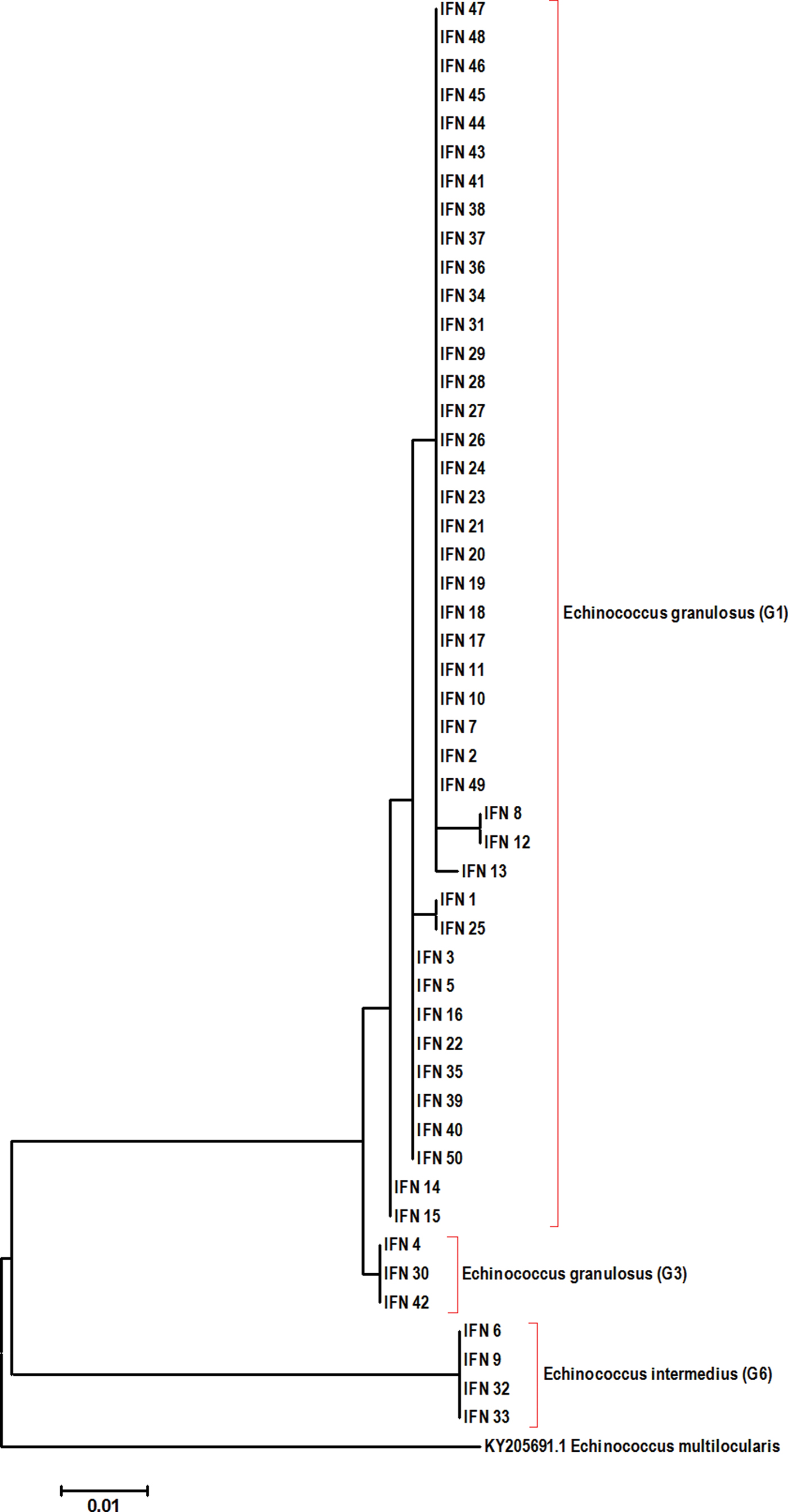
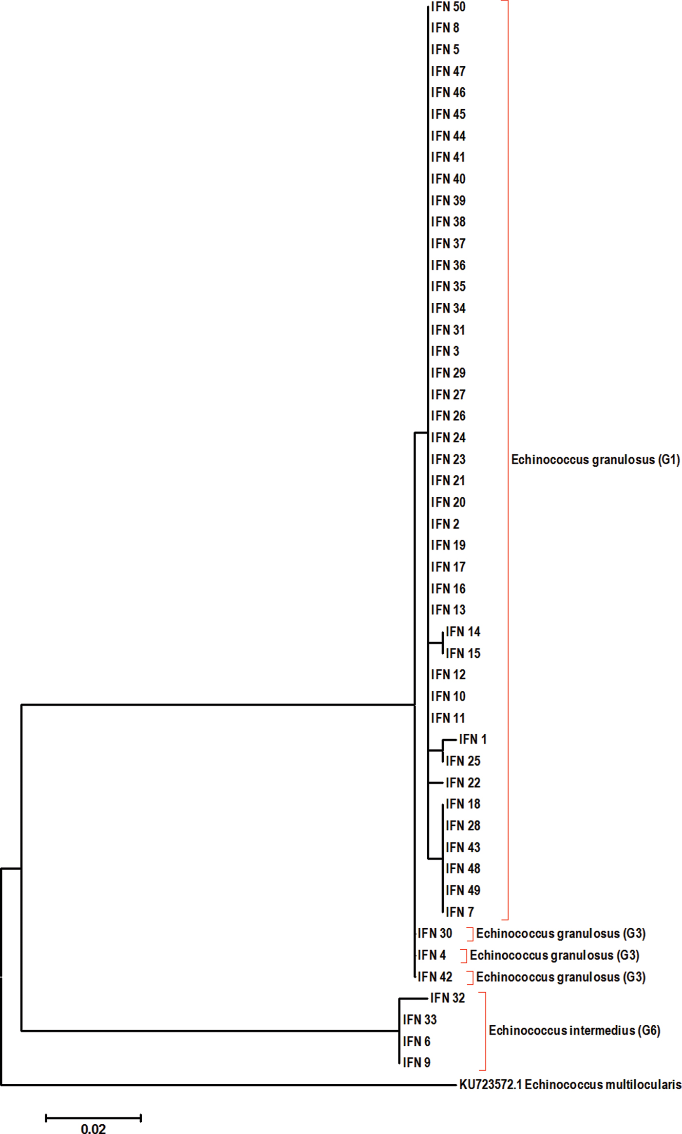
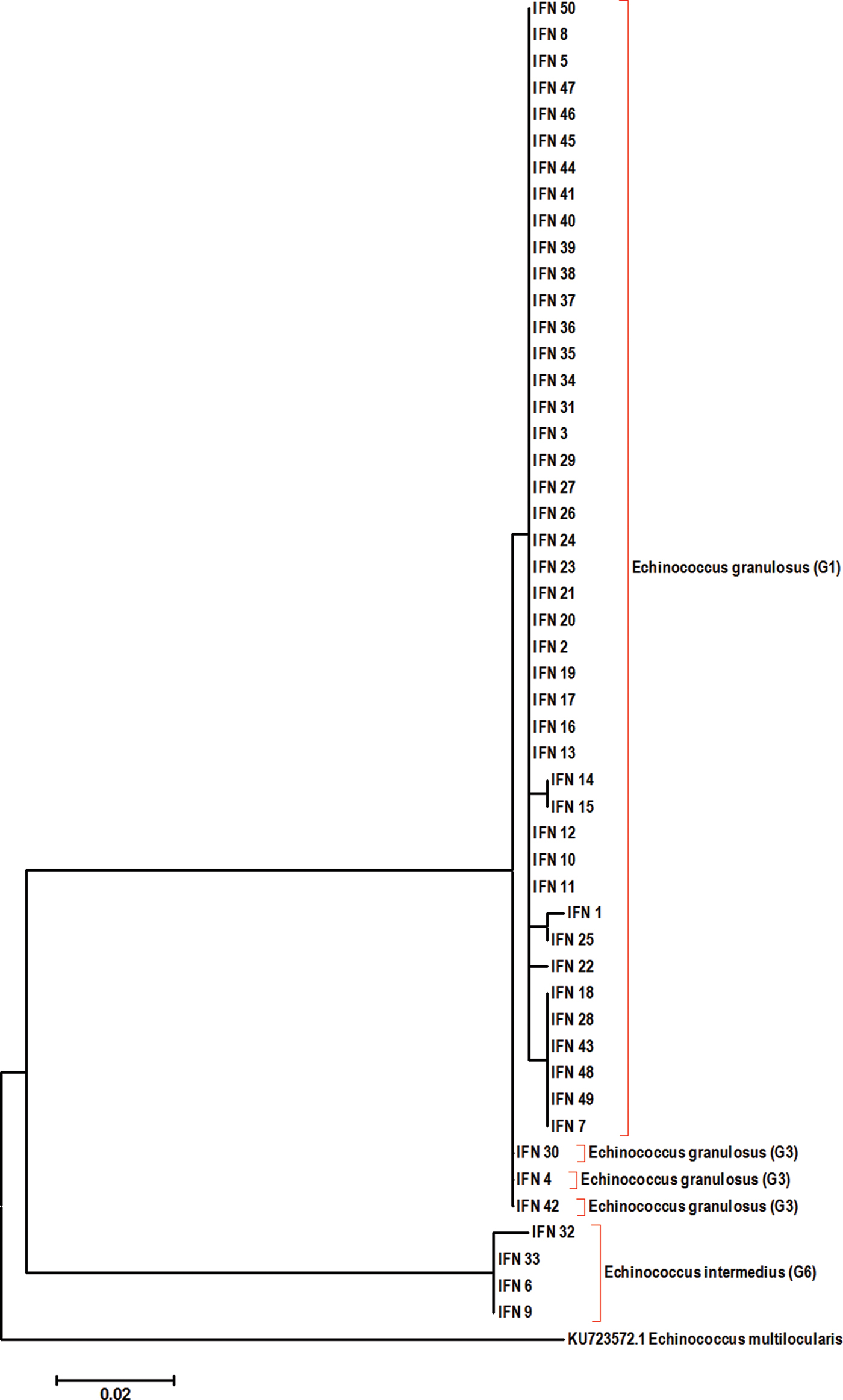
Considering the cox1 gene, six haplotypes in six polymorphic sites were identified in G1 strains; meanwhile, G3 and G6 isolates did not exhibit any intra-genotype polymorphism (fig. 1). Similar to cox1, for nad1 six haplotypes with five polymorphic sites were identified in G1 strains; however, unlike cox1, two G6 haplotypes (two polymorphic sites) were observed (fig. 2).
Phylogenetic analysis of E. granulosus isolates using cox1 genes. The evolutionary history was inferred using the Maximum Likelihood method according to the Tamura–Nei model (Tamura & Nei, Reference Tamura and Nei1993).The tree with the highest log likelihood (−783.2610) is shown. Initial trees for the heuristic search were obtained automatically by applying the neighbour-joining and the BioNJ algorithms to a matrix of pairwise distances estimated by using the maximum composite likelihood (MCL) approach, then selecting the topology with the superior log likelihood value. The phylogenetic tree is drawn to scale, with lengths of the branch measured in the number of substitutions per site. The analysis involved 51 nucleotide sequences. There were a total of 385 positions in the final dataset. Evolutionary analyses were conducted in MEGA7 software (Kumar et al., Reference Kumar, Stecher and Tamura2016). Echinococcus multilocularis (KY205691.1) was used as the outgroup.

Phylogenetic analysis of E. granulosus isolates using nad1 gene sequence by Maximum Likelihood method. The evolutionary history was inferred using the Maximum Likelihood method according to the Tamura–Nei model (Tamura & Nei, Reference Tamura and Nei1993). The tree with the highest log likelihood (−787.8991) is shown. Initial trees for the heuristic search were obtained automatically by applying the neighbour-joining and the BioNJ algorithms to a matrix of pairwise distances estimated by using the MCL approach, then selecting the topology with the superior log likelihood value. The phylogenetic tree is drawn to scale, with lengths of the branch measured in the number of substitutions per site. The analysis involved 51 nucleotide sequences. There were a total of 346 positions in the final dataset. Evolutionary analyses were conducted in MEGA7 software (Kumar et al., Reference Kumar, Stecher and Tamura2016). Echinococcus multilocularis (KU723572.1) was used as the outgroup.
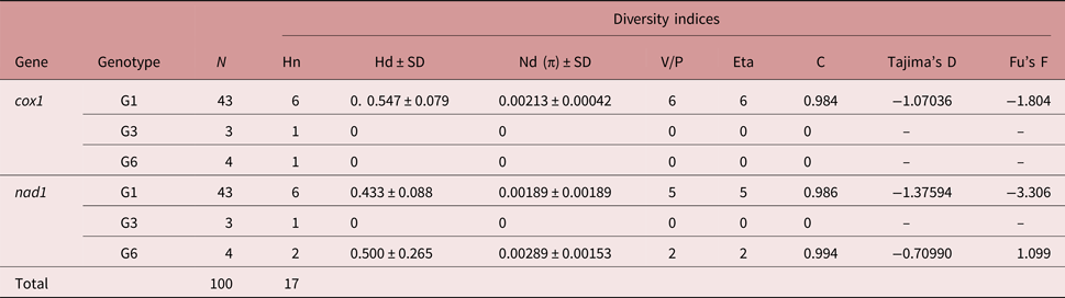
For G1 strains, the haplotype diversity index was higher for the cox1 gene (0.547) compared with nad1 (0.433). Neutrality indices of the nad1 and cox1 genes in E. granulosus G1 were negative values (cox1, Tajima's D = −1.07036, Fu's F statistic = −1.804; and nad1, Tajima's D = −1.37594, Fu's F statistic = −3.306) (table 2).
Diversity indices of E. granulosus (G1 and G3) and E. intermedius (G6) based on DNA sequences of cox1 and nad1 genes. N, number of isolates; Hn, number of haplotypes; Hd, haplotype diversity; Nd, nucleotide diversity; Eta, total number of mutations; C, sequence conservation; V/P, number of variable/polymorphic sites.

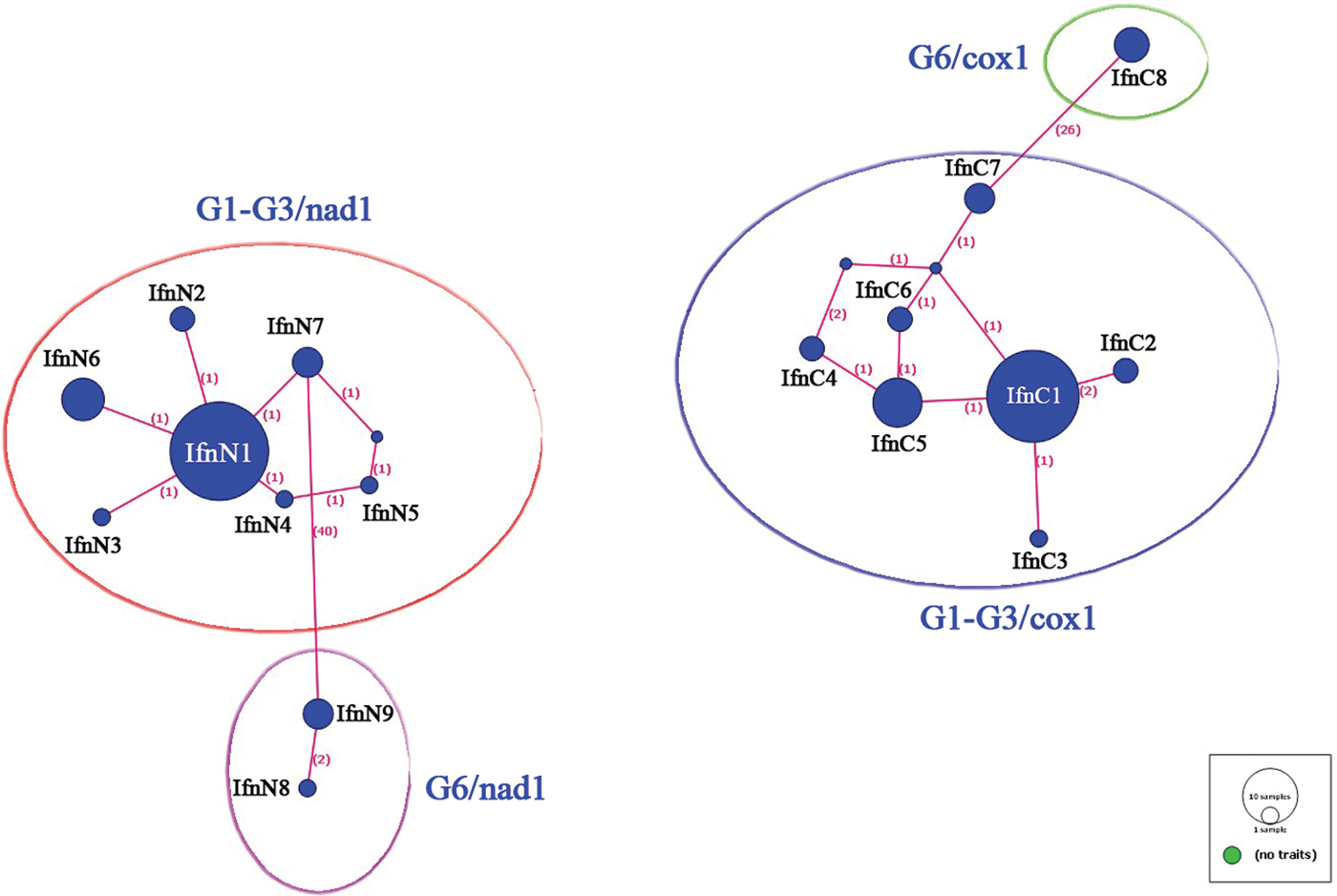
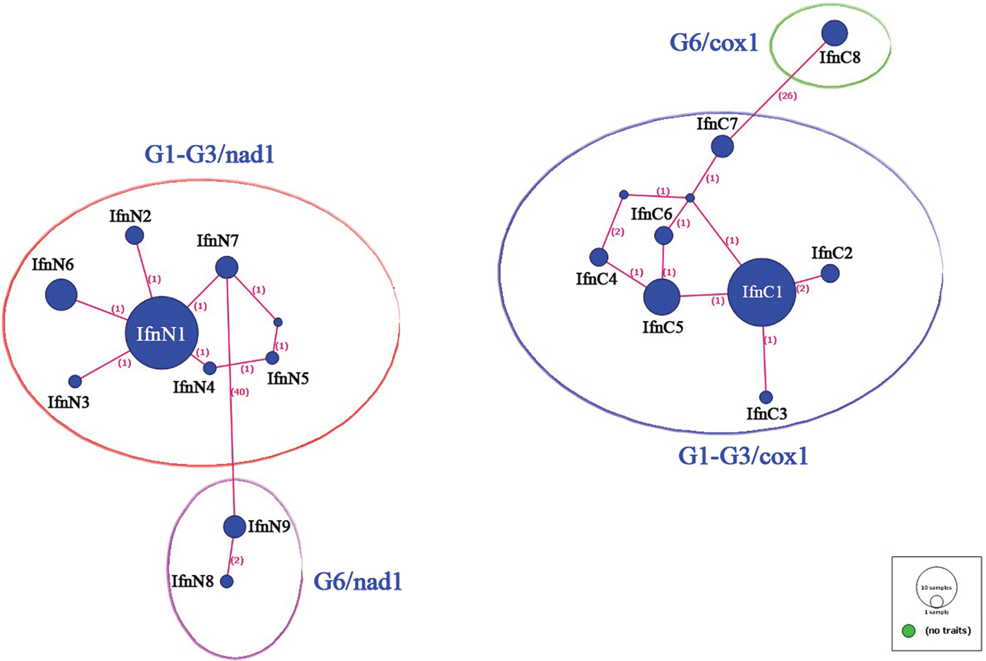
The frequency of the six haplotypes of the 43 E. granulosus (G1) isolates based on cox1 gene analysis were as follows: IfnC1, 28 isolates (65.11%); IfnC2, 2 isolates (4.65%); IfnC3, 1 isolate (2.32%); IfnC4, 2 isolates (4.65%); IfnC5, 8 isolates (18.6%) and IfnC6, 2 isolates (4.65%). Moreover, the frequency of the six haplotypes among the 43 E. granulosus (G1) isolates based on nad1 gene analysis were as follows: IfnN1, 32 isolates (74.41%); IfnN2, 2 isolates (4.65%); IfnN3, 1 isolate (2.32%); IfnN4, 1 isolate (2.32%); IfnN5, 1 isolate (2.32%) and IfnN6, 6 isolates (13.95%) (fig. 3). The haplotypes IfnN3 and IfnN5 were unique to the present study, and the reference strains with the highest identity (99%) for these were KY495928.1 for IfnN3 with a mutation at T187 G and KX298249.1 for IfnN5 with a mutation at A351G. The frequency and the accession numbers of the reference sequences most identical to the haplotypes are shown in table 1.
cox1 and nad1 DNA sequence haplotype network in the studied E. granulosus isolates using TCS networks. The numbers represent mutations.
Considering the cox1 gene data, no deletions or insertions were observed in the DNA sequences. Moreover, 380 (from 386) sites were invariable (monomorphic), and six were variable (polymorphic) sites, with six mutations in one singleton variable site (position 164) and five parsimony-informative sites (positions 4, 5, 46, 56, 101). Among three E. granulosus (G3) and four E. intermedius (G6) isolates, no polymorphism was seen in cox1 DNA sequences (table 1). The percentage identity and divergence of the G1 isolates are shown in supplementary fig. S1.
Considering the nad1 DNA sequences, no deletions or insertions were observed. Moreover, 341 (out of 346) sites were invariable (monomorphic), and five were variable (polymorphic) sites with five mutations in two singleton variable sites (positions 75, 244) and three parsimony-informative sites (positions 38, 50, 170). Among three E. granulosus (G3) isolates, no polymorphism was observed with the cox1 gene. Furthermore, among four E. intermedius (G6) isolates, 344 sites were invariable and two variable, with two mutations in two singleton variable sites (positions 230, 326).
The multiple alignments of cox1 gene amino acid sequences among G1 genotype E. granulosus samples showed amino acid substitutions at three parsimony-informative sites and at one singleton site. In total, there were four variable and 114 conserved sites. The amino acid sequence alignment of the cox1 gene in E. granulosus (G1) is available in supplementary fig. S2. The multiple alignments of nad1 gene amino acid sequences for the G1 genotype E. granulosus samples showed amino acid substitutions at four parsimony-informative sites and at one singleton site. In total, there were five variable and 103 conserved sites. The amino acid alignment reflecting the nad1 gene is available in supplementary fig. S3.
Discussion
Differences among E. granulosus infections have been recognized for many years and have been described as differences related to host specificity as well as biochemical, morphological and other biological differences (Alvarez Rojas et al., Reference Alvarez Rojas, Romig and Lightowlers2014). With the application of molecular tools to E. granulosus, a number of largely host-adapted species of the parasite and genotypes have been observed, which are maintained in separate cycles of transmission. The Echinococcus taxonomy has been a matter of controversy for many years. Many of the species described more than 50 years ago and later invalidated have now been shown to be valid, as a result of a large number of molecular epidemiological studies carried out all around the world (Thompson & McManus, Reference Thompson and McManus2002; Thompson, Reference Thompson2008).
The latest revision of the molecular classification of E. granulosus divided it into different species and genotypes, G1–G10 (Thompson, Reference Thompson2008; Cucher et al., Reference Cucher, Macchiaroli, Baldi, Camicia, Prada, Maldonado, Avila, Fox, Gutierrez, Negro, Lopez, Jensen, Rosenzvit and Kamenetzky2016; Thompson, Reference Thompson2017). The genetic characterization of E. granulosus is critical to understanding the pattern of transmission of the parasite between definitive and intermediate hosts, including humans (Alvarez Rojas et al., Reference Alvarez Rojas, Romig and Lightowlers2014). The existence of G1, G2, G3, G5, G6 (Sharafi et al., Reference Sharafi, Rostami-Nejad, Moazeni, Yousefi, Saneie, Hosseini-Safa and Yousofi-Darani2014) and G7 (Fadakar et al., Reference Fadakar, Tabatabaei, Borji and Naghibi2015) genotypes has been reported in Iran. Furthermore, the G1, G3, G6 (Sharafi et al., Reference Sharafi, Rostami-Nejad, Moazeni, Yousefi, Saneie, Hosseini-Safa and Yousofi-Darani2014) and G2 (Hosseini-Safa et al., Reference Hosseini-Safa, Mohag Hegh, Pestechian, Ganji, Mohammadi, Mahmoudi Lamouki and Rostami-Nejad2016) genotypes have been reported from the Isfahan area. The reported genotypes from human CE isolates in Iran are limited to G1, G2, G3 and G6 strains (Rostami et al., Reference Rostami, Shariat Torbaghan, Dabiri, Babaei, Ali Mohammadi, Sharbatkhori and Fasihi Harandi2015).
The majority of the E. granulosus isolates of the present study belonged to the G1 genotype. It is generally known that E. granulosus (G1) is responsible for most human cases of CE; however, other genotypes are also known to be infective to humans (Alvarez Rojas et al., Reference Alvarez Rojas, Romig and Lightowlers2014). Previous studies indicated the presence of G1 and G6 genotypes in human cases of CE in Isfahan (Kia et al., Reference Kia, Rahimi, Sharbatkhori, Talebi, Fasihi Harandi and Mirhendi2010; Shahnazi et al., Reference Shahnazi, Hejazi, Salehi and Andalib2011); however, the G3 genotype has not previously been reported here. This is the first report of E. granulosus G3 genotype isolated from human CE in Isfahan.
Rostami et al. (Reference Rostami, Shariat Torbaghan, Dabiri, Babaei, Ali Mohammadi, Sharbatkhori and Fasihi Harandi2015) studied the genetic characteristics of E. granulosus from 200 formalin-fixed, paraffin-embedded tissue samples of human CE in Iran. They used cox1 gene sequencing and reported that 54.4%, 0.8%, 1% and 40.8% of their studied samples belonged to the G1, G2, G3 and G6 genotypes, respectively. Their findings are very interesting considering the large number of G6 isolates that they reported. They inferred that the G1 genotype is the most prevalent genotype involved in human cases of CE in Iran. They also suggested that the high prevalence of the G6 genotype entails high infectivity for humans. However, it appears that the support for this hypothesis is limited. They also reported the first human case of CE caused by E. granulosus (G2) (Rostami et al., Reference Rostami, Shariat Torbaghan, Dabiri, Babaei, Ali Mohammadi, Sharbatkhori and Fasihi Harandi2015). According to the above-mentioned data, E. granulosus (G1) is the predominant cause of human CE in Iran and Isfahan, followed by E. intermedius (G6). However, in some regions, such as north African countries, e.g. Mauritania and Sudan, E. intermedius (G6) is the most common genotype found in sheep, camels, cattle and humans (Bardonnet et al., Reference Bardonnet, Piarroux, Dia, Schneegans, Beurdeley, Godot and Vuitton2002; Omer et al., Reference Omer, Dinkel, Romig, Mackenstedt, Elnahas, Aradaib, Ahmed, Elmalik and Adam2010; Sharbatkhori et al., Reference Sharbatkhori, Tanzifi, Rostami, Rostami and Fasihi Harandi2016).
Reports from Iran agree that CE in the country is mostly attributable to the G1 genotype. As an example, in one investigation all of the 30 human CE isolates belonged to the G1 genotype in Isfahan (Kia et al., Reference Kia, Rahimi, Sharbatkhori, Talebi, Fasihi Harandi and Mirhendi2010). Another study, carried out in the Ardabil province in north-western Iran, reported the sheep strain (G1) as the most prevalent in humans, goats, sheep and cattle. They also reported the buffalo strain (G3) not only in sheep and cattle, but also – and for the first time – in two human cases (Pezeshki et al., Reference Pezeshki, Akhlaghi, Sharbatkhori, Razmjou, Oormazdi, Mohebali and Meamar2013).
In an interesting report from Tehran, E. intermedius (G6) was isolated from all eight formalin-fixed, paraffin-embedded tissues studied from cases of cerebral CE. It has been claimed that E. intermedius (G6) has a higher affinity for the human brain (Sadjjadi et al., Reference Sadjjadi, Mikaeili, Karamian, Maraghi, Sadjjadi, Shariat-Torbaghan and Kia2013). In the present study, no cases of cerebral CE were investigated, and the four G6 genotype-positive samples were all sourced from CE affecting the liver.
In the present study, a considerable genetic diversity among identified E. granulosus (G1) isolates was observed, reflecting six haplotypes; yet, no intra-genotypic genetic diversity was observed among the three identified E. granulosus (G3), using cox1 and nad1 DNA sequences. Considering the diversity indices, such as nucleotide and haplotype diversity indices, these were higher based on the cox1 gene; on the other hand, the nad1 gene enabled differentiation of E. intermedius (G6) haplotypes with higher resolution.
In a recent study, the genetic variability of 79 and 39 E. granulosus isolates in different geographical populations of Iran was investigated by studying cox1 gene data. Samples were obtained from humans, camels, dogs, goats, sheep and cattle of different geographical sub-populations of Iran. The reported isolates belonged to genotypes G1 (92 isolates), G6 (14 isolates), G3 (8 isolates) and G2 (1 isolate), and the authors found 50 unique haplotypes. A high haplotype diversity index among E. granulosus complex was observed in their study, while, similar to the present study, nucleotide diversity was low across all populations. Furthermore, similar to the findings of the present study, the neutrality index values based on cox1 (Tajima's D and Fu's F) were negative in populations from the west, north-west, north and south-east of Iran (Spotin et al., Reference Spotin, Mahami-Oskouei, Harandi, Baratchian, Bordbar, Ahmadpour and Ebrahimi2017). The large number of haplotypes in the latter study, compared with that of the present study, may be a result of sampling from a broader geographical area.
The multiple alignment of the amino acid sequences of the identified G1 genotypes reflected the fact that all mutations in the various haplotypes caused changes in the distinct amino acid. This shows that all the variations among G1 genotype samples caused amino acid substitution in five different sites, producing six distinct haplotypes in both cox1 and nad1 genes.
In conclusion, according to the findings of the present study, human infection with the three genotypes of E. granulosus G1, G3 and E. intermedius (G6) is documented in Isfahan, central Iran. The G3 genotype in human samples of CE was reported for the first time in Isfahan. The G1 genotype is the most predominant genotype associated with human cases of CE, and the presence of G6 could indicate that camels constitute a reservoir for human infection in Isfahan. Moreover, comparison of cox1 gene data might yield higher diversity indices for population-based studies of E. granulosus compared with comparison of nad1 gene data.
Supplementary material
To view supplementary material for this article, please visit https://doi.org/10.1017/S0022149X17000967.
Acknowledgements
The authors would like to acknowledge the Vice-chancellor of Research and Technology, Isfahan University of Medical Sciences, Isfahan, Iran, for approval of this study. This paper is derived from a PhD thesis in parasitology.
Financial support
This study was supported financially by the Vice-chancellor of Research and Technology, Isfahan University of Medical Sciences, Isfahan, Iran.
Conflict of interest
None.