Introduction
The mean age of the population of Japanese inpatients with schizophrenia has been gradually increasing (Oshima et al., Reference Oshima, Mino and Inomata2007). Although the risks of cognitive impairment and gait disturbances generally increase with age, they are also typical symptoms of older patients with schizophrenia treated with antipsychotics (Schultz et al., Reference Schultz, Miller, Oliver, Arndt, Flaum and Andreasen1997). Thus, it is difficult to distinguish age-dependent mental and physical dysfunction from symptoms of cognitive impairment and antipsychotic-induced extrapyramidal symptoms in older patients with schizophrenia.
Among various disorders of the elderly population manifesting as dementia (psychomotor slowing and impaired attention, executive, and visuospatial dysfunction), gait disturbance (shuffling, magnetic, and wide-based), and urinary incontinence, iNPH is becoming noteworthy (Hakim and Adams, Reference Hakim and Adams1965; Shprecher et al., Reference Shprecher, Schwalb and Kurlan2008). The risk of developing iNPH also increases with age (Brean and Eide, Reference Brean and Eide2008). Pathologically, iNPH has been linked to the reduction of compliance between the cerebrospinal compartments and to disruption of CSF absorption (Hamlat et al., Reference Hamlat, Adn, Sid-ahmed, Askar and Pasqualini2006). However, the exact pathogenesis of iNPH remains unclear. The 10-meter walking test, TUG for an abnormal gait and MMSE for cognitive impairments are often used to assess iNPH symptoms (Shprecher et al., Reference Shprecher, Schwalb and Kurlan2008). However, negative symptoms and cognitive impairments of schizophrenia include psychomotor slowing and impaired attention and executive function, and antipsychotics often induce gait disturbance with Parkinsonism. Thus, iNPH symptoms mimic cognitive impairment and antipsychotic-induced Parkinsonism.
To our knowledge, only a few case reports of NPH in patients with schizophrenia are available (Maurizi et al., Reference Maurizi1987; Mishra et al., Reference Mishra, Sarkar, Mishra, Praharaj, Mahapatra and Sinha2011), and there has been no prospective survey regarding iNPH in older patients with schizophrenia. The present study investigated the prevalence and symptoms of iNPH in older patients with schizophrenia.
Materials and methods
Patients
We enrolled 21 patients (N = 21, mean age = 70.5 ± 5.9) with schizophrenia who were hospitalized in Futaiwa Hospital, Ehime, Japan from September 2014 to March 2015. The diagnosis of schizophrenia was established according to DSM-5 criteria by at least two expert psychiatrists on the basis of extensive clinical interviews and a review of medical records. The diagnosis of possible or probable iNPH was established according to the criteria proposed by the guidelines (Mori et al., Reference Mori2012). Patients who had a history of brain injury or stroke, and those who used wheelchairs were excluded. During the study, medications were not changed. All patients were of unrelated Japanese origin and signed written informed consent forms approved by the institutional ethics committees of Futaiwa Hospital and Ehime University Graduate School of Medicine.
Evaluation of symptoms of schizophrenia and iNPH
Symptoms of schizophrenia were evaluated using the 18-item BPRS (Rhoades and Overall, Reference Rhoades and Overall1988). The DIEPSS was used to evaluate antipsychotic-induced extrapyramidal symptoms (Inada, Reference Inada2009). Patients and caregivers were interviewed to assess iNPHGS (Kubo et al., Reference Kubo2008).
The gait disturbances were evaluated by TUG (Podsiadlo and Richardson, Reference Podsiadlo and Richardson1991) and the 10-meter walking test (Miyoshi et al., Reference Miyoshi2002). The gait disturbances were also assessed by the GSS (Kubo et al., Reference Kubo2008). Cognitive impairments were evaluated by the MMSE (Folstein et al., Reference Folstein, Folstein and McHugh1975). Neuropsychiatric symptoms were evaluated by the NPI assessed by caregivers (Cummings et al., Reference Cummings, Mega, Gray, Rosenberg-Thompson, Carusi and Gornbein1994).
Imaging data were obtained using a computed tomography scanner (TSX-021B/6I; Toshiba, Japan); we defined results for Evans index determinations of > 0.3 as abnormal values (Mori et al., Reference Mori2012).
CSFTT
Lumbar puncture was performed in the iNPH patients; CSF pressure was measured and 30–40 ml of CSF was removed. After the puncture, iNPHGS, TUG, and MMSE were evaluated again. The change of gait was evaluated two days after the tap, while change of cognition and urination was evaluated at one week. Patients having an improvement of one point or more on the iNPH grading scale (each domain and their total), or more than 10% improvement in time on TUG, or more than three points improvement in the MMSE were regarded as CSFTT-positive (Ishikawa et al., Reference Ishikawa, Hashimoto, Mori, Kuwana and Kazui2012). After these examinations, three patients were diagnosed as possible iNPH (Mori et al., Reference Mori2012).
Statistical analysis
Statistical analyses were conducted with SPSS 22.0 software (IBM Japan, Tokyo, Japan). Comparisons between no-iNPH and possible-iNPH patients were the Mann–Whitney U test. Age differences were analyzed using the Fisher's exact test. Correlations for each parameter were analyzed by Spearman's rank correlation coefficient. Statistical significance was defined at the 95% level (p = 0.05).
Results
No-iNPH versus possible-iNPH groups
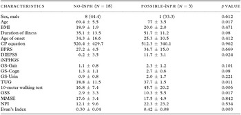
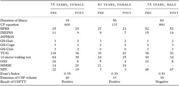
Patients’ characteristics and results of their examinations are shown in Table 1. In total, three (14.3%) patients were diagnosed as possible iNPH and two (9.5%) as probable iNPH. Patients with possible iNPH were significantly older than those without iNPH (p = 0.036). GS-Gait, GS-Cognition, and GS-Urine were not significantly changed in the possible iNPH group, whereas TUG (p = 0.011), 10-meter walking test (p = 0.006), and GSS (p = 0.017) were significantly increased in those patients. BPRS, MMSE, and NPI were not significantly changed in the possible iNPH group. In subscale of DIEPSS, Gait was significantly higher in possible iNPH group than in no-iNPH group (p = 0.024, data not shown). Results of CSFTT in the possible iNPH patients are shown in Table 2, and CT data of these patients are shown in Figure 1.
Demographic and clinical data of patients with possible iNPH and without iNPH (no iNPH). Comparisons between no-iNPH and possible-iNPH patients were conducted with Mann–Whitney U test. Age differences were analyzed using the Fisher's exact test

Values denote number (%) or mean ± standard deviation.
SC, schizophrenia; iNPH, idiopathic normal pressure hydrocephalus; BMI, Body Mass Index; CP equation, chlorpromazine equation; BPRS, Brief Psychiatric Rating Scale; DIEPSS, Drug Induced Extra-Pyramidal Symptoms Scale; iNPHGS, idiopathic Normal-Pressure Hydrocephalus Grading Scale; GS-Gait, iNPHGS for gait; GS-Cogn, iNPHGS for cognition; GS-Urin, iNPHGS for urinary function; TUG, Timed Up-and-Go test; GSS, Gait Status Scale; MMSE, Mini-Mental State Examination; NPI, neuropsychiatric inventory.
Results of CSFTT in patients with possible iNPH

iNPH, idiopathic normal pressure hydrocephalus; CP equation, chlorpromazine equation; BPRS, Brief Psychiatric Rating Scale; DIEPSS, Drug Induced Extra-Pyramidal Symptons Scale; iNPHGS, idiopathic Normal-Pressure Hydrocephalus Grading Scale; GS-Gait, iNPHGS for gait; GS-Cogn, iNPHGS for cognition; GS-Urin, iNPHGS for urinary function; TUG, Timed Up-and-Go test; GSS, Gait Status Scale; MMSE, Mini-Mental State Examination; NPI, neuropsychiatric inventory; CSF, cerebrospinal fluid; CSFTT, cerebrospinal fluid tap test.
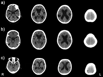
Computed tomography reveals disproportionately enlarged subarachnoid space hydrocephalus in (a) a 75-year-old female, (b) an 81-year-old female, and (c) a 75-year-old male. The features include tight high convexity and medial subarachnoid spaces and an enlarged Sylvian fissure associated with ventriculomegaly. Cerebrospinal fluid is distributed disproportionately between the superior and inferior subarachnoid spaces.

Correlations
The Evans index was significantly positively correlated with the NPI (r = 0.49, p = 0.03), 10-meter walking test (r = 0.463, p = 0.04), BPRS (r = 0.49, p = 0.02), and DIEPSS (r = 0.49, p = 0.03). The MMSE was significantly negatively correlated with GS-Cogn (r = −0.64, p < 0.01) and DIEPSS (r = −0.50, p = 0.03). GS-Gait was significantly positively correlated with GS-Cogn (r = 0.52, p = 0.02), TUG (r = 0.71, p < 0.01), 10-meter walking test (r = 0.64, p < 0.01), GSS (r = 0.65, p < 0.01), and DIEPSS (r = 0.44, p = 0.05).
Discussion
The first of our two major findings was that among 21 older inpatients with schizophrenia, we found three (14.3%) with possible iNPH. Because the prevalence of possible iNPH was estimated to be 2.9% in Japanese residents aged 65 years or older (Iseki et al., Reference Iseki2009), the prevalence of possible iNPH in older patients with schizophrenia seems to be five-fold higher than that of the older population without mental illness. To our knowledge, there are only a few published case-control studies regarding risk factors for iNPH; in these, hypertension, diabetes mellitus, and a low serum level of high-density lipoprotein cholesterol were identified as significant risk factors for iNPH (Casmiro et al., Reference Casmiro, D’Alessandro, Cacciatore, Daidone, Calbucci and Lugaresi1989; Kahlon et al., Reference Kahlon, Sundbärg and Rehncrona2002; Bergsneider et al., Reference Bergsneider, Black, Klinge, Marmarou and Relkin2005; Hashimoto et al., Reference Hashimoto, Ishikawa, Mori and Kuwana2010). Because these are well-known risk factors for vascular diseases and also well-known co-morbidities among patients with schizophrenia (Pratt et al., Reference Pratt, Roughead, Salter and Ryan2012; Papanastasiou, Reference Papanastasiou2013), vascular changes may be involved in the pathogenesis of iNPH in schizophrenia. In this study, patients with possible iNPH were significantly older than patients without iNPH (p = 0.036). Because older age is known to be independent of the response to CSF shunt surgery (Marmarou et al., Reference Marmarou2005), we should detect iNPH symptoms as early as possible and offer shunt surgery to older patients with schizophrenia if needed.
Our second major finding was that among the evaluations for schizophrenia utilized, only DIEPSS was significantly higher in the possible-iNPH patients. Furthermore, only the Gait and Global subscales of DIEPSS were significantly changed in the possible-iNPH group. Interestingly, other extrapyramidal symptoms, such as tremor and rigidity, did not significantly differ in the possible-iNPH patients. Thus, we might be able to distinguish symptoms of iNPH from drug-induced extrapyramidal symptoms by assessing gait disturbance and walking speed. Interestingly, the Evans index was significantly positively correlated with NPI, BPRS, the 10-meter walking test, and DIEPSS, which means this index is associated with both neuropsychiatric symptoms and gait disturbances. The Evans index is generally thought to reflect ventriculomegaly (Raftopoulos et al., Reference Raftopoulos1994); however, enlargement of the lateral ventricles occurs with non-specific global brain atrophy associated with aging in healthy participants and with neurodegeneration (Apostolova et al., Reference Apostolova2012). The progression of schizophrenia also induces overall brain atrophy and enlargement of ventricular volumes (Delisi et al., Reference DeLisi, Sakuma, Tew, Kushner, Hoff and Grimson1997). Thus, a higher Evans index in older patients with schizophrenia may be common and, thus, not useful to distinguish iNPH. As shown in Figure 1, we should pay more attention to other distinctive brain morphologies, such as narrowing of the sulci with focal dilation and narrowing of subarachnoid spaces over the high convexity, when evaluating brain morphology of schizophrenia patients for possible iNPH.
This study had several limitations. One limitation is that the observed prevalence of iNPH in older patients with schizophrenia cannot be generalized due to the small sample size and selection bias. A second limitation is that we did not have normal control patients. A third limitation is that the past history of brain traumatic event may be inaccurate and we could not exclude the possibility of secondary NPH. Lastly, in this preliminary study with a small sample size, we did not correct for multiple comparisons, which raise the chances of type I error. Thus, our results must be treated with caution until they are replicated in larger numbers of patients.
Conflict of interest
None.
Description of the authors’ roles
Y. Yoshino designed the study, wrote the protocol, managed the literature, searches and analyses, and undertook the statistical analysis. T. Yoshida managed the literature, searches, and analyses. T. Mori designed the study, wrote the protocol, and managed the literature. S. Hirota managed the literature. J. Iga managed the literature, searches, and analyses, undertook the statistical analysis. S. Ueno designed the study, wrote the protocol, managed the literature, searches and analyses, and undertook the statistical analysis.
Acknowledgments
We wish to thank Ms. Hiromi Onishi for technical assistance. We thank the staff of Futaiwa Hospital, Yawatahama, Ehime, Japan for understanding the purpose of this study. This work was partially supported by a Health and Labor Science Research Grant from the Japanese Ministry of Health, Labour and Welfare, a Grant-in-Aid for Scientific Research from the Japanese Ministry of Education, Culture, Sports, Science and Technology, and the Ehime Graduate University of Medicine's Good Practice Fund.