


Hyperketonaemia (once known as primary ketosis) in dairy cows often occurs within a few days after parturition (Mann and McArt, Reference Mann and McArt2023). This metabolic deviation is characterised by a negative energy balance (NEB) with an increase in non-esterified fatty acids (NEFA) and β-hydroxybutyrate (BHB) and a decrease in glucose concentration in the blood. Because hyperketonaemia is related to the clinical disease ketosis, a distinction is made between subclinical ketosis (SCK; 1.2 ≤ [BHB]blood ≤ 2.9 mmol/l) and clinical ketosis (CK; [BHB]blood ≥ 3.0 mmol/l). The nomenclature of ‘ketosis 1’ and ‘ketosis 2’ will not be used here (see also Mann and McArt, Reference Mann and McArt2023).
The traditional understanding of the pathogenesis of hyperketonaemia is related to the NEB at the onset of lactation or antepartum, the mobilisation of reserves such as NEFA from the fat depots and the uptake of NEFA by the liver, which eventually overwhelm hepatic metabolic capacity, leading to hepatic lipidosis and production of ketone bodies (KB) such as BHB. Hepatic lipidosis with <1% of triglyceride (TG) of wet weight is considered normal, 1–5% mild, 5–10% moderate and >10% severe (Bobe et al., Reference Bobe, Young and Beitz2004).
The conventional explanation of hyperketonaemia has been questioned in recent publications. Horst et al. (Reference Horst, Kvidera and Baumgard2021) discuss ‘the influence of immune activation on the transition cow health and performance – a critical evaluation of traditional dogmas’. They argue ‘that changes in circulating NEFA, ketones, and calcium are simply reflective of either (1) hormonal homeorhetic adjustments that healthy, high-producing cows use to prioritise milk synthesis, or (2) the consequence of immune activation and its sequelae’. In line with the possible role of the immune system is the review by Mezzetti et al. (Reference Mezzetti, Cattaneo, Passamonti, Lopreiato, Minuti and Trevisi2021): ‘Recent work on the transition period of dairy cows has highlighted the pivotal role of the immune function in affecting the severity of metabolic challenges the animal faces when approaching calving. This suggests that the immune system may play a role in the etiology of metabolic diseases.’ There is a growing body of evidence that the immune system is weakened by the transfer of IgG to the colostrum and the resulting decline in blood concentrations, leading to reduced defence against infections (Herr et al., Reference Herr, Bostedt and Failing2011; Nonnecke et al., Reference Nonnecke, Kimura, Goff and Kehrli2003; Van Knegsel et al., Reference Van Knegsel, de Vries Reilingh, Meulenberg, van den Brand, Dijkstra, Kemp and Parmentier2007). Furthermore, both the adaptive and innate immune systems are impaired postpartum in cows with hepatic lipidosis (Breukink and Wensing, Reference Breukink and Wensing1997; Wentink et al., Reference Wentink, Rutten, van den Ingh, Hoek, Müller and Wensing1997; Zerbe et al., Reference Zerbe, Schneider, Leibold, Wensing, Kruip and Schuberth2000), which is considered to be a major health risk with an increase in the incidence of so-called ‘production diseases’ and of early culling (Bobe et al., Reference Bobe, Young and Beitz2004; Melendez and Pinedo, Reference Melendez and Pinedo2024). Hence, hepatic lipidosis is a key factor that needs to be considered for a more accurate understanding of health and diseases.
This narrative overview aims to outline and propose the development of the pathophysiology of the hyperketonaemia, including (a) the known discrepancy between dry matter intake (DMI) and the requirements for nutrients for milk synthesis; (b) the resulting NEB, mobilisation of NEFA and its ectopic deposition; (c) the impairment of liver function with oxidative stress and inflammation; (d) the weakening of the immune system; (e) the significance of growth hormone (GH) and insulin-like growth factor (IGF-1) as a GH-IGF-1 axis for metabolic adaptation, and of the uncoupling process of this axis for metabolic alteration; and (f) an approach for integrating hepatic lipidosis into homeorhetic regulation.
Negative energy balance
Energy requirement and DMI
Around parturition, a dairy cow undergoes a transient period of NEB, maybe even a few days antepartum, but up to 2 months or more postpartum. The NEB is the consequence of reduced DMI antepartum and of an insufficient uptake of DMI postpartum in relation to the rapid increase in copious milk production. The reasons for this discrepancy between requirement of nutrients and DMI are unclear, but a novel hormone that inhibits feed intake, the growth differentiation factor GDF-15, could contribute to this reduced DMI (Giesy et al., Reference Giesy, Butler, Overton, Rial, Giordano and Boisclair2025). Vernon and Pond (Reference Vernon and Pond1997) proposed a conceivable (biological) explanation some time ago: ‘The inappetence around parturition is probably a throwback to the wild state when mothers would need to remain at the nest for a period of time and would be unable to feed.’ Knight (Reference Knight2001) emphasises this biologically based suggestion and points to some advantages of a reduced DMI: ‘Energetically, mobilization confers approximately 80% efficiency, whilst digestion is only 60% efficient,’ and ‘there is less need for physical activity (foraging)’. ‘Perhaps, the real conundrum is why so much effort is exerted by agricultural nutritionists and dairy farmers in trying to persuade the early-lactation cow to eat more. She knows better!’ These biological assumptions warrant the nutrition and protection of the newborn calf independent of an adequate DMI. Undernutrition is not a challenge for the metabolism of the cow because the production of milk solely for the nutrition of a calf is low and the deficit can easily be compensated for by mobilising reserves. The appetite of the calf is limited and is thus an ideal negative feedback mechanism for limitation of milk yield (MY) and consequently, NEB. Indeed, beef cattle at low milk production, sufficient for a calf, exhibited an increase in body weight (BW) postpartum (Hart et al., Reference Hart, Bines, Balch and Cowie1975). By contrast, Holstein Friesian cows lost BW for 11 weeks postpartum (Hart et al., Reference Hart, Bines, Balch and Cowie1975). The magnitude and duration of NEB postpartum hence appear to be a function of MY. This relationship has been clearly demonstrated by Friggens et al. (Reference Friggens, Berg, Theilgaard, Korsgaard, Ingvartsen, Løvendahl and Jensen2007), who observed a deeper nadir and longer duration of NEB with increasing MY: Danish Holstein > Danish Red > Jersey. The NEB becomes more negative within each breed with increasing number of lactations and milk volume, with the authors concluding that ‘evidence of genetically driven body energy change’ was occurring. The MY is the driving force of NEB (Bar-Peled et al., Reference Bar-Peled, Aharoni, Robinzon, Bruckental, Lehrer, Maltz, Knight, Kali, Folman, Voet, Gacitua and Tagari1988). In an experiment, the authors changed the milking frequency postpartum: (a) machine milking three times daily, (b) machine milking 6 times daily or (c) machine milking three times daily combined with three times suckling by a calf. The cows under condition (c) had the highest MY, the highest NEFA, the lowest glucose concentration, the lowest DMI and, consequently, the deepest NEB with the highest loss of BW. Interestingly, DMI is negatively correlated with the oxytocin concentration in the blood. The magnitude of NEB almost linearly correlates with MY. Brandt et al. (Reference Brandt, Pabst, Schulte-Coerne and Gravert1985) determined the energy balance postpartum during the first lactation. The duration of NEB was 11 weeks, and the mean NEB 1445 MJ net energy for lactation (NEL) or 15.3% of the requirement during this time. The NEB increases with increasing MY: <25 kg/d = 580 MJ NEL, 25–30 kg/d = 1323 MJ NEL and >30 kg/d = 1956 MJ NEL. The increase in DMI is only 0.11 kg/kg MY and agrees very well with the studies of Gruber et al. (Reference Gruber, Pries, Schwarz, Spiekers and Staudacher2006), with an increase of 0.1 kg DMI/kg MY. The gap between DMI and nutrient requirement per kg milk explains the correlation between increasing MY and increasing NEB; it also fits together with the data of Khan et al. (Reference Khan, Hosseini, Burrel, Rocco, McNamara and Loor2013). High genetic-merit cows have higher rates of lipolysis, and increasing MY correlates with the length of NEB (Brade, Reference Brade2013).
Furthermore, Karacaören et al. (Reference Karacaören, Jaffrézi and Kadarmideen2006) and Liinamo et al. (Reference Liinamo, Mäntysaari and Mäntysaari2012) described a negative genetic correlation between MY and DMI at the onset of lactation. The possible consequences of this genetic background have been succinctly expressed by Buttchereit et al. (Reference Buttchereit, Stamer, Junge and Thaller2011): ‘continued selection for high milk production will lead to a further increase in the postpartum energy deficit’.
NEB is not unusual in animals. Horst et al. (Reference Horst, Kvidera and Baumgard2021) argue that many mammals – such as bears, seals and whales – rely totally on reserves for postpartum milk production and experience a long-lasting NEB, but do not develop hyperketonaemia (Jebb and Hiller, Reference Jebb and Hiller2018). It is worth mentioning that the milk of seals and sea lions does not contain lactose or lactose-based oligosaccharides (Oftedal, Reference Oftedal2012), which supports the significance of this sugar for the pathogenesis of hyperketonaemia in dairy cows. A comparison of bears or seals, for example, with the dairy cow appears to be unsatisfying because the former mammals belong to those animals that can engage in ‘adaptive fasting’ (Martinez and Ortiz, Reference Martinez and Ortiz2017; Secor and Carey, Reference Secor and Carey2016). Adaptive fasting is ‘an inherent survival tactic and adaptation and is distinct from starvation’ (Martinez and Ortiz, Reference Martinez and Ortiz2017). This cannot be paralleled with the NEB of the dairy cow, which Vernon (Reference Vernon1998) considers as a ‘chronic undernutrition’. A deep and long NEB is usual in mammals that engage in ‘adaptive starvation’, but this biological adaptation should not be related to the insufficient DMI of the dairy cow for explaining the current NEB, which is a human-made deficit as a side effect of selection for high MY.
Undernutrition postpartum is mainly related to the energy deficit, but there is a shortage of protein too (Bell et al., Reference Bell, Burhans and Overton2000). They calculated that for the synthesis of glucose at least 500 g/d protein must be mobilised early postpartum. It is therefore not surprising that the 3-methyl-histidine concentration in blood as an indicator of muscle breakdown is higher at d 14 postpartum and positively correlated with NEFA at parturition in cows with hyperketonaemia (>200 μmol/l BHB in milk) (Akamatsu et al., Reference Akamatsu, Saitoh, Serizawa, Miyake, Ohba and Nakashima2007).
NEB, NEFA and hepatic lipidosis
The energy deficit is primarily compensated by mobilisation of fat from the abdominal and subcutaneous reserves, which leads to a slight increase in NEFA concentration in blood just before parturition and to the steep upsurge postpartum. NEFA are taken up by the liver proportional to their concentration in blood (Reynolds et al., Reference Reynolds, Aikman, Lupoli, Humphries and Beever2003) via the fatty acid receptor and transporter CD36 (cluster antigen 36) (Zhao et al., Reference Zhao, Li, Liu, Chen, Zhu, Gao, Du, Song, Liu, Lei and Li2023). NEFA are (a) metabolised by oxidation in the mitochondria and peroxisomes; (b) directed to the synthesis of KB, particularly BHB, when there is a shortage of oxaloacetate; or (c) converted to TG. The TGs are stored in the liver as droplets or assembled with cholesterol and apolipoproteins as very-low-density lipoprotein (VLDL), which serve as important carriers of TG out of hepatocytes. It is worth noting that the apolipoproteins B and A-I in serum are decreased in dairy cows with hepatic lipidosis (Marcos et al., Reference Marcos, Mazur, Cardot and Rayssiguier1990a), and the mRNA of apolipoprotein B in the liver is lower at the beginning of lactation (Marcos et al., Reference Marcos, Mazur, Cardot and Rayssiguier1990b) and in dairy cows with hyperketonaemia (1.2–3.0 mmol/l BHB) (Zhou et al., Reference Zhou, Chen, Meng, Ma, Xie, Shen, Li and Chang2023). Because apolipoprotein B contributes to VLDL export, these alterations may be responsible for the limited extrusion of VLDL in ruminants (Grummer, Reference Grummer1993). Katoh (Reference Katoh2002) has meticulously outlined the possible contribution of apolipoproteins to VLDL export and hepatic lipidosis, and mentions that hepatic lipidosis impairs VLDL transport. These data fit together with the conclusion of Gruffat et al. (Reference Gruffat, Durand, Chilliard, Williams and Bauchart1997): ‘Hepatic steatosis in cows during early lactation appeared to be associated with a dramatic decrease in intrahepatic apo B and a slight decline in apo B mRNA.’
The metabolic effects of NEFA uptake into the liver have been compellingly substantiated by Arshad and Santos (Reference Arshad and Santos2022). Data from four experiments with 329 dairy cows were analysed. The TG of the liver at d 8 postpartum is linearly correlated with the NEFA concentration in blood. In addition, the TG content in the liver is linearly correlated with the BHB concentration in the blood. This means that hyperketonaemia with 1.2 mmol/l BHB (former SCK) accounts for approximately 10–12% TG or severe hepatic lipidosis. Cows with the highest energy-corrected milk production (ECM, kg/d) have the highest TG content in the liver, numerically lower DMI and the lowest energy balance during the first 105 d postpartum (Arshad and Santos, Reference Arshad and Santos2022). In line with these data are the observations of Gross and Bruckmaier (Reference Gross and Bruckmaier2015). Cows with the highest milk production have lower DMI, the deepest NEB, the highest NEFA and BHB (>1.2 mmol/l = SCK), and the lowest glucose concentration early postpartum. This correlation has been indirectly confirmed in a recent study by Ma et al. (Reference Ma, Kok, Burgers, Bruckmaier, Goselink, Gross, Kemp, Lam, Minuti, Saccenti, Trevisi, Vossebeld and van Knegsel2024). In this study, cows with a severe NEB were found to have the highest MY and low DMI, exhibiting the highest NEFA and BHB and reduced glucose concentration during the first 6 weeks postpartum. The liver content of TG was not determined, but it is very likely increased (>10% TG), with a BHB concentration of ca. 1.50 mmol/l (interpolated from figure 4 at week 3; Ma et al., Reference Ma, Kok, Burgers, Bruckmaier, Goselink, Gross, Kemp, Lam, Minuti, Saccenti, Trevisi, Vossebeld and van Knegsel2024). These findings agree with the early observation of McNamara and Hilllers (Reference McNamara and Hilllers1986) that high genetic-merit dairy cows exhibit an increased sensitivity of adipose tissue for lipolysis, as well as with the conclusion of Friggens and Newbold (Reference Friggens and Newbold2007) that the degree of mobilisation in early lactation is largely genetically driven.
Hepatic lipidosis and impairment of liver functions
Oxidative stress and inflammation
The accumulation of TG in the liver causes histological and metabolic changes. Reid (Reference Reid1973) observed in the livers of lactating cows with starvation-induced hyperketonaemia an ‘increase in the size of hepatocytes, a decrease in the volume of the cell occupied by glycogen, a decrease in the number of mitochondria per hepatocyte with an enlargement of individual mitochondria and a massive increase in the volume of the cell occupied by liposomes and lipid droplets’. These substantial morphometric alterations of the liver cells affect the organ's functions. Bobe et al. (Reference Bobe, Young and Beitz2004) analysed this possible association from data in the relevant literature. Important metabolic impairments of hepatic lipidosis are, among other parameters, a reduction in gluconeogenesis, a decline in ureagenesis and of oxidation in peroxisomes. The damage to the liver cells is accompanied by increased activity of liver enzymes – such as alkaline phosphatase, aminotransferase or glutamate dehydrogenase – in the blood. It is also worth mentioning that the ‘strength of association increases with increasing concentrations of triacylglycerol’ (Bobe et al., Reference Bobe, Young and Beitz2004). It is therefore unsurprising that the functions impaired by hepatic lipidosis are associated with the incidence of metabolic disorders and inflammation (Bobe et al., Reference Bobe, Young and Beitz2004). The metabolic load, stress and ultimate damage to liver cells can further be deduced from studies investigating autophagic activity, apoptosis and necroptosis of liver cells.
Autophagy, ‘self-eating’, is an intracellular physiological process of proteolysis of proteins, which are delivered to the lysosomes for degradation and recycling. Dairy cows with mild hepatic lipidosis (1–5% TG) have enhanced autophagy 3–9 d postpartum in the liver (Chen et al., Reference Chen, Loor, Zhai, Liang, Yu, Du, Shen, Fang, Shi, Wang, Zhu, Song, Liu, Li, Wang and Li2020), which supports the assumption of a stressed metabolism of the liver and suggests an approach to compensate for this stress. However, severe hepatic lipidosis in dairy cows (>10% TG at 3–9 d postpartum) impairs autophagy and ‘may lead to cellular damage and inflammation’, with increased concentration of haptoglobin and serum amyloid A in the blood (Du et al., Reference Du, Liu, Loor, Fang, Bucktrout, Yang, Ye, Shi, Shen, Wang, Peng, Zhao, Bin, Xing, Zhu, Li and Li2018a). Impaired autophagy is, however, not restricted to liver cells. Cows with hyperketonaemia (>1.2 mmol/l BHB) exhibit an activated autophagy pathway in adipose tissue with enhanced lipolysis (Yue et al., Reference Yue, Fan, Chen, Jiang, Loor, Aboragah, Zhang, Bai, Fang, Shen, Wang, Song, Li, Liu, Li and Du2022a).
Macrophages are part of the innate immune system and can digest foreign compounds or cell debris. These cells exist in two forms: the pro-inflammatory M1, capable of secreting tumour necrosis factor (TNF)-α, interleukin (IL)-6 or IL-12, and the M2, which can inhibit inflammation via arginase 1 or IL-10 (for details, see Sun et al., Reference Sun, Gao, Chang, Jia, Xu, Hongdou, Xu, Mauck, Loor, Li and Xu2024). The polarisation to the pro-inflammatory M1 is promoted by high concentrations of NEFA via impairing autophagy (Sun et al., Reference Sun, Gao, Chang, Jia, Xu, Hongdou, Xu, Mauck, Loor, Li and Xu2024). This polarisation with increased M1 has also been observed in the adipose tissue of ketotic cows (Zhao et al., Reference Zhao, Li, Zhang, Chang, Wang, Zhao, Yang, Usman, Loor and Xu2025), with enhanced expression of lipolytic enzymes and inflammatory markers (IF; TNF-α, IFN-γ, TLR4, caspase 1) and NFκB-signalling activity.
Cell death, or apoptosis, is a physiological mechanism involved in remodelling tissues and is induced by a genetically programmed ‘suicide’ process. Dairy cows with hyperketonaemia (>1.2 mmol/l BHB at 5–6 d postpartum) exhibit increased apoptotic marker M30 in blood, increased caspase activity and augmented nuclear DNA fragmentation in the liver. ‘Based on these results, ketotic cows display severe hepatic apoptosis’ (Du et al., Reference Du, Chen, Huang, Peng, Bucktrout, Yang, Zhu, Wang, Li and Liu2017) and, in addition, oxidative stress and inflammation. This ties in with the earlier observation by Tharwat et al. (Reference Tharwat, Takamizuawa, Hosake, Endoh and Oikawa2012) of augmented hepatocyte apoptosis after parturition, ‘which may result from lipotoxicity in hepatocytes’.
High concentrations of BHB (2.0 mmol/l BHB for 8 h in vitro) inhibit apoptosis of bovine neutrophils, which may contribute to a low-level chronic and systemic inflammation of periparturient dairy cows with hyperketonaemia (Song et al., Reference Song, Wang, Loor, Jiang, Yang, Jiang, Liu, He, Feng, Du, Lei, Gao, Liu and Li2022). This finding is in agreement with recent studies by Wang et al. (Reference Wang, Liu, Zhang, Li, Ren, Jiang, Du, Lei, Gao, Wang, Li and Song2025). BHB promotes the degranulation of neutrophils. The authors conclude that degranulation by BHB is involved in the inhibition of neutrophil apoptosis in dairy cows with SCK, as this ‘enhances neutrophil survival and contributes to systemic inflammation’.
The damage to the liver is further corroborated by an increase in hepatic necroptosis. Necroptosis is a non-apoptotic programmed cell death and ultimately leads to the release of a damage-associated molecular pattern to the extracellular space (for details, see Liu et al., Reference Liu, Gu, Zhao, Li, Kong, Du, Qi, Li, Gao, Du, Song, Liu, Gao, Li and Lei2025), which triggers an inflammatory response. Dairy cows with clinical hyperketonaemia (>3 mmol/l BHB at 3–9 d postpartum) exhibit increased levels of necroptosis-related proteins in the liver. Histological ‘examination revealed lytic necrosis and inflammatory cells’ (Liu et al., Reference Liu, Gu, Zhao, Li, Kong, Du, Qi, Li, Gao, Du, Song, Liu, Gao, Li and Lei2025), which is in line with increased blood concentrations of ALT, AST, GLDH and GGT and an increased abundance of the mRNA of inflammatory cytokines, such as IL-6, TNF-α and IL-1B, in the liver. Furthermore, in vitro studies with isolated liver cells show that TNF-α activates necroptosis signalling (Liu et al., Reference Liu, Gu, Zhao, Li, Kong, Du, Qi, Li, Gao, Du, Song, Liu, Gao, Li and Lei2025).
The in vivo observation of necroptosis in cows with hyperketonaemia has been confirmed in vitro. In one experiment, isolated liver cells were incubated with a high concentration of NEFA and, again, oxidative stress and inflammation were observed (Liu et al., Reference Liu, Gu, Zhao, Li, Kong, Du, Qi, Li, Gao, Du, Song, Liu, Gao, Li and Lei2025). The data agree with the study of Li et al. (Reference Li, Zhao, Zu, Fan, Qi, Kong, Liu, Gao, Li, Gao, Du, Song, Liu, Li, Feng and Lei2025). In vitro incubation of isolated liver cells with 1.2 mmol/l NEFA for 12 h causes the accumulation of lipids and induces an inflammatory response. It supports the assumption of an induction of sterile inflammation in the absence of microorganisms, which is associated with damaged and necrotic cells (Chen and Nuñez, Reference Chen and Nuñez2010).
The data obtained in the studies mentioned above suggest that the physiological mechanisms of autophagy, apoptosis and necroptosis are overwhelmed by the metabolic load and are evidently impaired by hepatic lipidosis. This confirms early observations by Katoh (Reference Katoh2002) relating to an increase in the inflammatory cytokines haptoglobin and serum amyloid A in cows with hepatic lipidosis. Indeed, cows with hyperketonaemia (>1.2 mmol/l BHB in blood or urine test >15 mg/dl acetoacetate) had increased biomarkers of inflammation: serum amyloid A, haptoglobin and lipopolysaccharide-binding protein (Abuajamieh et al., Reference Abuajamieh, Kvidera, Fernandez, Nayeri, Upah, Nolan, Lei, DeFrain, Schoenberg, Trout and Baumgard2016).
The association of histological alterations with hepatic lipidosis and inflammation has been confirmed in a recent study. Cows with hepatic lipidosis to a mild (TG ≥1% –<5%) or moderate (TG ≥5%–<10%) degree have an increased proportion of collagen type I (fibrosis) in the extracellular matrix (Zhang et al., Reference Zhang, Shao, Liu, Wang, Loor, Jiang, Cuan, Li, Wang, Li, He, Huang, Liu and Lei2023). These cows have a greater percentage of liver cells with apoptosis and increased oxidative stress and inflammation. The data are in general agreement with the study of Lapointe et al. (Reference Lapointe, Roy, Lavoie, Bergeron, Beaudry, Blanchet, Petit and Palin2015).
NEB with hepatic lipidosis is not only a health risk. According to the recommendation of the German Society of Nutrition (Gesellschaft für Ernährungsphysiologie (GfE), 2023), 30 MJ metabolizable energy (ME) are mobilized per kg loss of BW (≥second lactation). The regain of the lost BW requires 38 MJ ME and consequently, milk production during the period of an NEB is very uneconomical, and the deficit of energy between gain and regain should be included in the calculation of feed efficiency.
Alterations antepartum
In a retrospective study, Mezzetti et al. (Reference Mezzetti, Cattaneo, Passamonti, Lopreiato, Minuti and Trevisi2021) observed inflammatory conditions antepartum in cows with hyperketonemia (>1.2. mmol/l BHB) in early lactation, which suggests that hepatic lipidosis can be initiated antepartum. There is a growing body of evidence for antepartum changes. Hammon et al. (Reference Hammon, Stürmer, Schneider, Tuchscherer, Blum, Engelhard, Genzel, Staufenbiel and Kanitz2009) studied metabolic parameters ante- and postpartum in dairy cows. Cows with a more pronounced hepatic lipidosis at both 7 d antepartum and 7 d postpartum had a lower DMI, higher ECM and a deeper NEB postpartum with corresponding metabolic alterations: high NEFA and BHB and lower glucose concentrations. At 7 d postpartum, the BHB concentration was >1.5 mmol/l, indicating an SCK and a severe hepatic lipidosis. A diminished DMI is even evident at around 13 d antepartum and continues until parturition in dairy cows with a body condition score (BCS) > 4.0 (Grummer et al., Reference Grummer, Mashek and Hayirly2004) and is consequently associated with a longer and deeper NEB. Furthermore, decreased DMI before parturition predisposes to SCK postpartum (Goldhawk et al., Reference Goldhawk, Chapinal, Veira, Weary and von Keyserlingk2009). Thus, over-conditioned cows (BCS 4, range 4–5) at the end of pregnancy increase lipolysis and liver TG, which ‘may alter the hepatic gene expression to a pro-inflammatory state’ (Pascottini et al., Reference Pascottini, Koster, Nieuwerburgh, Van Poucke, Peelman, Fievez, Leroy and Opsomer2021) and ‘cows with a dry BCS ≥ 4.0, or that lost one or more BCS unit across the transition to lactation period, had greater BHB’ (Rathbun et al., Reference Rathbun, Pralle, Bertics, Armento, Cho, Do, Weigel and White2017). Additionally, the NEFA concentration antepartum predisposes the liver TG content following calving (Grummer et al., Reference Grummer, Mashek and Hayirly2004).
The importance of BCS, DMI, MY and energy balance ante- and postpartum has been carefully studied by Casaro et al. (Reference Casaro, Pérez-Báez, Bisinotto, Chebel, Prim, Gonzalez, Carvalho Gomes, Tao, Toledo, Do Amaral, Bollati, Zenobi, Martinez, Dahl, Santos and Galvão2024). ‘A moderate BCS (3.25–3.75) 21 d before calving was associated with intermediate DMI and EB (energy balance) ante- and postpartum but greater MY compared with thinner and fatter cows. Our findings indicate that a moderate BCS is ideal for ensuring a successful lactation.’ Indeed, the prepartum period decides the postpartum outcome (Litherland et al., Reference Litherland, Dann and Drackley2025). During the far-off (FO) period (d −60 to −25) and close-up (CU) period; (d −24 until parturition) cows were fed either ad libitum or a restricted diet (80% of National Research Council, USA, recommendations). Cows fed the restricted diet FO had greater gluconeogenesis rates in vitro both prepartum and postpartum, but restricted DMI during the CU period had minimal effects. By contrast, overfeeding of dairy cows antepartum during the dry period tended to lead to higher TG in the liver and resulted in greater evidence for inflammation postpartum (Janovick et al., Reference Janovick, Trevisi, Bertoni, Dann and Drackley2023). These findings agree with the study of Ametaj et al. (Reference Ametaj, Bradford, Bobe, Nafikov, Lu, Young and Beitz2005). In their work, hepatic lipidosis was induced by feeding, in addition to the TMR, 8 kg of cracked corn 4 weeks before expected parturition. This led to an increase in total lipid content in the liver postpartum and an increased concentration of NEFA and IF such as serum amyloid A and haptoglobin. These data confirm the ‘long-held principle in dairy cattle husbandry that managing BCS and minimizing hyperketonaemia optimizes health and performance of dairy cattle’ (Krogstad and Bradford, Reference Krogstad and Bradford2025).
Transition and the immune system
Both the innate and the adaptive immune system are impaired during the transition period. Nonnecke et al. (Reference Nonnecke, Kimura, Goff and Kehrli2003) observed a progressive decline in IgG antepartum, which agrees with the data of Herr et al. (Reference Herr, Bostedt and Failing2011). The authors describe a decline antepartum in IgG in the blood of healthy cows of ca. 50% until parturition and a slow increase postpartum, which corresponds with the tendency of a reciprocal concentration of natural antibodies between blood and colostrum to occur (Van Knegsel et al., Reference Van Knegsel, de Vries Reilingh, Meulenberg, van den Brand, Dijkstra, Kemp and Parmentier2007). The nadir of IgG concentration ensues at parturition (Herr et al., Reference Herr, Bostedt and Failing2011) and indicates reduced protection mechanisms via IgG against infections.
The physiological decline in IgG around parturition is associated with a suppressed adaptive immune response under conditions of hepatic lipidosis. Cows with hepatic lipidosis that have been vaccinated with tetanus toxoid d 3 postpartum have lower titres of tetanus toxoid-specific IgG d 14 after vaccination (Wentink et al., Reference Wentink, Rutten, van den Ingh, Hoek, Müller and Wensing1997). The adaptive immune system, IgG, is evidently weakened at parturition and early postpartum and predisposes the cows to infectious diseases at this time. Furthermore, the functional properties of polymorphonuclear neutrophilic granulocytes of the innate immune system are reduced postpartum in cows with hepatic lipidosis (Zerbe et al., Reference Zerbe, Schneider, Leibold, Wensing, Kruip and Schuberth2000), which may explain a decreased clearance of endotoxins in dairy cows with hepatic lipidosis (Breukink and Wensing, Reference Breukink and Wensing1997). Furthermore, there is evidence for a general reduction in leukocytes’ functions during the parturition period (Nonnecke et al., Reference Nonnecke, Kimura, Goff and Kehrli2003), with the authors concluding that ‘results from the present study confirm the occurrence of a generalized suppression of the adaptive-arm of the immune system of the periparturient dairy cow’. The interactions between metabolic factors and periparturient immunity has been expertly and comprehensively summarised by Abuelo et al. (Reference Abuelo, Mann and Contreras2023).
It is also worth mentioning that an increase in inflammation and a decrease in immune competence is associated with the degree of NEB at parturition and postpartum (Figure 2 in Trevisi et al., Reference Trevisi, Cattaneo, Piccioli-Cappelli, Mezzetti and Minuti2025). This fits together with the statement of Ronca et al. (Reference Ronca, Gerussi, Collins, Parente, Oa and Invervizzi2025) that the liver is a central ‘hub’ of the immune system and ‘an aberrant adaptive or innate immune response in the liver is the basis of all liver diseases’.
GH and IGF-1
GH-IGF-1 axis and hyperketonaemia
Parturition is linked with the transition of the anabolic metabolism antepartum to the catabolic status postpartum and is mainly mediated by an increase in the concentration of GH, as well as a decline in insulin and IGF-1 concentration, both ante- and postpartum (Doepel et al., Reference Doepel, Lapierre and Kennelly2002; Piechotta et al., Reference Piechotta, Maysegades, Ligges, Lilientahl, Hoeflich, Miyamoto and Bollwein2015). The low insulin is combined with a GH-dependent insulin resistance (IR) in adipose tissue and muscle. This framework of hormones permits the catabolic metabolism and milk production in conditions of insufficient DMI and NEB postpartum (Bauman and Currie, Reference Bauman and Currie1980; Butler et al., Reference Butler, Marr, Pelton, Radcliff, Lucy and Butler2003; Collier et al., Reference Collier, Bauman and Baumgard2025; Du et al., Reference Du, Zhu, Peng, Cui, Zhang, Shi, Guan, Sha, Shen, Yang, Li, Wang, Li and Liu2018b; Herdt, Reference Herdt2000; Lucy, Reference Lucy2004; Lucy et al., Reference Lucy, Jiang and Kobayashi2001, Reference Lucy, Verkerk, Whyte, Macdonald, Burton, Cursons and Holmes2009) have all found similar results.
- Increase in lipolysis by GH
- Stimulation of glucose output from the liver by GH
- Increase in milk production by GH
- Stimulation of IGF-1 production in the liver by GH
- Feedback by IGF-1 and inhibition of GH release (negative feedback)
- GH-dependent IR
- Reduced glucose uptake in adipose tissue and muscle due to IR
- Reduced lipogenesis in adipose tissue due to IR
- Reduced inhibition of gluconeogenesis in the liver by low insulin concentration
Hence, the catabolic metabolism postpartum is a result of a stimulation of catabolic mechanisms combined with a reduction in possible anabolic steps. The catabolic metabolism is regulated and limited by the GH-dependent stimulation of IGF-1 synthesis in the liver, which is activated by GH binding to the growth hormone receptor A1 (GHRA1) in liver cells. The GH-dependent release of IGF-1 into the blood serves as a negative feedback mechanism and inhibits the release of GH from the pituitary gland and prevents a catabolic (lipolytic) GH-dependent overreaction. The sequence ‘GH release and IGF-1 feedback’ is called the GH-IGF-1 axis. Unfortunately, this negative feedback system by the action of IGF-1 is uncoupled from increasing milk production by reduced GHR-1A expression in the liver (Lucy et al., Reference Lucy, Verkerk, Whyte, Macdonald, Burton, Cursons and Holmes2009), but it is still intact in beef cattle (Jiang et al., Reference Jiang, Lucy, Crooker and Beal2005) and depends on insulin (Fritzen and da Silva, Reference Fritzen and da Silva2025). Infusion of insulin on d 10 postpartum for 96 h together with glucose (hyperinsulinemic-euglycemic clamp) increases the expression of GHR-A and IGF-1 mRNA in the liver and thus, insulin ‘appears a key metabolic signal in coupling GH-IGF-1 – axis’ (Butler et al., Reference Butler, Marr, Pelton, Radcliff, Lucy and Butler2003). The elimination of the negative IGF-1 feedback system increases the GH concentration and its lipolytic activity, as shown in vitro. ‘In bovine adipocytes, GH induced lipolysis and inflammatory response’ (Gao et al., Reference Gao, Yu, Shi, Song, Fang, Gao, Lei, Song, Li, Du and Liu2026). Furthermore, Fritzen and da Silva (Reference Fritzen and da Silva2025) conclude that the somatotropic axis ‘is well known for the regulation of inflammatory tone’.
In fact, GH, NEFA and BHB concentrations are significantly higher in cows with hyperketonaemia (1.2 mmol/l), with GH positively correlated with NEFA and BHB and negatively with glucose (Mohsin et al., Reference Mohsin, Zhou, Huiru, Du, Shen, Pierzchala, Sobiech, Mietkiewska, Pareek, Huang and He2022). There is a growing body of evidence that hyperketonaemia in dairy cows (>3.0 mmol/l BHB; 3–6 d in milk [DIM]) and very likely hepatic lipidosis exhibits an uncoupled GH-IGF-1 axis (Du et al., Reference Du, Zhu, Peng, Cui, Zhang, Shi, Guan, Sha, Shen, Yang, Li, Wang, Li and Liu2018b). These cows have a high concentration of NEFA and BHB and a reduced glucose concentration and the typical hormonal changes with an uncoupled GH-IGF axis: high GH and low IGF-1, and a decreased expression of hepatic GHR-1A. Treatment of calf hepatocytes in vitro with NEFA or BHB reduced the GHR1A mRNA expression (Du et al., Reference Du, Zhu, Peng, Cui, Zhang, Shi, Guan, Sha, Shen, Yang, Li, Wang, Li and Liu2018b). Hence, the effects of an uncoupled GH-IGF-1 axis, high NEFA and BHB aggravate the parameters of hyperketonaemia. A possible role of IGF-1 for the pathogenesis of hyperketonaemia (clinically diagnosed in combination with ketone bodies in urine) is also indicated by studies from Piechotta et al. (Reference Piechotta, Maysegades, Ligges, Lilientahl, Hoeflich, Miyamoto and Bollwein2015). The authors observed an association between low IGF-1 concentrations antepartum (perhaps due to an uncoupled GH-IGF-1 axis even antepartum) and the incidence of hyperketonaemia postpartum. This finding supports the onset of hyperketonaemia antepartum mentioned above.
The metabolic impairment of an uncoupled GH-IGF-1 axis and its health risks have been well expressed by Lucy (Reference Lucy2004): ‘The consequences of inadequate GHR 1A expression are serious. The liver remains unresponsive to GH and various GH-dependent processes (including gluconeogenic mechanism) are not initiated. This may predispose the cow to hepatic lipidosis and ketosis and preclude the normal hepatic mechanism for nutrient partitioning during increased lactation.’ Fritzen and da Silva (Reference Fritzen and da Silva2025) suggest an ‘interaction between the uncoupling of the somatotropic axis and the reduction in IGF-1 levels with the dysregulation of the postpartum inflammatory resolution’. The crucial significance of an intact GH-IGF-1 axis for the health and performance of dairy cows has been emphasised by Barret et al. (Reference Barret, Steele and Overton2014): ‘Optimising the somatotropic axis of IGF-1, GH and GHR1A around calving is a major factor in maintaining health and productivity in early lactation in high yielding dairy cows.’ Hence, the uncoupled GH-IGF-1 axis should be included in any discussion of the pathogenesis of hyperketonaemia and inflammation.
Uncoupled GH-IGF-1 axis and hepatic lipidosis in mice
In humans, non-alcoholic fatty liver disease, which has now also been termed metabolic dysfunction-associated steatotic liver disease, is a globally widespread chronic liver disease with unclear causes. A possible explanation could be a diminished effect of GH on the liver. Corresponding studies in mice with ablation of hepatic GHR receptors (knockout [KO] mice = uncoupled GH-IGF-1 axis) provide results that should be addressed in the discussion of the pathogenesis of hepatic lipidosis in cows. Mice without a hepatic GHR receptor have increased GH and decreased IGF-1 concentrations in their blood, just like cows with hyperketonaemia (>3.0 mmol/l at 3–6 DIM; Du et al., Reference Du, Zhu, Peng, Cui, Zhang, Shi, Guan, Sha, Shen, Yang, Li, Wang, Li and Liu2018b). The enhanced NEFA concentration in blood is associated with a higher TG content in the liver. The hepatic lipidosis correlates with increased oxidised lipids in the liver (oxidative stress); furthermore, expression of the inflammatory cytokines TNF-α, IL-1β, IL-6 and IL-18 increases in the livers of KO mice (Liu et al., Reference Liu, Cordoba-Czhacon, Kineman, Cronstein, Muzumbar, Gong, Werner and Yakar2016). The results of these model studies are remarkable in their resemblance to the findings in cows with hepatic lipidosis and point to a health risk arising from an uncoupled GH-IGF-1 axis (loss of negative feedback on GH release). There is very likely a further agreement with the incidence of MASDL in humans. The pathophysiology of this human disease includes hepatic lipidosis with non-alcoholic steatohepatitis (an inflammatory condition), lipoperoxidative stress and, finally, hepatic injury (Musso et al., Reference Musso, Gambino and Cassader2009).
GH-IGF-1 axis and hepatic uptake of NEFA
The released NEFA are taken up by the liver in relation to the blood concentration (Reynolds et al., Reference Reynolds, Aikman, Lupoli, Humphries and Beever2003) by the carrier CD36. Studies in mice have demonstrated that the expression of CD36 is reduced by GH (Barclay et al., Reference Barclay, Nelson, Ishikawa, Murray, Kerr, McPhee, Powel and Waters2011). An increase in CD36 mRNA has been observed in the liver of dairy cows in early lactation (McCarthy et al., Reference McCarthy, Waters, Kenny, Diskin, Fitzpatrick, Patton, Wathes and Morris2010) and in cows with an increased NEB postpartum (Wathes et al., Reference Wathes, Cheng, Salavati, Buggiotti, Takeda, Tang, Becker, Ingvartsen, Ferris, Hostens and Crowe2021). These authors discuss the implication of increased CD36 for the enhanced uptake of NEFA by the liver at NEB. The importance of CD36 for NEFA uptake has also been confirmed by Zhao et al. (Reference Zhao, Li, Liu, Chen, Zhu, Gao, Du, Song, Liu, Lei and Li2023). Cows with clinical hyperketonaemia (>3.0 mmol/l BHB, median DIM: 9) display an increased abundance of CD36 and consequently, a heavy load of hepatic lipid, which is probably related to the uncoupled GH-IGF-1 axis in cows with hyperketonaemia (Du et al., Reference Du, Zhu, Peng, Cui, Zhang, Shi, Guan, Sha, Shen, Yang, Li, Wang, Li and Liu2018b). In vitro, overexpression of CD36 in calves’ liver cells and a high NEFA load causes an inflammatory response. These alterations have been attenuated by silencing the expression of CD36 (Zhao et al., Reference Zhao, Li, Liu, Chen, Zhu, Gao, Du, Song, Liu, Lei and Li2023).
Homeorhesis and hepatic lipidosis
Regulation and partitioning of nutrients in late pregnancy and early lactation is controlled by homeorhesis (Bauman and Currie, Reference Bauman and Currie1980). This is a complex alteration and (a) includes a chronic regulation, (b) involves many organ systems and (c) is associated with a change of a set point as the pronounced IR. However, the homeorhetic regulation of MY in early lactation does not include a direct negative feedback system. By contrast, negative feedback and, hence, limitation of MY is indirectly manifested through the appetite of the calf.
These responses are associated with a decline in insulin antepartum (Doepel et al., Reference Doepel, Lapierre and Kennelly2002) and an increase in GH postpartum (Etherton and Bauman, Reference Etherton and Bauman1998), which causes an IR (Vernon, Reference Vernon1989). As a consequence, the catabolic metabolism antepartum is transformed into a catabolic one and is characterised ‘as the orchestrated or coordinated changes of metabolism of body tissue necessary to support a physiological state’ – i.e. ‘coordination of metabolism in various tissues’. Further, ‘nature has accorded a high priority to the functions pregnancy and milk secretion allowing them to proceed at the expense of other metabolic processes even to the point that a disease state is created’ (Bauman and Currie, Reference Bauman and Currie1980). In other words, the ‘physiological state’ of milk production has homeorhetic priority and, as a result, other physiological functions are secondary and can be restricted, which involves possible health risks in early lactation.
The rapid increase in milk production in the absence of sufficient DMI, combined with the loss of BW early postpartum, mirrors the homeorhetic regulation. Priority for the ‘physiological state’ MY at inadequate DMI leads to a restriction of a second ‘state’ with a loss of BW. Hence, the homeorhetic ‘physiological state’, priority for (high) milk production, is accompanied by NEB with a possible hepatic lipidosis. Damage and impairment of the liver by hepatic lipidosis ultimately triggers hyperketonaemia and inflammation – in other words, homeorhesis – ‘even to the point that a disease state is created’ (Bauman and Currie, Reference Bauman and Currie1980). This possible health risk is an overlooked and unwanted side effect of homeorhesis and related to the priority of the ‘physiological state’ at increased milk production. This conclusion is very likely justified because an NEB later in lactation during positive NEB (no homeorhetic period) does not induce hyperketonaemia, as Baird (Reference Baird1977) emphasised: ‘The cow will attempt to maintain milk production despite food deprivation and as a result will become ketotic. Later in lactation milk yield falls rapidly in response to food deprivation, and ketosis is avoided.’ Thus ‘cows are only susceptible to the disorder [primary ketosis, the author] during early lactation, when the homeorhetic stimulus to lactate is at a maximum’ (Baird, Reference Baird1981). Hence, negative side effects of homeorhesis have already been suggested years ago and recently confirmed by Van Saun (Reference Van Saun2023). ‘Excessive lipid mobilisation and hepatic fatty liver infiltration’ is considered to be a ‘dysregulation of these homeorhetic mechanisms’ and is related to inflammation and immunosuppression. It appears to be justified to describe hepatic lipidosis and hyperketonemia as a homeorhetic disease, because it is directly correlated with the homeorhetic regulated state, MY. Remarkably, McArt and Oetzel (Reference McArt and Oetzel2023) integrate Ca metabolism in the process of homeorhetic regulation: ‘The large and constant calcium draw at the initiation of lactation might lead to a period of calcium homeorhesis rather than homeostasis.’
Synthesizing new perspectives on hepatic lipidosis
MY and inadequate DMI
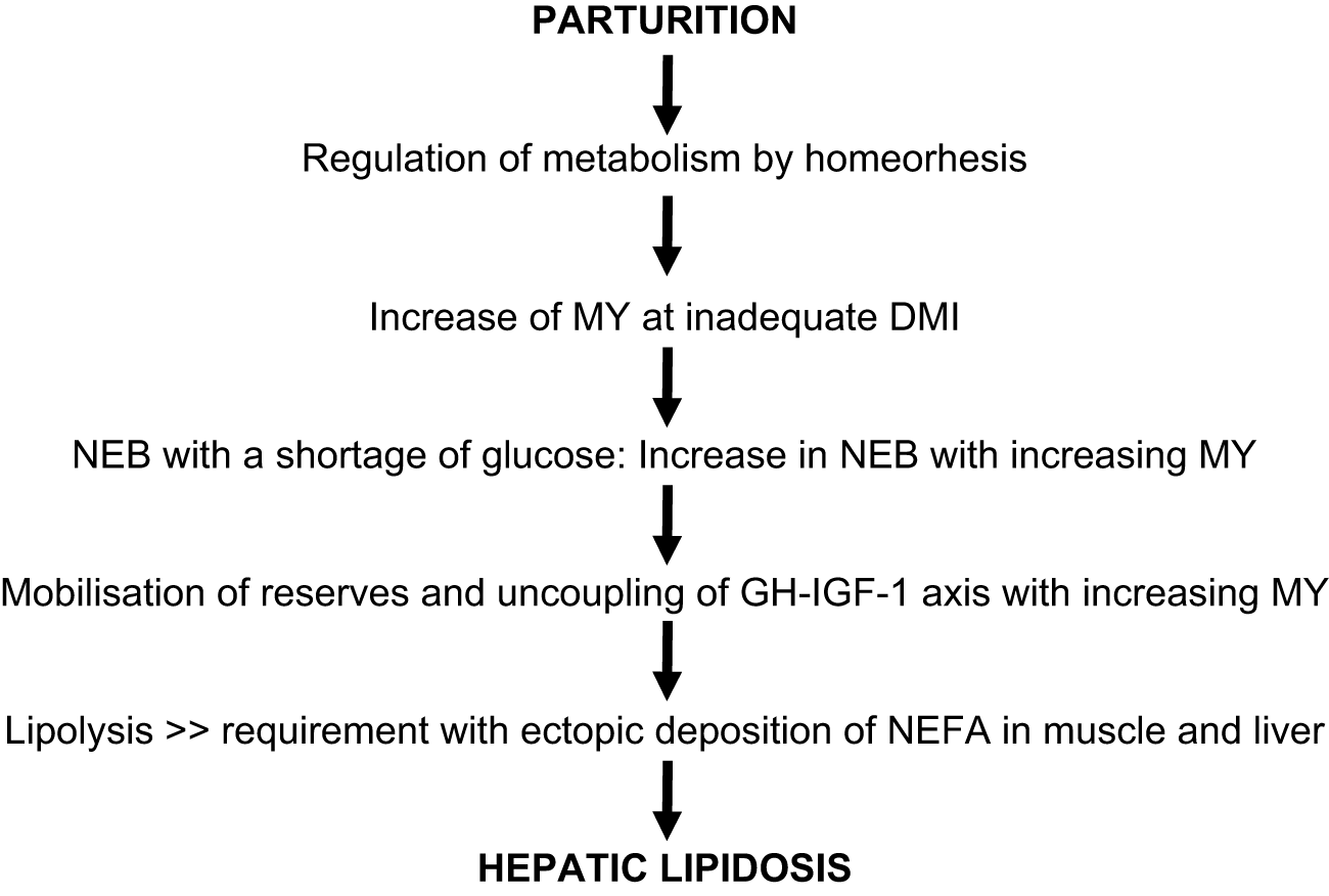
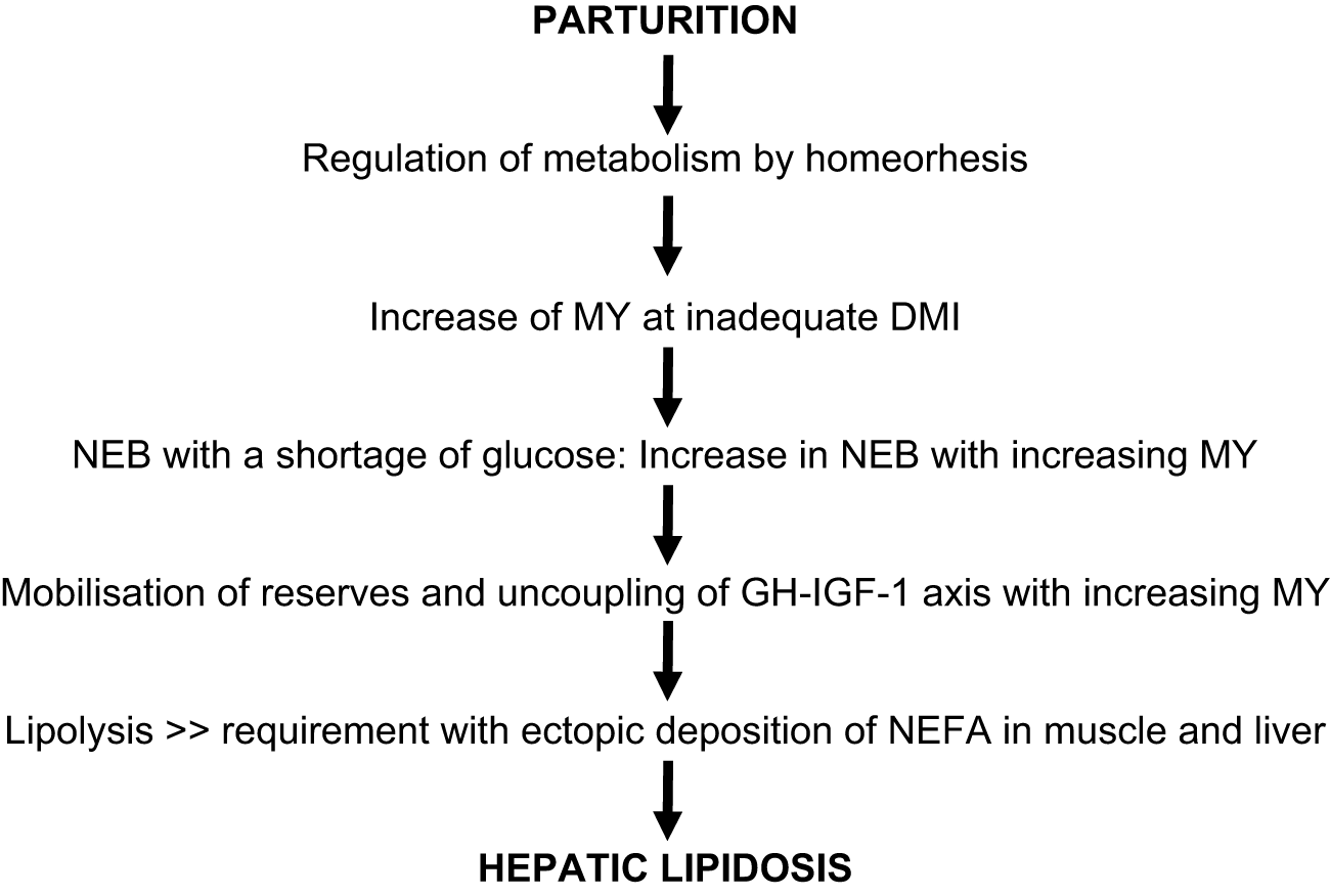
The dairy cow experiences undernutrition postpartum, with the result being a negative genetic correlation between MY and energy balance (Buttchereit et al., Reference Buttchereit, Stamer, Junge and Thaller2011) and, unsurprisingly, a genetic correlation between MY and hyperketonaemia (Simianer et al., Reference Simianer, Solbu and Schaeffer1991; Uribe et al., Reference Uribe, Kennedy, Martin and Kelton1995). The author is unaware of any studies specifically looking at NEB in beef cattle in early lactation, which is probably short and very limited in extent, but nowadays the NEB in dairy cows varies from 3 or 4 weeks to 2 or 3 months (Coffey et al., Reference Coffey, Simm, Oldham, Hill and Brotherstone2004) or more than 100 d (Bulang et al., Reference Bulang, Kluth, Engelhard, Spilke and Rodehutscord2006), with a loss of 1000–2000 MJ NEL or even more (Coffey et al., Reference Coffey, Simm, Oldham, Hill and Brotherstone2004; Sutter and Beever, Reference Sutter and Beever2000). The characterisation of the current NEB in high-producing cows as ‘chronic undernutrition’ (Vernon, Reference Vernon1998) has been well described. The association between MY and inadequate DMI permits the following sequence to be proposed:
- Increase in MY with inadequate DMI (genetic correlation)
- Increasing NEB with increasing MY and mobilisation of reserves
- Uncoupled GH-IGF-1 axis with increasing MY
- Increased GH and decreased IGF-1 with enhanced lipolysis and risks of inflammation
- Lipolysis above the requirement, metabolic capacity and increase in NEFA
- Ectopic deposition of NEFA in muscle and liver
- NEFA uptake by the liver and enhanced expression of CD36 at NEB
- Metabolism of NEFA and resynthesis of NEFA in the liver to TG and VLDL
- Limited export of VLDL and accumulation of TG: hepatic lipidosis
- Hepatic lipidosis with damage to structure and functions of the liver cells, i.e.:
o cell swelling, decrease in the number of mitochondria
o leakage of enzymes into the blood
o inhibition of gluconeogenesis and ureagenesis
o production of BHB
o reduced synthesis of apolipoproteins and VLDL export
o impairment of autophagy, apoptosis and necroptosis
o immunosuppression
o oxidative stress and inflammation
The sequence of this cascade begins with milk production postpartum, but it is very likely that the pathogenesis of hepatic lipidosis can be traced back in many cases to the prepartum period with high BCS and reduced DMI. Unfortunately, no information can be given about the length of time between the beginning of the NEB and overt hyperketonaemia (≥1.2 mmol/l BHB) and, finally, inflammation. Starvation-induced hyperketonaemia at d 5–14 postpartum by 50% feed restriction shows little evidence for the involvement of inflammation (Drackley et al., Reference Drackley, Dann, Bertoni and Trevisi2025), which supports the assumption that antepartum alterations perhaps activate the postpartum outcome. The sequence of postpartum alterations is summarised in Figure 1.
Schematic and putative diagram of the pathophysiology of metabolism in a high-producing cow postpartum.
Relief of NEB
The sequence of MY and insufficient DMI, NEB and metabolic changes can be interrupted or at least mitigated. Goff et al. (Reference Goff, Kimura and Horst2002) observed no hypocalcaemia or hyperketonaemia in mastectomised cows postpartum. Lacasse et al. (Reference Lacasse, Vanacker, Ollier and Ster2018) have summarised various methods to reduce the sudden metabolic burden postpartum and recommend one milking frequency per day for 5–6 d postpartum on the basis of their observation of an increase in glucose and a decrease in NEFA and BHB concentration. The usual prophylaxis of hyperketonaemia with propylene glycol is a supplement for glucose production and increases the supply of the key metabolite glucose. Similar results are obtained by feeding rumen-protected glucose (RPG). RPG ‘supplementation downregulates metabolites of tissue mobilization and inflammatory response’ (Zhang et al., Reference Zhang, Wang, Tang and Xiong2025) and its effects agree with the data from a previous study by Li et al. (Reference Li, Tan, Jiao, Long, Zhou, Yi, Liu, Kang, Wang, Duan, Tang, He and Han2019). RPG reduces IF in transition cows. It is worth mentioning that in human medicine, ‘intravenous administration of glucose induces removal of TAG (TG) from the liver and lessens lethal effects of endotoxaemia’ (Hinshaw et al., Reference Hinshaw, Peyton, Archer, Black, Coalson and Greenfield1974). Obviously, an increase in glucose (or its precursor) intake interrupts the cascade between NEB and the pathophysiological consequences and, vice versa, clearly shows the association between these parameters under the condition of a shortage of glucose.
Evidence for systemic inflammation
Oxidative stress and inflammation are not restricted to the liver. Cows with overwhelming and protracted lipolysis proceed to these alterations in adipose tissue (Zachut and Contreras, Reference Zachut and Contreras2022; Zhao et al., Reference Zhao, Li, Zhang, Chang, Wang, Zhao, Yang, Usman, Loor and Xu2025) and in their mammary glands too (Yue et al., Reference Yue, Pu, Loor, Jiang, Dong, Shen, Li, Gao, Lei, Du, Song, Liu and Li2022b). In bovine adipocytes, GH-induced lipolysis and inflammatory response, with upregulated mRNA of TNF-α and IL-1B, have also been observed (Gao et al., Reference Gao, Yu, Shi, Song, Fang, Gao, Lei, Song, Li, Du and Liu2026), supporting the assumption of a generally low-level chronic inflammation.
Hepatic lipidosis and a vicious circle
The sequence mentioned above tends to exacerbate and accelerate the pathogenesis of hepatic lipidosis, thereby generating a vicious circle:
- Apolipoprotein B in the liver of ketotic cows is lower and very likely contributes to a reduced VLDL export and accumulation of TG, enhancing the risk of hepatic lipidosis (Zhou et al., Reference Zhou, Chen, Meng, Ma, Xie, Shen, Li and Chang2023).
- Vice versa, hepatic lipidosis reduces VLDL export (Katoh, Reference Katoh2002).
- Pro-inflammatory cytokines have been suggested as mediators of reduced DMI in the hypothalamus (Kuhla, Reference Kuhla2020).
- BHB infusion in sheep (Schlumbohm and Harmeyer, Reference Schlumbohm and Harmeyer2004) and cows (Zarrin et al., Reference Zarrin, Matteis, Vernay, Wellnitz, van Dorland and Bruckmaier2013) reduces blood glucose concentration; BHB inhibits glucose production in sheep hepatocytes in vitro (Demigné et al., Reference Demigné, Yacoub, Rémésy and Fafournoux1986).
- Severe hepatic lipidosis in dairy cows (>10% TG at 3–9 d postpartum) impairs autophagy and ‘may lead to cellular damage and inflammation’ (Du et al., Reference Du, Liu, Loor, Fang, Bucktrout, Yang, Ye, Shi, Shen, Wang, Peng, Zhao, Bin, Xing, Zhu, Li and Li2018a), and cows with hyperketonaemia (>1.2 mmol/l BHB) exhibit an activated autophagy pathway in adipose tissue with enhanced lipolysis (Yue et al., Reference Yue, Pu, Loor, Jiang, Dong, Shen, Li, Gao, Lei, Du, Song, Liu and Li2022b).
- Ketotic cows display severe hepatic apoptosis and, in addition, oxidative stress and inflammation (Du et al., Reference Du, Liu, Loor, Fang, Bucktrout, Yang, Ye, Shi, Shen, Wang, Peng, Zhao, Bin, Xing, Zhu, Li and Li2018a).
- Dairy cows with clinical hyperketonaemia (> 3 mmol/l BHB at 3–9 d postpartum) exhibit increased levels of necroptosis-related proteins in the liver, with lytic necrosis and inflammatory cells, and an increased abundance of the mRNA of inflammatory cytokines such as IL-6, TNF-α and IL-1B in the liver (Liu et al., Reference Liu, Gu, Zhao, Li, Kong, Du, Qi, Li, Gao, Du, Song, Liu, Gao, Li and Lei2025).
- The infusion of the cytokine TNF-α increases the TG content of the liver and decreases DMI at 7 d by 19%; the data at the molecular level are consistent with an inhibition of gluconeogenesis (Bradford et al., Reference Bradford, Mamedova, Minton, Drouillard and Johnson2009) and an exacerbation of GH-induced lipolysis (Gao et al., Reference Gao, Yu, Shi, Song, Fang, Gao, Lei, Song, Li, Du and Liu2026).
- An uncoupled GH-IGF-1 axis is associated with increased MY (Lucy, Reference Lucy2004), with enhanced lipolysis (Gao et al., Reference Gao, Yu, Shi, Song, Fang, Gao, Lei, Song, Li, Du and Liu2026) and inflammation (Fritzen and da Silva, Reference Fritzen and da Silva2025).
- NEFA or BHB reduce the expression of the GHR-1A mRNA in liver cells in vitro (vicious circle via uncoupling of the GH-IGF-1 axis) (Du et al., Reference Du, Zhu, Peng, Cui, Zhang, Shi, Guan, Sha, Shen, Yang, Li, Wang, Li and Liu2018b).
- Positive correlations exist between NEFA and IF and negative correlations between IF and glucose and insulin (Cincovic et al., Reference Cincovic, Stojanovic, Djokovic, Majkic, Staric, Petrovic and Kovacevic2025).
- This correlation agrees with recent findings in non-pregnant and ‘dry’ cows. Infusion of TG emulsions increases NEFA, BHB, and IF, alters cytokine profiles and leads to IR (Lacasse et al., Reference Lacasse, Beaudoin, Thibault and Ster2026).
- The oxidation capacity of fatty acids is decreased in the livers of ketotic cows (Ch et al., Reference Ch, Liu, Li, Xia, Zhang and Wang2010).
- Possible infections activate the immune system and increase demand for glucose (Kvidera et al., Reference Kvidera, Horst, Abuajamieh, Mayorga, Sanz Fernandez and Baumgard2017).
- BHB has a strong negative effect on the formation of an extracellular neutrophils trap (NET) and hence bactericidal activity, with NET formation being impaired in hyperketonaemic animals (Grinberg et al., Reference Grinberg, Elazar, Rosenshine and Shpigel2008), which increases the risk of infectious diseases. Furthermore, 2.5 and 5.0 mmol/l BHB impair reduced neutrophil responses against Staphylococcus aureus, including extracellular traps, neutrophil burst, phagocytosis and chemotaxis in vitro (Quiroga et al., Reference Quiroga, Burgos, Henríquez, Carretta, Alarcón, Mella, Guíñez and Morán2026), which could contribute to the association between hyperketonaemia and infectious diseases such as mastitis and metritis (Galvão et al., Reference Galvão, Flaminio, Brittin, Sper, Fraga, Caixeta, Ricci, Guard, Butler and Gilbert2010; Swartz et al., Reference Swartz, Bradford and Mamedova2021).
These changed parameters intensify the clinical signs of hepatic lipidosis.
Hepatic lipidosis and general health risks
Hepatic lipidosis is a health risk and associated with diseases such as displacement of the abomasum, metritis and mastitis (Bobe et al., Reference Bobe, Young and Beitz2004). These associations have since been confirmed by Arshad and Santos (Reference Arshad and Santos2022) (Table 1).
Association between liver triglyceride (TG = % of wet wt.), metabolites and diseases
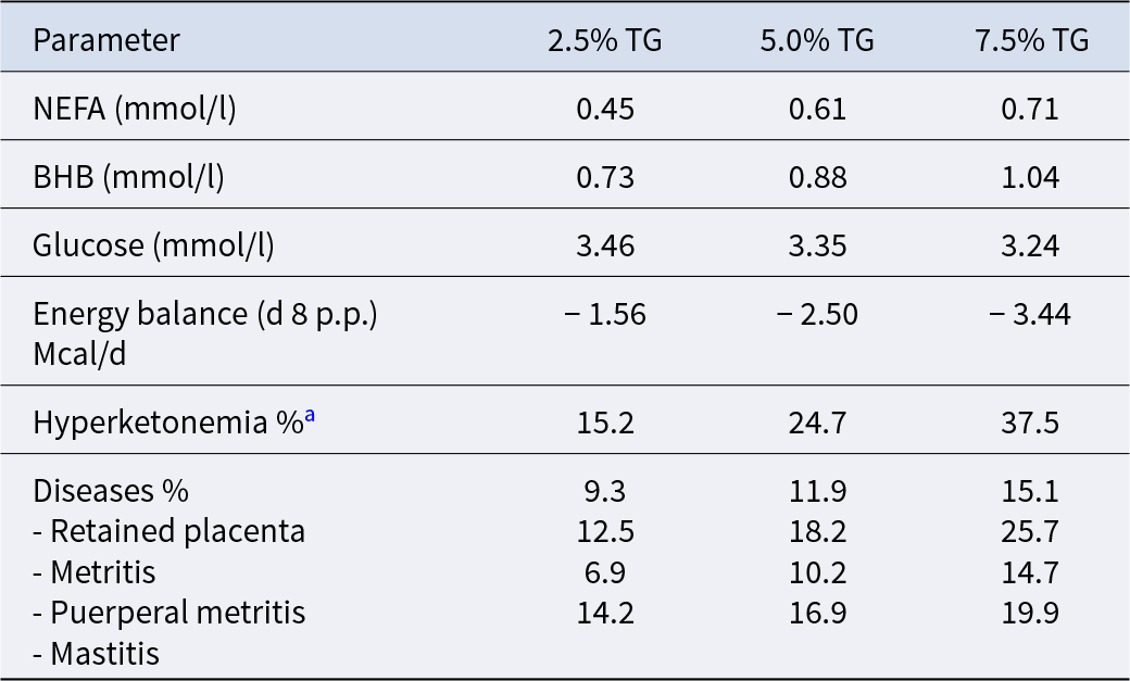
Table 1 Long description
Parameters are listed across three liver triglyceride levels, 2.5 percent TG, 5.0 percent TG, and 7.5 percent TG. NEFA in mmol per liter is 0.45, 0.61, and 0.71. BHB in mmol per liter is 0.73, 0.88, and 1.04. Glucose in mmol per liter is 3.46, 3.35, and 3.24. Energy balance at day 8 postpartum in Mcal per day is minus 1.56, minus 2.50, and minus 3.44. Hyperketonemia percent is 15.2, 24.7, and 37.5. Diseases percent lists four conditions: retained placenta is 9.3, 11.9, and 15.1; metritis is 12.5, 18.2, and 25.7; puerperal metritis is 6.9, 10.2, and 14.7; mastitis is 14.2, 16.9, and 19.9.
Source: Modified from Arshad and Santos (Reference Arshad and Santos2022).
a BHB > 1.2 mmol/l.
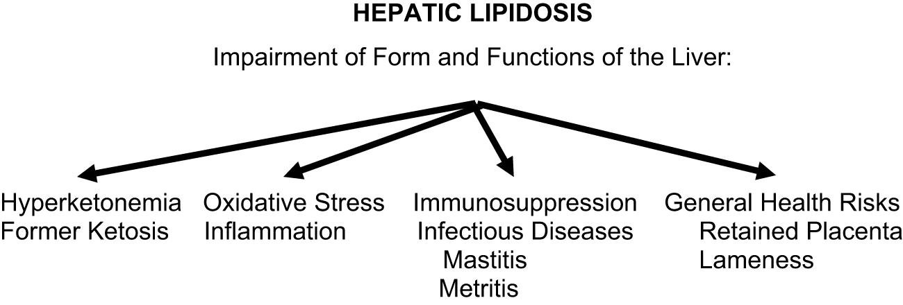
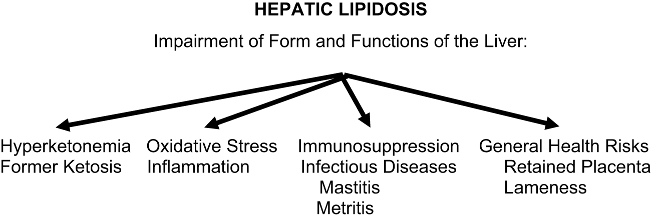
Hence, hepatic lipidosis is associated with so-called ‘production’ diseases. These health risks include hyperketonaemia, infectious diseases like metritis and mastitis and consequently inflammation. Hyperketonaemia appears to be a marker of a disease, as proposed by Mann and McArt (Reference Mann and McArt2023), and is probably not the cause of a disease, which is very likely hepatic lipidosis. Therefore, the suggestion here is that hepatic lipidosis should be considered as a major threat and evidence of a predisposition to diseases. The correlation with hyperketonaemia is quite evident, but the current knowledge permits a reliable relationship to be drawn with oxidative stress and inflammation, immunosuppression and infectious disease, as well as further health risks (Figure 2).
Schematic association between hepatic lipidosis and hyperketonaemia, oxidative stress, inflammation, immunosuppression and further health risks.
The drop in IgG at parturition (Herr et al., Reference Herr, Bostedt and Failing2011: Van Knegsel et al., Reference Van Knegsel, de Vries Reilingh, Meulenberg, van den Brand, Dijkstra, Kemp and Parmentier2007) and the impairments to both the adaptive (Wentink et al., Reference Wentink, Rutten, van den Ingh, Hoek, Müller and Wensing1997) and the innate (Zerbe et al., Reference Zerbe, Schneider, Leibold, Wensing, Kruip and Schuberth2000) immune system suggest that immunosuppression is occurring. This could be a reason for the high incidence of infectious diseases postpartum and for a transition into a systemic inflammation.
Apparent contradictions
The sequence between NEB and eventual hepatic lipidosis and associated health risks appears to conflict with recent findings. Ketotic cows (≥1.20 mmol/l BHB at enrolment) exhibit increased biomarkers of inflammation (haptoglobin, serum amyloid AS, cytokine IL-4, MIP-1α and TNFα) (Chirivi et al., Reference Chirivi, Cortes-Beltran, Munsterman, O'Connor and Contreras2023). These authors suggest ‘a link between CK (clinical ketosis) and systemic inflammation, indicating that CK may be a consequence of systemic inflammation rather than a stand-alone metabolic disorder solely derived from NEB’. The parallel observation of hyperketonaemia and IF fits together with the sequence of the pathophysiology mentioned above and the positive correlation between NEFA and IF (Cincovic et al., Reference Cincovic, Stojanovic, Djokovic, Majkic, Staric, Petrovic and Kovacevic2025). The observations of Chirivi et al. (Reference Chirivi, Cortes-Beltran, Munsterman, O'Connor and Contreras2023) are in agreement with previous studies that emphasise inflammation and, as a consequence, various diseases (Abuajamieh et al., Reference Abuajamieh, Kvidera, Fernandez, Nayeri, Upah, Nolan, Lei, DeFrain, Schoenberg, Trout and Baumgard2016; Ametaj, Reference Ametaj2005; Ametaj et al., Reference Ametaj, Bradford, Bobe, Nafikov, Lu, Young and Beitz2005; Horst et al., Reference Horst, Kvidera and Baumgard2021; Ma et al., Reference Ma, Kok, Burgers, Bruckmaier, Goselink, Gross, Kemp, Lam, Minuti, Saccenti, Trevisi, Vossebeld and van Knegsel2024; Mezzetti et al., Reference Mezzetti, Cattaneo, Passamonti, Lopreiato, Minuti and Trevisi2021; Trevisi et al., Reference Trevisi, Cattaneo, Piccioli-Cappelli, Mezzetti and Minuti2025). This reasonable assumption can be true, but the concomitant appearance of hyperketonaemia and inflammation could also be in line with the progression proposed above. However, a systemic inflammation due to cases of severe mastitis, metritis, lameness or subacute rumen acidosis cannot be excluded as possible causes for the pathogenesis of hepatic lipidosis, but it appears to be a secondary health risk because the cows are already sick and exhibit reduced DMI. For example, Goff et al. (Reference Goff, Kimura and Horst2002) observed initial displacement of the abomasum and then hyperketonemia – very likely due to the reduced DMI. However, a further discussion of this complex is beyond the scope of this review.
Future directions
Bottlenecks in production and health in early lactation are an outcome of the inadequacy of DMI. Indeed, this has been called the Gordian knot of dairy cow husbandry (Martens, Reference Martens2023). Consequently, improved DMI should be included in the breeding index. The NEB causes a lipolysis and an increase in NEFA postpartum above the requirement for energy production and milk fat synthesis. The ectopic deposition of NEFA is the key mechanism of hepatic lipidosis. The question is whether it is possible to select cows with an adequate mobilisation without or only a slight increase in NEFA?
Conclusion
Selection for a raised MY with frequent milking is not accompanied by an equivalent DMI postpartum, causes an uncoupled GH-IGF-1 axis and eliminates the negative feedback for GH release. The desired increase in MY with an expansion of the NEB postpartum for up to 2 or 3 months and with an unwanted and neglected increase in the magnitude up to 1000–2000 MJ is a human-made problem with a genetic background. The resulting ‘chronic undernutrition’ is compensated by a mobilisation of reserves, particularly NEFA, above the requirement and the cow's metabolic capacity, which results in an ectopic deposition in muscle and liver tissue and ultimately leads to a hepatic lipidosis. The impairment to the structure and functions of the liver cells in hepatic lipidosis is definitively correlated with hyperketonaemia, oxidative stress and inflammation. The impairment to both the adaptive and the innate immune system around parturition favours a low-level chronic and systemic inflammation, which is not restricted to the liver and includes the mammary gland and the adipose tissue and makes entry into a vicious circle more likely. The NEB with the sequences mentioned above is a side effect of the homeorhetic reaction of high-producing cows to prioritise milk synthesis at inadequate DMI which, unfortunately, at increased MY leads to the pathophysiological state known as hepatic lipidosis. This conclusion is supported by data from mastectomised cows with a relief of the metabolic stress and the mitigating effects provided by a supplementation of propylene glycol or RPG on the metabolic load and on inflammation. However, this cascade does not exclude inflammation initiated by primary infectious diseases such as metritis, mastitis, lameness or subacute rumen acidosis.
Acknowledgements
The complex regarding production and diseases has been intensively discussed over many years with colleagues F. Menn, A. Gruber and W. Brade. No AI technologies were used in the writing or editing or proofreading of this manuscript.
Declaration of interest
There are no conflicts of interest.
Data availability statement
Not applicable.
Ethics and approvals
Data from the literature were used. There was thus no need for a formal ethical approval to be obtained.
Funding statement
I am very grateful for the long-lasting support of the Margarete Markus Foundation.
List of abbreviations
- ALT
Alanine-amino-transferase
- AST
Aspartate-amino-transferase
- BCS
Body condition score
- BHB
β-Hydroxybutyric acid
- CD36
Cluster of Distinction 36 Fatty acid transporter
- CK
Clinical ketosis
- DAMP
Damage-associated molecular pattern
- DIM
Days in milk
- DMI
Dry matter intake
- GDF-15
Growth differentiation factor-15
- GGT
Gamma glutamyl-transferase
- GH
Growth hormone
- GLDH
Glutamate dehydrogenases
- GHR-A1
Growth hormone receptor A1
- IF
Inflammatory marker
- IGF-1
Insuli-like growth factor 1
- IgG
Immunoglobulin G
- IL-6
Interleukin 6
- IL-1B
Interleukin 1B
- IFN-γ
Interferon gamma
- IR
Insulin resistance
- MJ
Megajoule
- MY
Milk yield
- NEFA
Non-esterified fatty acids
- NEB
Negative energy balance
- NFκB
Nuclear factor-κB
- SCK
Subclinical ketosis
- TG
Triglyceride
- TLR-4
Toll-like receptor 4
- TNF-α
Tumour necrosis factor
- VLDL
Very-low-density lipoproteins


 Open access

