Introduction
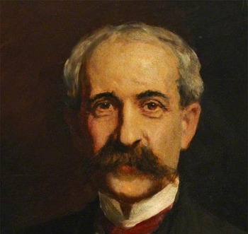
This historical article explores the contribution of the Semon Lectures as a backbone to the development of modern ENT. Named in honour of Sir Felix Semon (Figure 1), lectures have occurred annually (excluding the War years) since 1913. ENT has progressed to become an independent clinical specialty. Forty-two published articles describe the 44 lectures between 1913 and 1970. Forty were published in The Journal of Laryngology & Otology (formerly The Journal of Laryngology, Rhinology, and Otology). A new website (semonlectures.org) contains additional historical and biographical material. The corpus of these lectures provides a remarkable overview of current trends within British ENT between 1913 and 1970. The themes discussed include the development and professionalisation of ENT, research and practice, and the way practice has changed over time.
Sir Felix Semon (1849–1919).

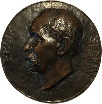
This published material is of much more than antiquarian interest. Several of the individual lectures give descriptions of clinical issues of lasting contemporary importance (e.g. Brown Kelly (1926), who described the Kelly–Paterson syndromeReference Kelly1). Through Semon's legacy, these lectures highlight key professional clues to the development of otolaryngology. Semon was a cornerstone of the modern profession, and many lecturers discuss his legacy. The Semon lecturers are an international collaboration, with 21 from England, 4 from Scotland, 13 from Europe and 6 from North America, between 1913 and 1970. Most lecturers chose as a subject the state of the art within their chosen area of expertise. All were male and each received, as a token of esteem, the Semon Medal (Figure 2).
The Semon Medal. Courtesy of Mr John Watkinson.

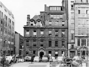
Sir Felix Semon was born in 1849 in Germany. He made significant progress within ENT and accumulated several famous private patients, including Queen Victoria and Winston Churchill. As a laryngologist, Semon contributed significantly through research and publications. Of note, he drew our eye to Semon's Law, thyroid surgery and the singing voice. Morell Mackenzie provided much of the inspiration for Semon becoming a laryngologist, establishing the first specialist ENT hospital in 1865, Golden Square Hospital (Figure 3), which was later incorporated into Gray's Inn Road and was where Semon worked.Reference Watkinson2
The ENT Hospital, Golden Square, London, circa 1950s. Courtesy of Mr Navnit Shah.

When Semon retired (circa 1911), the working class had to access healthcare either through voluntary hospitals funded by charities and philanthropists, or public hospitals funded via the 1834 Poor Law – principally for inmates of the workhouses.Reference Keay3 David Lloyd George introduced the National Insurance Act in 1911, which enabled workers to pay a set amount of money each week, and in return they could see a general practitioner and have hospital admission costs covered.Reference Williams4 This scheme was available only to the low-paid. Others had to pay and many struggled to afford the cost of care.
By the time the Ministry of Health was founded in 1919, medicine had progressed to such a level that hospital-based medicine became necessary for many. Payment for the voluntary hospitals was now means-based so nobody was denied care because of poverty. Payment for the upper and middle classes who did not qualify for the reduced rates of the working class was at a set rate, and many struggled to make ends meet.Reference Gosling5
Doctors frequently used the prestige they gained from working in voluntary and teaching hospitals to bolster their private practice. This meant patients were able to receive good care regardless of where they were seen, especially in large cities. Felix Semon split his time between private practice, and voluntary and specialist hospitals. His career was stunted at the beginning, being a German-speaking Jew with little English; later, as his language skills improved, he was able to enhance both his progression and reputation. Anti-Semitism became much more prominent after World War I and he mentions in his autobiography how ostracised he felt at the end of his life.Reference Stevenson6 Patients were well acquainted at this time with seeing specialists for a particular ailment, and conditions such as hoarseness were commonly seen by the specialist laryngologist. Although methods relating to disease diagnostics and treatment were available, the understanding of the pathology was not always accurate.Reference Stevenson6
Professionalisation of ENT
The Semon Lectures were established with the intention of developing ENT professionally, and this theme runs through all 44 orations. Semon himself was involved in determining the guest list for the initial lectures, and invited the Presidents of the Royal Societies and other medical societies. This was an attempt to counteract the low status of ENT in the late nineteenth and early twentieth centuries – an issue raised by many lecturers. McBride (1913) vividly portrayed the low status of laryngology circa 1880, with many London hospitals lacking departments, and the dogged reluctance of medical schools to add the subject to teaching curricula.Reference McBride7 Similar reflections were provided by Capps (1957), with laryngology portrayed as a ‘secondary responsibility’ of junior general physicians.Reference Capps8
The initiation of the lecture series followed the establishment of many major international ENT journals. The Annals of Otology, Rhinology and Laryngology was founded in 1892 and The Laryngoscope in 1896.Reference Birkett9 Congresses and societies were established first in America: the New York Laryngological Society was formed in 1873 by Clinton Wagner, and the American Laryngological Association in 1878 when several physicians (who identified themselves as laryngologists) felt this was necessary ‘for the advancement of the special department of surgery in which they were chiefly interested’.Reference Birkett9
England was slower to progress in this area, and it was a few years before ENT was properly recognised within medicine. Semon met the General Secretary of the International Medical Congress, Sir William MacCormac, and was able to secure a subsection for laryngology at a London meeting in 1881.Reference McBride7 What followed was the establishment of the Laryngological Society of London (1893), which brought together some of the best laryngologists in the country.Reference McBride7 McBride spoke favourably about Semon's achievements, stating: ‘This remarkably rapid rise to a position of influence becomes still more astonishing when it is remembered that at the time when it occurred laryngology was under a cloud, and that there was a tendency among the leaders of the profession in this country to think and also to speak disparagingly not only of the specialty but even by implication of those who practised it’.Reference McBride7
Another issue that plagued the professionalisation of ENT was the teaching of the specialty. The USA first formalised teaching in laryngology, both in Philadelphia by J Solis Cohen and at Harvard Medical School by H K Oliver in 1866, before it became more widespread.Reference Birkett9 It was 57 years later, in 1923, that the General Medical Council made laryngology and otology teaching compulsory for every medical student.Reference Turner10
Throughout the history of ENT, there have been heated debates over the boundaries of the specialty and which subspecialties it should encompass. Previously, otology, rhinology, laryngology and ophthalmology were all combined and practised by the same physician, especially in smaller hospitals (‘eye and ear’ hospitals), and as recently as 1887 ophthalmology was segregated to be its own specialty.Reference Birkett9 With the advent of bronchoscopy, the demarcations between the areas covered by thoracic surgeons, respiratory physicians and laryngologists became blurred. Before World War II, bronchoscopy was under the realms of laryngology. Mosher (1929) and Guisez (1924) spoke at length about how they looked with a bronchoscope to the distal oesophagus.Reference Mosher11,Reference Guisez12 After the War, the environment was different, and many of the bronchoscopy cases had been taken over by the thoracic surgeons, which many laryngologists felt was inappropriate given their lack of knowledge of the upper airways. Holinger (1960) provided the idea of joint working between the two specialties, in line with previous lecturers (McBride in 1913 and Huizinga in 1954).Reference Holinger13 Similarly, the borders within other specialties were explored, to include neurosurgery, general and endocrine surgery, as well as plastic surgery which specifically made advances in soft tissue reconstruction techniques, helping to treat injured soldiers, sailors, and pilots during the First and Second World Wars. Also around this time, Nager (1939) commented on the disagreement between the neurosurgeons and rhinologists over the best way to resect pituitary tumours, and whether to favour either a trans-frontal or trans-sphenoidal approach.Reference Nager14
After World War II, the impetus for social change that eventually culminated in the establishment of the National Health Service (NHS) by Aneurin Bevan in 1948 was years in the making. The Emergency Medical Service, which was set up in 1938, centralised the running of hospitals in the anticipation of a great number of casualties from World War II. It was this centralisation of services which is widely regarded as the starting point for the concept that would become the NHS.Reference Greengross, Grant and Collini15 In fact, people had been campaigning for a public medical service since the early 1900s. One of the major factors in gaining public support for this change was William Beveridge's report in 1941 on the co-ordination of social insurance and the role for a comprehensive national health service. Beveridge's idea that ‘The purpose of victory is to live in a better world than the old one’ resonated with the population amidst the ongoing war.Reference Delamothe16 When the NHS was set up, it comprised three main divisions: nationalised hospitals, general practitioners and community health services. These three divisions have remained to the current day, although the way they are managed has changed over the years.Reference Greengross, Grant and Collini15
ENT surgery as a combined specialty was established in the early twentieth century, when its components – otology, rhinology and laryngology – were amalgamated. Advancements in the areas of antibiotics, anaesthesia, and safely using radioactive material around this time led to drastic improvements and successes in the field of ENT. Since then, ENT has gone on to have significant overlaps with other specialties, such as neurosurgery, general surgery and plastic surgery.Reference Weir17 Indeed, operations like thyroid and parathyroid surgery are now performed primarily by ENT surgeons, rather than general and endocrine surgeons.Reference Smith, Watkinson and Shaha18 Similarly, there is a balance between plastic surgery and ENT and rhinoplasty surgery, although, nowadays, the further one goes away from the nose, the less the ENT surgeon is involved.Reference Weir17 Additionally, skull base surgery is usually carried out in conjunction with neurosurgeons.Reference Snyderman, Carrau and Kassam19
The training pathways have also changed over the years. Surgery was traditionally taught in the format of an apprenticeship, where surgeons would learn from a mentor over 12 to 14 years. Whereas, in recent years, the four surgical royal colleges have adopted a new training programme, whereby surgeons typically complete an eight-year training programme and relevant examinations before then becoming a consultant in their field.Reference Ribeiro20
The mastery of general surgery within ENT has proved indispensable. The first generation of laryngologists limited themselves to endolaryngeal methods, and surrendered every external operation to the general surgeon of the time. Only with some difficulty and considerable determination have the present and immediate past generation of otolaryngologists expanded to define specifically the boundaries over the last 50 years, establishing defined ENT surgical techniques. Battles have been won and lost when defining boundaries between general and endocrine surgery, ENT, maxillofacial surgery, and plastic surgery. Today, the faultless training in basic general surgery is a sine quâ non for the development of reliable laryngo-rhinologists, masters of their realm!
Research and practice
Advances were made in general anaesthesia, and following the discovery of antibiotics and radiotherapy. This meant that, by the 1960s, ENT was a specialty in its own right. Indeed, many of the operations described and instruments used at that time remain as a legacy to some of the previous Semon lecturers (Killian, Hayek, Mollison, Birkett and Negus to mention a few).
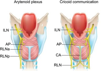
Throughout the lecture series, there are examples where continued research has led to a shift in accepted facts. One such example is ‘Semon's Law’, which states that if there is a lesion of the recurrent laryngeal nerve, then the abductor fibres will be affected before the adductor fibres. This Law was based on years of experimentation and research by Felix Semon. Although it had critics at the time, the Law was widely supported. Negus supported this Law, and in his 1930 lecture, he used comparative anatomy and physiology to show how the Law worked in practice and why it should be upheld.Reference Negus21 Just three years later, Burger gave his Semon Lecture disproving the Law, drawing results from several different researchers on the topic,Reference Burger22 followed up in 1957 by Capps, who also highlighted various issues with the Law.Reference Capps8 Recent publications have again addressed this issue and outlined what seems to be the definitive explanation of the anatomical innervation and nerve supply to the larynx (Figure 4).Reference Watkinson2 It looks likely that such advances side by side commenting on the physiology of the nervous innervation will follow.
The posterior relations of laryngeal anatomy, showing the anastomotic connections between the internal branch of the superior laryngeal nerve (ILN) above and the recurrent laryngeal nerve (RLN) below on the cricoid muscle (Galen's anastomosis). Also shown are the cross-anastomotic cricoid connections (CA) and the arytenoid plexus (AP). The letters (a) and (p) refer to the anterior and posterior branches, respectively. Courtesy of Elsevier.

Trends in practice
Throughout the decades, it is apparent that the Semon Lectures have changed ENT practice. There are some themes which stand out, in particular the periodic changes with regard to radical versus conservative treatment. Examples include the way primary disease (laryngeal, oropharyngeal and thyroid), together with skin malignancies and cervical metastases, have been managed, and the significant advances made by Butlin, Hayes Martin, Suarez, Bocca and Crile, together with Shah (2006), O'Brien (2008), Shaha (2013) and Watkinson (2019).Reference Watkinson2,Reference Lectures23–Reference Lectures25
Laryngeal cancer treatment was discussed in the lecture series (in 1927) when Colledge spoke about how laryngectomy could be a very effective treatment for laryngeal cancer. He did, however, warn of the importance of planning which operation was going to be attempted before the operation started, saying ‘This decision should be made before any operation was begun and not left to an exploratory investigation … If any doubt existed it would generally be found that laryngectomy was required’.Reference Colledge26 Colledge did concede that ‘no larynx should be sacrificed unnecessarily’,Reference Colledge26 but maintained his view that the appropriate surgery should be conducted to minimise the chance of disease recurrence. At this point, radiotherapy was a new treatment, and one which Colledge thought was disappointing and unsatisfactory.Reference Colledge26
A few years later, Harmer gave his Semon Lecture on the value of radiotherapy in treating laryngeal cancers. He spoke about the importance of having an expert team managing the radiotherapy treatment, with radiologists, surgeons, physicists and pathologists all working to ensure that the treatment was administered appropriately. The results he obtained in his clinic, he said, were just as good as those from surgery, and he spoke of a future where radiotherapy would be conducted in conjunction with surgery to achieve the best outcomes.Reference Harmer27
When Ormerod lectured in 1953, he spent time examining the changes in laryngeal cancer treatment, and highlighting the improvements made in other areas, such as with antibiotics and radiation. He then further classified the cancers by their anatomy to make the decision of whether surgery or radiotherapy were more appropriate, as well as additional features, such as the type of cancer. He also felt that partial laryngectomies were often unsuccessful, and it was necessary to be more radical with treatment.Reference Ormerod28
In 1968, Lederman reminisced on the issues addressed up to this point when deciding between surgery and radiotherapy, or a mixture, saying: ‘In the same way that the early laryngeal surgical disasters were overcome, so the early disasters of radiation therapy have by acting as an incentive to further endeavour resulted in the development of techniques that are today successful and reasonably free of undesirable complications’.Reference Lederman29 He played a large role in advocating radiotherapy in this instance, claiming that ‘Early laryngeal cancer can be equally successfully treated both by surgery or radiotherapy’.Reference Lederman29 Two years later, Som once again argued the case for the role of conservative surgery, and spoke of the risks of radiotherapy to the patient. He spoke at length on the characteristics of various cancers that made them more appropriate for conservative rather than radical management. He aligned his views on management with those held by Semon back in 1880, who advocated for partial laryngectomy at a time when total laryngectomy was the mainstay of treatment.Reference Som30
The tonsils have been discussed throughout the lecture series in a similar manner. In 1932, Kahler spoke of the changing trends during the previous fifty years over the necessity for tonsils, and what was felt to be the role of the tonsils, saying, ‘There is scarcely another problem that has been written and spoken about as much as the tonsil problem’.Reference Kahler31 The differing views over whether to remove the tonsils in the hope of avoiding infections or keep them to protect against infection, which he highlighted then, were like the issues raised by Wright in 1949. In his lecture, he equally spoke of the issue that so much time was spent removing the tonsils and deciding whether this was appropriate, when it often seemed to make little difference to patients.Reference Wright32
Over the last five decades, British otolaryngology has seen a significant expansion in its profile, together with committed diagnostic and surgical therapeutic armamentariums. There would be a further 51 Semon Lectures, including 5 given by women, of whom one was a speech therapist (Perry, 1995) and another an anatomist (Standring, 2014). Also included was one father and son (Hollinger, 1960 and 2003), as well as a convicted criminal (Wilson, 1964)!
The specialty has expanded, and has subspecialist fields that include: otology and skull base with implantation, rhinology and skull base and facial plastics, together with head and neck and endocrine surgery. The specialty embraces a formal training programme, with recognised exit exams, a commitment to multidisciplinary team working, adherence to guidelines and a formal approach to research in all fields. Significant advances have been made in the field of cochlear implantation, rhinology and skull base surgery, along with robotic transoral head and neck surgery. One hundred years on, Semon can be proud of his legacy and the subsequent contribution all the lecturers have made to British otolaryngology.
Conclusion
The Semon Lecture series has been fundamental both in establishing ENT as a respected specialty within the medical community and providing a platform through which the history of the specialty can be explored. By allowing lecturers free rein in choosing their topic, it becomes clear how trends and research develop over time and the effects that has over the specialty. Through collating this information and providing a dedicated website (semonlectures.org), it is hoped that the constantly evolving landscape of ENT can be further mapped throughout time for all to enjoy.
Acknowledgements
The authors would like to thank: Brendan Clarke (Department of Science and Technology Studies, University College London), the Get-A-Head Charitable Trust, University College London Department of Science and Technology Studies, and the Royal Society of Medicine Sections of Laryngology and Otology.
Competing interests
None declared