Neonates with aortic coarctation can still present in critical clinical condition, generally due to late diagnosis and to the presence of associated anomalies.Reference Tani, Minich and Hawkins1, Reference Quaegebeur, Jonas, Weinberg, Blackstone and Kirklin2 It has been shown that surgery carries a high risk when performed in such neonates with ventricular dysfunction, and that death can occur before any surgery is attempted.Reference Quaegebeur, Jonas, Weinberg, Blackstone and Kirklin2, Reference Fiore, Fischer and Schwartz3 Percutaneous dilation of the coarcted segment is rarely attempted in neonates because of the high rate of restenosis and complications. These are directly related to the small arterial access, leading to femoral injury, and to the fragility of ductal tissue, prompting formation of aneurysms.Reference Patel, Madani, Paris, Warner and Hijazi4–Reference Rao, Chopra, Koscik, Smith and Wilson6 Although surgery is generally preferred in most centres, even in the presence of left ventricular dysfunction,Reference Sudarshan, Cochrane, Jun, Soto and Brizard7 to the best of our knowledge there is only one report describing a favourable outcome in neonates with isolated coarctation and severe systemic hypoperfusion.Reference Fesseha, Eidem and Dibardino8
Our aim, therefore, was to evaluate our results in critically sick neonates with aortic coarctation not responding to medical management, referred to us with multiorgan failure, after the introduction of emergency aortic balloon dilation as a bridge to surgery, comparing the findings with those achieved in a comparable period prior to the introduction of balloon dilation.
Population and methods
We studied neonates referred over a period of 6 years with aortic coarctation and left ventricular dysfunction, with or without multiorgan failure. Criterions for exclusion were infants older than 1 month, those with associated malformations apart from patency of the arterial duct, ventricular septal defect, a non-stenotic parachute mitral valve, and a non-stenotic bifoliate aortic valve. We also excluded patients with left ventricular hypoplasia, defined in accordance with the criterions established by Rhodes and colleagues.Reference Rhodes, Colan, Perry, Jonas and Sanders9
During the period of the study, our departmental policy required different plans for treatment. During the first half of the period, from January, 2001 to December, 2003, all patients were medically treated and referred to surgery after stabilization of their clinical and haemodynamic condition. Medical treatment consisted of mechanical ventilation, infusion of prostaglandin1 at a dose of 0.1 microgram/kilogram/minute, and infusion of dobutamine at a dose of 10 to 15 microgram/kilogram/minute. Patients with ventricular dysfunction and multiorgan failure were not referred for surgery. During the second half of the period, from January, 2004, through January, 2007, patients with ventricular dysfunction and persisting multiorgan failure 24 hours after introduction of medical treatment as described above were referred for balloon dilation. Only patients in a stable clinical condition were then referred for subsequent surgery.
We identified patients with left ventricular dysfunction and multiorgan failure on the basis of:
• Clinical signs of systemic hypoperfusion, such as anuria, hepatomegaly, absence of peripheral pulses, or reduced radial pulses.
• Laboratory signs of systemic hypoperfusion and disseminated intravascular coagulation, such as pH less than 7, levels of lactates in the blood higher than 5 mmol/L, levels of alanine aminotransferase in the blood higher than 30 U/L, levels of glutamine aminotransferase in the blood higher than 30 U/L, levels of γ glutamiltransferase in the blood higher than 40 U/L, levels of urea nitrogen in the blood higher than 8 mmol/L, levels of creatinine in the blood higher than 90 μmol/L, prolongation of the prothrombin time to greater than 200%, decreased levels of platelets to less than 509/litre, decreased levels of fibrinogen in the blood to less than 4 g/L, decreased levels of factor V in the blood by less than 70%, and increased levels of fibrin split products to greater than 0.5 microgram/millilitre.
• Echocardiographic signs of depressed left ventricular function, such as left ventricular fractional shortening of less than 20% associated with moderate-to-severe mitral regurgitation.
The study represents a retrospective appraisal of our departmental policy. It was approved by the institutional review committee. Parents of all children gave informed consent. Subsequent to 2004, all parents accepted emergency interventional catheterization.
We reviewed the hospital records, catheterization, surgical files, and documents concerning the last follow-up to identify clinical and laboratory characteristics at presentation, associated malformations, complications occurring during and after aortic balloon dilation, and/or surgery. We recorded all complications occurred during medical treatment, balloon dilation and follow-up, seeking possible predictors of outcome. All children had clinical, echographic and haemodynamic evaluation at Necker Enfants Malades. Preoperative evaluation consisted of electrocardiogams, transthoracic echocardiograms, and blood tests.
Transthoracic echocardiogram. The mitral annulus was measured in the long axial view, and horizontal diameters of the ascending aorta were measured in the suprasternal view. Fractional shortening was calculated, and associated anomalies and the presence of mitral regurgitation, if present, were described. Systolic pulmonary pressure was estimated on the basis of direction and velocity of the flow through the patent arterial duct, or by evaluating the velocity of the regurgitant jet across the tricuspid valve.
Aortic balloon dilation. The technique of dilation of aortic coarctation remained unchanged during the period of study,Reference Rao, Chopra, Koscik, Smith and Wilson6 and was performed via the femoral artery by using a 3-French sheath. The peak-to-peak gradient was recorded on pullback, and an aortogram was obtained in lateral and antero-posterior views. The balloon diameter was chosen in accordance with the size of horizontal aorta, and was not larger than 2.5 times the diameter of the coarcted segment. Assessment of hypoplasia of the aortic arch was performed by using the ratio of the diameter of the distal arch to that of the ascending aorta, as previously described.Reference Fiore, Fischer and Schwartz3 Haemodynamic measurements and angiography were repeated at the end of the procedure. Success was judged on the appearance of femoral pulses, and the decrease of the peak-to-peak gradient.
Surgical treatment. The coarcted segment was resected, and extended end-to-end anastomosis achieved in all patients,Reference Sudarshan, Cochrane, Jun, Soto and Brizard7 banding the pulmonary trunk in presence of a non-restrictive ventricular septal defect. After 2004, banding was performed using resorbable material in neonates in whom the ventricular septal defect was deemed to have the potential for spontaneous closure.Reference Bonnet, Patkai, Tamisier, Kachaner, Vouhe and Sidi10 The procedure was performed through a left posterolateral thoracotomy in the fourth intercostal space, paying attention to resect all ductal tissue, and extending the incision into the undersurface of the aortic arch as proximal as possible having resected the coarcted segment. In patients undergoing surgery after balloon dilation, the dilated area was inspected in order to identify intimal tears not evident at angiography.
Statistical analysis. We obtained descriptive statistics for the total population. Medians and ranges were described as continuous variables, with categorical variables summarized as frequencies.
Independent risk factors for morbidity and mortality were sought using stepwise multiple logistic regression analysis performed using SAS Institute Software (Version 8.02, Cary, North Carolina). Factors that were tested included the age and weight of the patients at diagnosis, their date of birth, any associated ventricular septal defect, multiorgan failure, and the percentage of left ventricular fractional shortening. A p value less than 0.10 was used for selection for entry into the model, with a p value less than 0.05 being necessary for retention. Comparison for individual parameters before and after balloon dilation was performed by using the two-tailed paired t test. A p value of less than 0.05 was considered statistically significant.
Results
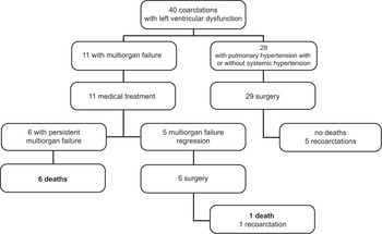
The First Half of the Period of Study (Figure 1)
The clinical characteristics of the patients are shown in Table 1. Those with ventricular dysfunction and multiorgan failure, having a median age of 15 days, with a range from 3 to 30 day, were significantly older than patients with ventricular dysfunction without multiorgan failure, their median age being 12 days, and with a range from 2 to 22 days (p = 0.02). At the time of diagnosis, 23 patients, accounting for 79% of those with left ventricular dysfunction but without multiorgan failure, had a small persistently patent arterial duct. The duct had closed in all the patients with ventricular dysfunction and multiorgan failure. Multiorgan failure was reversed under medical treatment in 5 patients (45%). The remaining 6 patients, who did not respond to medical management, all died.
Flow chart illustrating the clinical characteristics and treatment in patients born during the period from January, 2001, through December, 2003.

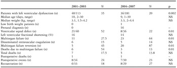
Comparative data.

Surgery, performed in 34 patients, after a median period of 6 days, and with a range from 1 to 20 days from diagnosis, included concomitant banding of the pulmonary trunk in 8, and banding using resorbable material in 3. Of these patients, 2 had a ventricular septal defect closed using extracorporeal circulation. There was 1 postoperative death due to congestive heart failure in one of the patients with a ventricular septal defect.
The mean duration of aortic clamping was 26.5 plus or minus 8.4 minutes, and the mean duration of ventilation was 2.5 plus or minus 1.3 days. We documented 8 major adverse events in the postoperative period, specifically 1 death, 1 instance of multiorgan failure, 3 instances of congestive cardiac failure, and individual episodes of severe pulmonary hypertension, sepsis, and supraventricular arrhythmia.
During the period of follow-up, 5 patients required reoperations, for closure of the ventricular septal defect in 4, and removal of the pulmonary arterial band in the other. Percutaneous dilation of recoarctation was required 2 to 30 months after surgery in 6 patients (17%). There were no late deaths.
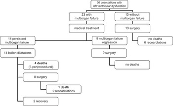
The Second Half of the Period of Study (Figure 2)
The clinical characteristics of the patients are again shown in Table 1. Patients with ventricular dysfunction and multiorgan failure, having a median age of 13 days, and a range from 1 to 30 days, were again significantly older than patients with ventricular dysfunction without multiorgan failure, whose median age was 10 days, with a range from 1 to 21 days (p = 0.03). At time of diagnosis, 28 patients (78%) with left ventricular dysfunction without multiorgan failure had a small persistently patent arterial duct, the duct being closed in all those with ventricular dysfunction and multiorgan failure. Medical treatment reversed multiorgan failure in 9 patients. Surgery was undertaken in 22 patients without multiorgan failure, or in whom multiorgan failure had regressed after medical treatment. The remaining 14 were referred for balloon dilation.
Flow chart illustrating clinical characteristics and treatment in patients born during the period from January, 2004, through January, 2007.

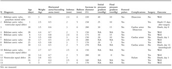
The clinical characteristics and haemodynamic data for those undergoing balloon dilation are shown in Table 2. In one patient with a ventricular septal defect, who developed severe ventricular dysfunction and multiorgan failure during the first hours of life, possibly due to prenatal closure of the arterial duct, balloon dilation was performed at 24 hours of life.
Clinical, angiographic, and haemodynamic parameters of patients referred to balloon angioplasty.

NA: not measured.
Median procedural and fluoroscopy times were, respectively, 30 minutes with a range from 25 to 90 minutes, and 10 minutes with a range from 4 to 25 minutes. It proved possible to dilate the aortic isthmus in all but 1 neonate. In this patient, the procedural time was 90 minutes, it proving impossible to obtain vascular access. He died in the periprocedural period. The diameter of the coarcted site increased from 2.1 ± 0.4 to 3.8 ± 0.6 millimetres (p less than 0.001). The peak-to-peak gradient decreased from 29.4 ± 5.8 to 8.8 ± 4.4 mmHg (p less than 0.001). Balloon dilation reopened the arterial duct in 6 patients. Fractional shortening, evaluated 24 hours after angioplasty, increased from 14.2 ± 4.3 to 31 ± 12.9% (p less than 0.001). There were 5 early complications, and 3 periprocedural deaths, giving 21% early mortality. Multiorgan failure regressed in 11 patients.
In 1 patient, who had recovered a normal left ventricular function after balloon dilation, and in whom systemic hypertension had regressed, death was related to a subdural haemorrhage, confirmed at echography, occurring 14 days after balloon dilation. We speculate that this death was due to a late complication of the cardiac arrest that had occurred during balloon dilation.
It proved possible to proceed to elective surgical intervention in 8 patients at a median time of 10 days following balloon dilation. Concomitant closure of a ventricular septal defect was undertaken in one of these patients. Surgery in these patients was performed to relieve early recoarctation in 5, and because of persistence of the clinical gradient between the arms and legs in the other 3. Thus, balloon angioplasty proved incomplete, or else permitted early recoarctation, in 73% of the patients.
Intimal tears were found at surgical inspection in 3 patients. The median time of aortic clamping was 12 minutes, with a range from zero to 20 minutes. The median duration of mechanical ventilation was 1 day, with a range from zero to 6 days. We detected transient systemic hypertension in 3 patients. Death occurred in 1 patient 15 days after surgical repair of the coarctation due to persistent severe pulmonary hypertension.
In the patients undergoing surgery as a first treatment, the mean time from diagnosis to surgery was 8 days, with a range from 2 to 24 days. In 3 patients with ventricular septal defects, we performed classic banding in 1, and banding using absorbable material in the other 2. The median time of clamping was 14 minutes, with a range from zero to 25 minutes, and the postoperative median duration of mechanical ventilation was 1.5 days, with a range from zero to 17 days. There were 7 major adverse events, with 1 patient dying, 2 suffering transient postoperative pulmonary hypertensions, individual episodes of transient systemic hypertension and transient hepatic failure, and 2 instances of convulsions, albeit without sequels, after regression of the multiorgan failure.
During the period of follow-up, reoperation was needed in 3 patients to close ventricular septal defects. An additional 2 patients with localized coarctation and good morphologic and haemodynamic results have yet to undergo surgery, at 24 and 10 months, respectively, following the balloon angioplasty. In 8 patients (26%), percutaneous dilation of recoarctation was necessary 3 to 36 months after surgery. There have been no late deaths
Comparative results. This data is illustrated in Table 1. The patients in the 2 groups had similar ages, weights, and left ventricular fractional shortening. A larger number of patients was referred after 2004. The percentage of those with ventricular septal defect was lower after 2004, as well as those with ventricular dysfunction. Ventricular dysfunction associated with multiorgan failure was more frequent after 2004. Global mortality diminished after 2004, but not significantly so. The number of deaths due to ventricular dysfunction associated with multiorgan failure, however, diminished significantly, while reversion of multiorgan failure significantly increased during the second period of the study. The number of major postoperative events, of postoperative deaths, and the incidence of recoarctation, all did not change.
Multivariate analysis of the variance showed that independent risk factors for mortality were date of birth, age at diagnosis, ventricular dysfunction associated with multiorgan failure, and presence of a ventricular septal defect.
Discussion
To the best of our knowledge, ours is the first report illustrating the impact of percutaneous treatment of aortic coarctation in a series of consecutive neonates with ventricular dysfunction and multiorgan failure, persisting despite medical treatment. We have shown that:
• emergency balloon angioplasty reverses systemic hypoperfusion in the majority of patients, and significantly diminishes the mortality of multiorgan failure.
• balloon dilation in critically ill patients is still burdened by high mortality, morbidity, and the need for reintervention.
• surgery has a very low risk when performed in patients in stable clinical condition.
Aortic coarctation can still present with catastrophic clinical condition. In accordance with the findings of other authors, we have confirmed that the date of birth, age at diagnosis, and the presence of a ventricular septal defect all constitute risk factors for multiorgan failure and death.Reference Quaegebeur, Jonas, Weinberg, Blackstone and Kirklin2 Indeed, prenatal diagnosis is performed in a minority of patients,Reference Head, Jowett, Sharland and Simpson11 and delivery in tertiary referral centres is rarely advised. We can hypothesize that, in our series, the increased referral, and the diminished incidence of left ventricular dysfunction, observed during the second period of the study was due to the increasing use of prenatal echocardiography, as previously reported.Reference Franklin, Burch, Manning, Sleeman, Gould and Archer12 Patients presenting with multiorgan failure generally had had a late diagnosis, and were referred from other centres.
Percutaneous dilation of discrete coarctation in neonates and infants in stable haemodynamic condition has previously been reported.Reference Fiore, Fischer and Schwartz3–Reference Rao, Chopra, Koscik, Smith and Wilson6 In these series, morbidity and the rates of complications were lower with transcatheter compared to surgical therapy,Reference Rao, Chopra, Koscik, Smith and Wilson6 but intimal tears were a very frequent event, and restenosis occurred in half the patients.Reference Park, Lucas, Sklansky, Kashani and Rothman5 Indeed, the utility of balloon angioplasty for the initial treatment of aortic coarctation in neonates and infants remains controversial. Concerns include early restenosis, formation of aneurysms, and the potential for early and late ischaemic injury of the legs.Reference Fiore, Fischer and Schwartz3, Reference Park, Lucas, Sklansky, Kashani and Rothman5, Reference Galal, Schmaltz, Joufan, Benson, Samatou and Halees13, Reference Redington, Booth, Shore and Rigby14 Only a minority of patients do not require further reinterventions, such as surgery or redilation of recoarctation.
In accordance with previous reports,Reference Fiore, Fischer and Schwartz3, Reference Park, Lucas, Sklansky, Kashani and Rothman5, Reference Rao, Chopra, Koscik, Smith and Wilson6 we confirmed a high rate of restenosis in our patients, probably related to elastic recoil of ductal tissue surrounding the site of coarctation. Although hypoplasia of the aortic isthmus proximal to the coarcted segment is considered a relative contraindication to balloon angioplasty in neonates, due to impairment of flow into the dilated area,Reference Fiore, Fischer and Schwartz3 we did not observe any differences in outcome in patients with or without hypoplasia of the horizontal aorta.
Trauma of the femoral vessels is a complication reported by most authors.Reference Fiore, Fischer and Schwartz3–Reference Park, Lucas, Sklansky, Kashani and Rothman5, Reference Redington, Booth, Shore and Rigby14 We did not experience any acute femoral arterial trauma, since we used 3-French sheaths, as previously described.Reference Agnoletti, Boudjemline and Largen15 Late occlusion, however, remains an additional concern, potentially causing impairment of growth of the leg on the side of arterial access. An antegrade venous approach, or an axillary or carotid approach, could possibly be used in infants with cardiogenic shock.
The duration of cross-clamping is a critical factor in the presence of depressed left ventricular function. The potential advantages of balloon angioplasty over surgery in sick patients are the brief period of aortic flow interruption during dilation, and the absence of neurological damage to the spinal chord.Reference Fiore, Fischer and Schwartz3 In our series of severely sick patients, nonetheless, even a brief period of aortic occlusion was poorly tolerated, and lead to cardiac arrest.
Neurological damage is a well known complication of dilation of the aortic isthmus. It can be attributed to embolic events, or temporary impairment of already compromised cerebral flow. In our series, neurological damage was often fatal.
Some authors have occasionally used balloon dilation as a palliative treatment of severely ill patients.Reference McMahon, Alromani and Nihill16 Standard coronary or metal absorbable stents have been used to treat elastic recoil, without preventing an adequate later surgical repair of coarctation.Reference Radtke, Waller, Hebra and Bradley17, Reference Schranz, Zartner, Michel-Behnke and Akinturk18 We do not utilize stents in this setting, but we can speculate that percutaneous left ventricular assistance could have allowed improvement of left ventricular function in some patients.Reference Khan and Gazzaniga19
To the best of our knowledge, there is but one publication concerning a large series of patients with aortic coarctation and severe left ventricular dysfunction.Reference Quaegebeur, Jonas, Weinberg, Blackstone and Kirklin2 In this retrospective multi-institutional study, patients were included with complex mitral valvar disease, left ventricular hypoplasia, and complex obstruction to the left ventricular outflow tract. As in our series, preoperative deaths were described in severely ill patients, and surgery was burdened by early and late mortality.Reference Quaegebeur, Jonas, Weinberg, Blackstone and Kirklin2 Indeed, left ventricular dysfunction, severe pulmonary hypertension, and coexistence of additional congenital heart disease or non-cardiac lesions remain major factors associated with a higher operative risk.Reference Quaegebeur, Jonas, Weinberg, Blackstone and Kirklin2, Reference Sudarshan, Cochrane, Jun, Soto and Brizard7, Reference Uddin, Haque, Salama, Uthman, Abushaban and Shuhaiber20, Reference Li, Bengur, Ungerleider, Herlong and Sanders21
In our series, surgery performed after stabilization of clinical condition, or after balloon angioplasty, was generally uneventful, and mortality was extremely low. Despite careful resection of ductal tissue, recoarctation was a frequent event, as previously reported.Reference Sudarshan, Cochrane, Jun, Soto and Brizard7 We can speculate that the high rate of recoarctation in children undergoing surgical repair after percutaneous treatment could be due to persistent hypoplasia of the horizontal aorta.
Our study has several potential limitations. First, it is a retrospective study, comparing patients treated during different periods of time. Patients with systemic hypoperfusion were not randomized for surgery versus balloon treatment, but rather we compared the results in 2 series of consecutive patients. Second, the number of patients with ventricular dysfunction and multiorgan failure was small. Although we tried to narrow our criterions for selection, the degree of ventricular dysfunction and of peripheral ischaemic damage was variable.
Despite these caveats, we can conclude that aortic coarctation, when encountered in neonates with severe left ventricular dysfunction and systemic hypoperfusion, has a bad prognosis. Emergency dilation offers good palliation in those patients who do not respond to medical treatment. It significantly diminishes the mortality of multiorgan failure, and can provide a bridge to surgery in seriously ill patients. Indeed, surgery can safely be performed after stabilization of the clinical condition. The technique of balloon dilation, nonetheless, has to be considered palliative. Because of this, attentive follow-up of the patients undergoing such interventions is mandatory.