Implications
In weaned piglets, decreased gut health is responsible for decreased performance, higher mortality and adds to the excessive use of antimicrobials. Oxidative stress and the glutathione (GSH) redox status play a pivotal role in the regulation of the intestinal integrity. Determining the role and the capacity of the GSH redox system will contribute to the identification of new targets for remediation and thus has therapeutic significance in weaned piglets.
Introduction
Glutathione serves as the major endogenous antioxidant, acting as a free radical scavenger in every animal cell. Glutathione-dependent enzymes such as glutathione peroxidase (GPx) and GSH reductase significantly accelerate these chemical reactions. Cells tightly regulate the oxidation and reduction reactions of GSH, and keep GSH predominantly in a reduced status. The redox status of GSH can be expressed by its half-cell redox potential (GSH/GSSG Eh). In electrochemistry, this is calculated via the Nernst equation, based on the concentrations of GSH, its oxidized form glutathione disulphide (GSSG), and the standard electrode potential of the half-cell reaction. As a result, an increased GSH/GSSG Eh can be interpreted as a more oxidized cell environment (Jones and Sies, Reference Jones and Sies2015). The actions of GSH are considered to act beyond scavenging radical species. The redox status of a cell affects basic cell processes through regulating the function of a large number of redox sensitive proteins. For example, accumulating in vitro research demonstrates that altered GSH and GSSG levels modulate the cellular transition status, governing the decision between proliferation and apoptosis (Circu and Aw, Reference Circu and Aw2012). Particularly in the gut, these actions of GSH are of specific interest because the intestinal epithelium is prone to high cell turnover. Besides cell turnover, oxidative stress can also disturb the barrier function of the epithelial monolayer. Reactive oxygen species and lipid peroxidation products are able to disrupt the tight junction protein complex, the actin cytoskeleton and the adherens junctions. Here, GSSG accumulation is a crucial event in peroxide-induced increase in permeability (Rao, Reference Rao2008).
During the weaning transition, villus atrophy and impairment of the mucosal barrier function are commonly observed phenomena in the small intestine (Pluske et al., Reference Pluske, Hampson and Williams1997). However, little is known about the mucosal GSH redox status and how it interacts with the intestinal integrity in vivo. To investigate the role of GSH, an animal experiment was conducted in which the small intestine of weaned piglets was exposed to an oxidative challenge, that is, diets that were supplemented with primary oxidation products obtained by slow thermal treatment of linseed oil (Tres et al., Reference Tres, Bou, Codony and Guardiola2010; Liu et al., Reference Liu, Kerr, Chen, Weber, Johnston and Shurson2014a). These products, mainly hydroperoxides, are still highly reactive, and even after degradation to aldehydes like hexanal and 4-hydroxynonenal (Kanazawa and Ashida, Reference Kanazawa and Ashida1998), it is expected that these will initiate further oxidation. This model seems suitable because mucosal GSH also governs the elimination of luminal lipid peroxides by aid of GPx (Aw, Reference Aw2005). In broilers and finisher pigs, peroxidized lipids have shown to influence the histomorphology and barrier function of the small intestine, however, the GSH redox status was not investigated here (Dibner et al., Reference Dibner, Atwell, Kitchell, Shermer and Ivey1996; Liu et al., Reference Liu, Kerr, Weber, Chen, Johnston and Shurson2014b). Furthermore, Tsunada et al. (Reference Tsunada, Iwakiri, Noda, Fujimoto, Fuseler, Rhoads and Aw2003) could relate the effect of peroxidized lipids to the mucosal GSH redox status, but his results were generated in rats. In this experiment, we therefore aimed to study the role of the GSH/GSSG redox status in maintaining small intestinal integrity in weaned piglets. Preliminary results have been presented at a conference (Degroote et al., Reference Degroote, Wang, Vergauwen, Noten, Smet, Ginneken and Michiels2017).
Material and methods
Preparation of peroxidized linseed oil
Fresh, non-stabilized linseed oil (Mosselman, Ghlin, Belgium) was divided into two batches. The first batch was characterized by peroxide value (POV) of 0.59 mEq O2/kg oil. This batch was then stabilized by adding 100 mg/kg of butylhydroxytoluene and stored at −20°C. To the second batch, 143.5 mg/kg FeSO4.7H2O, 58.94 mg/kg CuSO4.5H2O, 600 mg/kg H2O2 and 3 mL/kg H2O were added, followed by heating the oil at 55°C for 276 h, under continuous agitation and air exposure (95/5 vv O2/CO2 at 0.25 bar). Daily samples were analysed for POV analysis, and thermal treatment was stopped when reaching a plateau of 225 mEq O2/kg oil for 2 consecutive days. The peroxidized oil batch was then cooled down and kept frozen (−20°C) until preparation of the experimental diets within 4 weeks.
Experimental animals and feeds
For this experiment 48 piglets (Topigs hybrid × Piétrain) were selected at birth from 16 litters at one farm. Uncastrated boars and gilts were included when the birth weight was within 1 SD of the mean litter birth weight. Selected piglets were weaned at 19.6 days of age, transported to the trial facilities and allocated to the pens and treatments. Four piglets were housed per pen (4.20 m²/pen) with full slatted floors, conventional ventilation scheme, starting ambient temperature at 30°C.
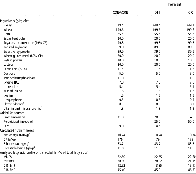
A basal weaner diet was prepared from which all three experimental diets were derived by adding fat sources, such a lard and linseed oil (whether or not peroxidized), according to Table 1. Gradual substitution of fresh linseed oil and lard by peroxidized linseed oil was used to induce a different content of lipid peroxides, while achieving a similar fatty acid profile of the added fat.
Ingredient composition and nutritional values of the experimental diets fed to piglets post-weaning

CON=pair-fed, 5% fresh linseed oil; ACON=ad libitum fed, 5% fresh linseed oil; OF1=pair-fed, 2.5% peroxidized linseed oil; OF2=pair-fed, 5% peroxidized linseed oil; MUFA=monounsaturated fatty acids.
1 The mineral and vitamin premix supplied as the following (per kilogram diet): vitamin A, 10 000 IU; vitamin D3, 2000 IU; vitamin E; 54 IU; vitamin K3, 1 mg; vitamin B1, 1.2 mg; vitamin B2, 3.7 mg; vitamin B3, 12 mg; vitamin B6, 2 mg; vitamin B12, 0.030 mg; niacine, 20 mg; folic acid, 0.8 mg; Fe2+, 100 mg Zn2+, 100 mg; Cu2+, 15 mg; Mn2+, 80 mg; Se6+, 0.3 mg; I, 1 mg; BHT, 75 mg; ethoxyquin, 37.7 mg.
2 Tailor made flavor additive with no antioxidant properties ascribed (Scentarom, Merchtem, Belgium).
3 According to Centraal Veevoeder Bureau (1997), Centraal Veevoederbureau, Lelystad, The Nederlands.
Experimental design
Four treatments were included in this experiment. A first treatment consisted of piglets fed the diet containing 5% fresh linseed oil ad libitum during the entire experiment (ACON). The remaining three treatments consisted of piglets either fed the diet with 5% fresh linseed oil (CON), 2.5% peroxidized linseed oil (OF1) or 5% peroxidized linseed oil (OF2), ad libitum until day 5 post-weaning and according to a pair-feeding meal scheme from days 6 to 28 post-weaning. The feed restriction was accomplished by offering three equal meals per day corresponding to a total daily intake of 32 g/kg BW. The selected piglets were allocated to the treatments according to litter, BW and gender, resulting in 12 pens with four animals per pen. Treatments CON and OF2 were replicated in four pens with sacrification of two pens, each at days 5 and 28 post-weaning. Treatments ACON and OF1 were replicated in two pens and these pigs were sacrificed at day 28 post-weaning.
Sample collection
Blood was collected by cranial caval vein puncture at days 2, 5, 12 and 26 post-weaning of all live animals. Pair-fed animals were sampled 3.5–4.0 h post-prandial. Ethylenediaminetetraacetic acid-tubes were centrifuged (3000×g, 15 min) and plasma was stored at −20°C until the analysis of the GPx activity, malondialdehyde (MDA) concentration, oxygen radical absorbance capacity (ORAC) and ferric reducing antioxidant power (FRAP). Erythocytes were isolated from heparinized blood, rinsed with milliQ and hemolized with metaphosphoric acid. Samples were snap frozen in liquid nitrogen and stored at −80°C pending the quantification GSH and GSSG.
Animals were anaesthetized by electrical stunning and during exsanguination, blood was collected and processed as described above. No fasting period was included for ad libitum fed piglets. Pair-fed piglets were sampled 3.5 to 4.0 h post-prandial. Subsequently, the small intestine was exposed and four sections were obtained at both 5% (duodenum) and 75% (distal jejunum) of the total length. Sections were rinsed with 0.9% saline and used for mucosa collection, Ussing chamber experiments, and histomorphology measurements. The first 20 cm section of each site was slit longitudinally and mucosal scrapings were obtained. A first subsample was used for an acid mucosal homogenate with a 10% perchloric acid solution for analysis of GSH and GSSG. A second subsample was turned into a mucosal homogenate using 1% triton-X-100 phosphate buffer. Aliquots were taken to determine total protein content before centrifugation (13 000×g, 15 min, 4°C). The supernatant was used for GPx and MDA analysis. Liver tissue was processed in an identical way as mucosa. All samples were snap frozen in liquid nitrogen and stored at −80°C.
Ussing chambers and histomorphology
Two duodenal and two jejunal segments of each animal were mounted into modified Ussing chambers (Mußler Scientific Instruments, Aachen, Germany) with opening area of 1.07 cm², for determination of the macro-molecular flux of 4-kDa fluorescein isothiocyanate dextran (FD4; Sigma-Aldrich, Overijse, Belgium) and 40-kDa horseradish peroxidase (HRP; type IV; Sigma-Aldrich) (Michiels et al., Reference Michiels, Missotten, Dierick, Fremaut and De Smet2010; Wang et al., Reference Wang, Degroote, Van Ginneken, Van Poucke, Vergauwen, Dam, Vanrompay, Peelman, De Smet and Michiels2016). The apparent permeability coefficient (Papp) was calculated for each marker from the following equation:
 $${\rm P}_{{{\rm app}}} {\equals}{\rm dc}\,/\,{\rm dt}\,{\times}\,V\,/\,\left( {A\,{\times}\,C_{0} } \right)\,\left[ {{\rm cm}\,/\,{\rm s}} \right]$$
where dc/dt is the change in concentration in the acceptor compartment between 20 and 100 min, (µg/ml per s) calculated from the slope of the concentration–time curve, V the buffer volume in the donor compartment (ml), C 0 the initial marker concentration in the donor compartment (µg/ml) and A the exposed tissue surface area (cm2).
For histomorphology, duodenal and distal jejunal segments (10 cm) were placed in 4% paraformaldehyde solution (phosphate buffered saline, pH 7.4) for 2 h at room temperature. After rinsing, the tissue was kept on ice-cold phosphate buffered saline prior to dehydration and paraffin embedding. Transverse 5 µm sections were stained with haematoxylin and eosin. Villus height, villus width and crypt depth were estimated in 30 well oriented villi and adjacent crypts using image analysis software in at least 10 sections per tissue sample (Olympus BX 61, analySIS Pros, Aartselaar, Belgium).
Analytical methods
Analysis of the fatty acid profile of linseed oil and lard was performed according to Raes et al. (Reference Raes, de Smet and Demeyer2001). The POV of linseed oil was measured by a redox titration method. Assessment of the GPx activity was determined spectrophotometrically (Hernandez et al., Reference Hernandez, Zomeno, Arino and Blasco2004). The thiobarbituric acid reactive substances method was employed to measure MDA concentrations (Grotto et al., Reference Grotto, Santa Maria, Boeira, Valentini, Charão, Moro, Nascimento, Pomblum and Garcia2007). The FRAP assay was done according to Benzie and Strain (Reference Benzie and Strain1996). The ORAC assay was based on Ou et al. (Reference Ou, Hampsch-Woodill and Prior2001). The biuret method was applied to determine the total protein content of tissue samples. The GSH and GSSG concentrations were quantified as the derivatives of 2,4-dinitrofluorobenzene by HPLC (Yoshida et al., Reference Yoshida1996), relative to γ-glutamyl-glutamate as internal standard. The mucosal Eh values for the GSH/GSSG redox couple were calculated by using the appropriate forms of the Nernst equation (mV), at pH 7.4 and temperature of 37°C, where concentrations of GSH and GSSG were expressed in molarity.
 $${\rm E}_{{\rm h}} {\equals}{\minus}264{\minus}\left( {61.5\,/\,2} \right){\times}\,{\rm log}\left( {{\rm GSH}^{2} \,/\,{\rm GSSG}} \right)$$
Fluorescence intensity of FD4 was measured at λ exc of 485 nm and λ em of 538 nm. The HRP activity was measured by a modification of the Worthington method (McKie et al., Reference McKie, Zammit and Naftalin1999), following the rate of increase in optical density at 460 nm.
Statistical analysis
The data were analysed by GLM procedures (SAS Enterprise Guide 6; SAS Institute, Cary, NC, USA) with the individual level as experimental unit. For single measurements the model only contained the fixed effect of dietary treatment. Repeated measurements were analysed for the fixed effects of treatment, days post-weaning and their interaction. Differences were considered significant at P<0.05, and were followed by a Tukey multiple comparison test. The linear effect of dietary inclusion of lipid peroxides was tested for pair-fed animals by linear regression. Bivariate Pearson correlation coefficients were considered significant at P<0.05, and were considered relevant when |r|>0.5.
Results
Growth performance
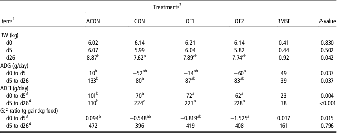
Table 2 summarizes the BW, average daily gain (ADG), average daily feed intake (ADFI) and gain to feed ratio (G:F) of the different treatments. During the first 5 days post-weaning, all animals were fed ad libitum. The ADFI was significantly reduced in treatments CON, OF1 and OF2 to 31%, 29% and 39% of ADFI in ACON, respectively. All treatments except ACON displayed a reduced BW on day 5 when comparing to day 0 post-weaning. This resulted in a negative ADG for these treatments, with the ADG of treatment OF2 being 70 g/day lower than treatment ACON (P<0.05). Hence, also the G:F ratio was significantly lower for treatment OF2, as compared to ACON, during the first 5 days post-weaning. After day 5, treatments CON, OF1 and OF2 were pair-fed by meal feeding. The ADFI ranged between 223 and 228 g/day, and was not different among groups as was instigated by pair-feeding (P=0.962), but was 27% lower than in the ad libitum fed group (ACON; P<0.001). Strangely BW was only significantly different between ACON and CON. As a result, also the ADG between days 5 and 26 post-weaning was significantly reduced with 40% for treatment CON, as compared to ACON (P=0.037). Feed efficiency was not affected during this phase.
Animal performance of piglets fed increasing amounts of peroxidized linseed oil

a,bWithin a row, least square means without a common superscript letter differ significantly (P<0.05).
1 ADG=average daily gain; ADFI=average daily feed intake; G:F=gain:feed ratio.
2 ACON=ad libitum fed, 5% fresh linseed oil; CON=pair-fed, 5% fresh linseed oil; OF1=pair-fed, 2.5% peroxidized linseed oil; OF2=pair-fed, 5% peroxidized linseed oil.
3 Statistical evaluation at the pen level, with two pens for treatments ACON and OF1, and four pens for treatments CON and OF2.
4 Feed intake for treatment ACON treatment was monitored at the pen level, resulting in two replicates for treatment ACON and eight replicates for treatments CON, OF1 and OF2.
Oxidative profile of blood plasma and erythrocyte glutathione redox status
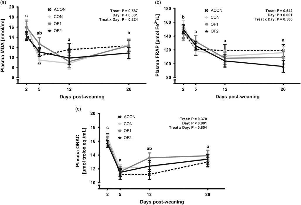
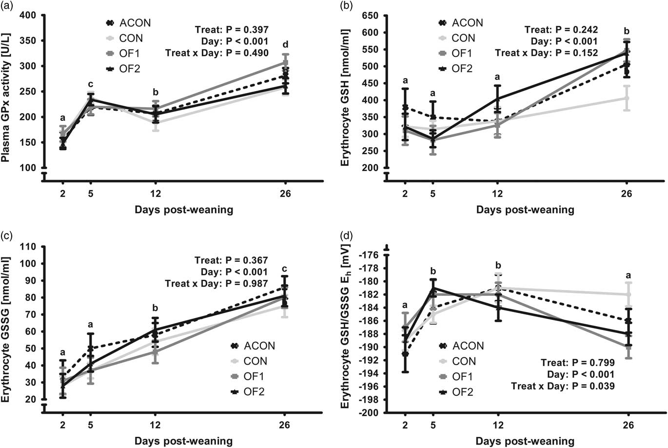
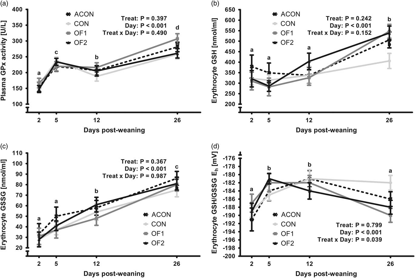
During the experiment, all live animals were subjected to blood sampling at days 2, 5, 12 and 26 post-weaning. These results are presented in Figures 1 and 2. Days post-weaning had a significant impact on all measured parameters in plasma or erythrocytes (P<0.001). Plasma MDA and FRAP decreased after weaning, whereas plasma ORAC values recovered on day 26 post-weaning after having shown a significant decrease on day 5. Plasma GPx and erythrocyte GSH and GSSG increased significantly from days 2 to 26 post-weaning. Treatment, however, did not affect ORAC, FRAP, MDA, GPx, GSH and GSSG measured in plasma or erythrocytes. Only a significant interaction was found between days post-weaning and treatment for GSH/GSSG Eh (P=0.039). Values significantly differed between the treatment groups at day 26, but not at days 2, 5 or 12. At day 26, OF1 and OF2 were, respectively, 6 and 7 mV lower than treatment CON (P<0.05). In addition, blood samples were collected during exsanguination, when animals were sacrificed at days 5 and 28 post-weaning. These results aligned with the results above. No effects were found on day 5 (data not shown). However, at day 28 post-weaning, GSH/GSSG Eh was decreased with 9 mV (P=0.007) and GSH was increased with 27% (P=0.014) in OF2 when comparing with ACON.
Effect of pair-feeding a diet containing 5% fresh (CON), 2.5% peroxidized (OF1) or 5% peroxidized linseed oil (OF2), or ad libitum feeding a diet containing 5% fresh linseed oil (ACON) on the plasma malondialdehyde (MDA) concentrations (a); the ferric reducing ability of plasma (FRAP) (b); and the plasma oxygen radical absorbance capacity (ORAC) (c) of weaner piglets at 2, 5, 12 and 26 days post-weaning. a,b,cRepresent differences between days post-weaning (P⩽0.05).

Effect of pair-feeding a diet containing 5% fresh (CON), 2.5% peroxidized (OF1) or 5% peroxidized linseed oil (OF2), or ad libitum feeding a diet containing 5% fresh linseed oil (ACON) on the plasma glutathione peroxidase (GPx) activity (a); the erythrocyte glutathione (GSH) concentrations (b); the erythrocyte glutathione disulphide (GSSG) concentrations (c); and erythrocyte glutathione redox status (GSH/GSSG Eh) (d) of weaner piglets at 2, 5, 12 and 26 days post-weaning. a,b,c,dRepresent differences between days post-weaning (P≤0.05).
Oxidative profile and glutathione redox status of the small intestinal mucosa
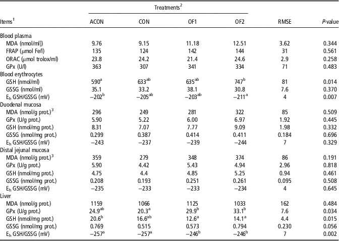
At days 5 post-weaning, eight animals were sacrificed from treatments CON and OF2. No significant differences were found for MDA, GPx, GSH, GSSG and GSH/GSSG Eh in small intestinal mucosa (data not shown). The results obtained at day 28 post-weaning are represented in Table 3. The different tissue levels of MDA were not affected by the treatments. However, it was found that the SD of the MDA concentrations in duodenal and jejunal mucosa of ACON pigs was twofold to fivefold higher as compared to other treatments (no equality of variance). When excluding ACON from statistical evaluation, treatment effects were found for pair-fed animals (CON, OF1 and OF2). Here, MDA tended (+29%; P=0.061) to be higher in duodenal mucosa of OF2, and was significantly higher (+34%; P=0.019) in jejunal mucosa of OF2, compared to CON. This treatment effect on mucosal MDA was found to be linear in both the duodenum (r=0.502; P=0.021) and jejunum (r=0.562; P<0.005). No treatment effects were found for the mucosal GPx activity, the GSH and GSSG concentration, or the GSH/GSSG Eh.
Markers of oxidative status and glutathione (GSH) redox status in piglets at 28 days post-weaning, fed increasing amounts of peroxidized linseed oil
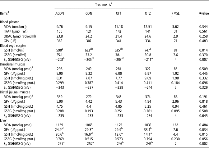
a,bWithin a row, least square means without a common superscript letter differ significantly (P<0.05).
1 MDA=malonddialdehyde; GPx=glutathione peroxidase activity; FRAP=ferric reducing ability of plasma; ORAC=oxygen radical absorbance capacity; GSSG=glutathione disulphide; Eh GSH/GSSG=glutathione redox status.
2 ACON=ad libitum fed, 5% fresh linseed oil; CON=pair-fed, 5% fresh linseed oil; OF1=pair-fed, 2.5% peroxidized linseed oil; OF2=pair-fed, 5% peroxidized linseed oil.
3 Homoscedasticity, assessed by Levene’s Test for Equality of Variances, was violated (P<0.05).
Oxidative profile and glutathione redox status of liver
Hepatic MDA concentrations were not affected by the treatment. However, liver GSH was significantly decreased in OF1 (−39%) and OF2 (−21%), relative to ACON (P=0.015) but not to CON. Furthermore, GSSG in liver was significantly higher in OF2 when comparing with CON (P=0.017). This resulted in a 11 mV increase of the GSH/GSSG Eh for both OF1 and OF2 when comparing to CON as well as ACON (P=0.002). This was accompanied with an increased hepatic GPx activity in OF1 (+47%) and OF2 (+63%), when compared to the CON treatment (P=0.034).
Small intestinal histomorphology and barrier function
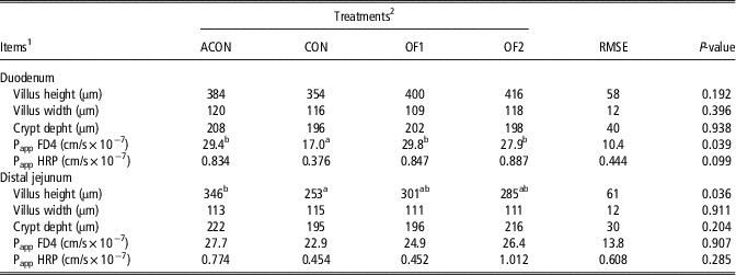
At day 5 post-weaning, no significant treatment differences were found regarding the small intestinal histomorphology or barrier function (P>0.05; data not shown). At day 28 post-weaning (Table 4), the jejunal villus height was significantly decreased for treatment CON, but not for OF1 and OF2, relative to treatment ACON. Regarding the barrier function at day 28, the duodenal FD4 flux was clearly affected (P=0.039) and was increased in OF1 (+75%), OF2 (+64%), and in ACON (+73%), when comparing to the CON treatment. Finally, also duodenal HRP fluxes tended (P=0.099) to be higher in treatments ACON, OF1 and OF2 (v. CON). The jejunal FD4 and HRP fluxes were not significantly different between treatments.
Markers of small intestinal histomorphology and barrier function in piglets at 28 days post-weaning, fed increasing amounts of peroxidized linseed oil

a,bWithin a row, least square means without a common superscript letter differ significantly (P<0.05).
1 Papp FD4=mucosal apparent permeability coefficient of fluorescein isothiocyanate dextran 4 kDa; Papp HRP=mucosal apparent permeability coefficient of horseradish peroxidase 40 kDa.
2 ACON=ad libitum fed, 5% fresh linseed oil; CON=pair-fed, 5% fresh linseed oil; OF1=pair-fed, 2.5% peroxidized linseed oil; OF2=pair-fed, 5% peroxidized linseed oil.
Correlation analysis
Since the largest treatment effects were found at day 28 post-weaning, correlations between the parameters at this age and only across pair-fed treatments CON, OF1 and OF2 were calculated. Here, it was observed that duodenal MDA concentration was positively associated with local GPx activity (r=0.731; P<0.001). Glutathione and GSSG concentrations in mucosa were not related to MDA or GPx. Furthermore, no significant or meaningful correlations (|r|>0.5; P<0.05) were found between the duodenal or jejunal histomorphology and the oxidative and redox profile (local MDA, GPx, GSH, GSSG and GSH/GSSG Eh levels). Regarding the barrier function, jejunal FD4 flux was linked to the local GSH (r=0.619; P<0.05) and GSSG (r=0.635; P<0.05) levels. Similarly, the jejunal HRP fluxes were positively correlated with the local GSH and GSSG concentrations, respectively, with r=0.733 (P<0.01) and r=0.586 (P<0.05). The duodenal HRP flux was only correlated with the hepatic GSSG level (r=0.650; P<0.01), as was also observed in the jejunum for hepatic GSSG (r=0.627; P<0.01) and GSH/GSSG Eh (r=0.547; P<0.05).
Discussion
The objective of this experiment was to determine if an altered GSH redox status, by imposing a dietary oxidative challenge, would affect the small intestinal integrity during the acute weaning phase (day 5 post-weaning) and after the adaptive weaning phase (day 28 post-weaning) (Burrin and Stoll, Reference Burrin and Stoll2003). To modulate the GSH redox status, dietary peroxidized linseed oil was chosen as a challenge model. However, several aspects of this model have to be taken into account. First, peroxidation of linseed oil caused a reduction of its C18:3n-3 content. Thus, lard was used to equalize the fatty acid profile of the added fat sources. Secondly, large amounts of dietary linseed oil (4.1% to 5.0%) may reduce feed intake (Solà-Oriol et al., Reference Solà-Oriol, Roura and Torrallardona2011). Moreover, De Rouchey et al. (Reference DeRouchey, Hancock, Hines, Maloney, Lee, Cao, Dean and Park2004) indicated that feeds with a POV of 2.40 mEq O2/kg feed or higher result in a reduced feed intake and growth of weaned piglets. Our experiment reached these thresholds. Hence, a pair-feeding system from day 6 was applied to standardize feed intake across treatments CON, OF1 and OF2. The observations on performance showed that ad libitum fed piglets (ACON) clearly outperformed pair-fed piglets that were fed restrictedly. No significant performance effects were observed when higher amounts of peroxidized linseed oil were incorporated in the diet (CON, OF1 and OF2).
To evaluate the effectiveness of the dietary challenge with peroxidized linseed oil, MDA was monitored in different tissues (Liu et al., Reference Liu, Kerr, Chen, Weber, Johnston and Shurson2014a and Reference Liu, Kerr, Weber, Chen, Johnston and Shurson2014b). Concerning pair-fed treatments, a linear increase of mucosal MDA concentrations upon increasing inclusion of peroxidized linseed oil was observed. This confirms that the small intestinal mucosa was more exposed to oxidants at day 28 post-weaning (Tsunada et al., Reference Tsunada, Iwakiri, Noda, Fujimoto, Fuseler, Rhoads and Aw2003). Still, MDA levels in plasma and liver remained unaffected, which does not correspond with several other studies (Yuan et al., Reference Yuan, Chen, Zhang and Yu2007; Lu et al., Reference Lu, Harper, Zhao, Estienne and Dalloul2014). Aw (Reference Aw2005) addressed that differences in mucosal GSH redox status between studies could conceivably explain these dissimilarities for plasma MDA, since mucosal GSH is highly involved peroxide scavenging. Our results offer further in vivo proof, and confirm the pivotal role of GSH in small intestinal handling of luminal peroxides. For instance, the duodenal GPx activity at day 28 linearly increased with the MDA concentration in the duodenum, and liver GPx activity was significantly higher when lipid peroxides were administered. This illustrates the biological role of the GSH redox system, as GPx is a key enzyme in intestinal handling of luminal lipid peroxides (Wingler et al., Reference Wingler, Muller, Schmehl, Florian and Brigelius-Flohe2000; Aw, Reference Aw2005). In these reactions, GSH is used as reducing agent, resulting in the generation of GSSG. Consequently, it was expected that mucosal GSH and GSSG concentrations would change as well. However, in the current study, GSH and GSSG in duodenal and jejunal mucosa were not affected, and were not correlated to the MDA concentration nor to GPx activity. According to the working model of intestinal elimination of luminal peroxides, formulated by Aw (Reference Aw2005), this would then imply that the mucosal GSH levels were sufficient to detoxify the lipid peroxidation products. As a result, the GSH redox status was maintained at −230 to −240 mV, indicating a fairly reduced homeostasis of the GSH/GSSG pool. Nevertheless, the hepatic GSH redox status was significantly increased with 11 mV in treatments OF1 and OF2, representing a shift towards more oxidative stress when lipid peroxides were administered. This change seemed to be driven by a significantly increased GSSG concentration and GPx activity in liver, in particular for treatment OF2. Furthermore, hepatic GSH concentrations seem slightly decreased, although this was not a significant effect between pair fed animals. Seemingly, an increased efflux of GSH and GSSG from the liver into blood and bile could have contributed to the altered hepatic GSH/GSSG Eh (Aw, Reference Aw1994). In our study, erythrocyte GSH concentrations were highest in treatment OF2. However, we did not investigate its levels in bile. However, it is plausible that the hepatic GSH redox system, through an increased hepatic GPx activity and biliary GSH efflux, has contributed to the elimination of luminal peroxides and maintenance of the mucosal GSH homeostasis.
Remarkably, it was found that ad libitum feeding (ACON), compared to restricted feeding (CON), resulted in an increased duodenal FD4 flux at day 28 post-weaning. A higher daily intake of C18:3n-3 for ad libitum fed piglets might have effectuated this. For example, it has been demonstrated that linseed oil in the maternal lactation diet increases the intestinal FD4 flux of their offspring (De Quelen et al., Reference De Quelen, Chevalier, Rolli-Derkinderen, Mourot, Neunlist and Boudry2012). Bearing this in mind, evaluating the effect of dietary lipid peroxides and GSH redox disturbance can only be valid when comparing the pair-fed treatments, in which the dietary fatty acid profile was equalized. Here, feeding 2.5% and 5% peroxidized linseed oil increased the paracellular permeability of the duodenum, and tended to increase the transcellular permeability, at 28 days post-weaning. Strangely, Liu et al. (Reference Liu, Kerr, Weber, Chen, Johnston and Shurson2014b) found no detrimental effect on paracellular transport in young pigs fed several sources of thermally oxidized lipids. However, it was previously reported that oxidative stress can affect epithelial permeability in vitro and in vivo (Rao, Reference Rao2008; Maeda et al., Reference Maeda, Miyazono, Ito, Hamada, Sekine and Horie2010). Maeda et al. (Reference Maeda, Miyazono, Ito, Hamada, Sekine and Horie2010) demonstrated the negative effects of methotrexate-mediated interference with the thiol metabolism on FD4 permeability in the rat duodenum. Co-administration of N-acetyl cysteine, as a precursor of GSH, could restore FD4 permeation to control levels. Unfortunately, the GSH redox status was not quantified in that study. Our current study allowed these possible correlations. Based on earlier evidence generated in Caco-2 cells, using mercaptosuccinate, diethyl maleate and carmustine as selective inhibitors of the GSH redox system, it was hypothesized that an increased GPx activity and GSSG accumulation are required to potentiate the effect of peroxides on intestinal permeability (Rao et al., Reference Rao2008). In the current study, no direct alterations of the duodenal GSH redox system were found, nor did this correlate to the local barrier function. Nevertheless, duodenal barrier disturbance did co-occur with an increased hepatic GPx activity and GSH/GSSG Eh in treatments OF1 and OF2. Duodenal HRP flux was also positively correlated to the hepatic GSSG level, further demonstrating the significant role of the hepatic GSH redox system in maintaining small intestinal integrity. This was also observed in the jejunum, where hepatic GSSG and GSH/GSSG Eh were correlated with the jejunal HRP flux. Conversely, although unaffected by treatment, the jejunal FD4 and HRP fluxes were both positively correlated to the local GSH and GSSG levels in pair-fed piglets. This suggests that, under oxidative stress conditions, barrier function is related to the local GSH redox system. In the duodenum, however, the hepatic GSH redox system seems to impact more on the mucosal integrity, conceivably through biliary GSH and GSSG efflux.
A vast amount of in vitro work supports the cause and effect relation between the cellular redox status and intestinal cell transition (Circu and Aw, Reference Circu and Aw2012). Tsunada et al. (Reference Tsunada, Iwakiri, Noda, Fujimoto, Fuseler, Rhoads and Aw2003) nicely demonstrated this in vivo in rats, and found that dietary lipid peroxides result in intestinal GSH redox imbalance, which interfered with enterocyte proliferation and apoptosis. Mucosal GSH/GSSG ratios were restored by GSH supplementation, and this alleviated the peroxide-induced suppression of intestinal cell turnover. However, Tian et al. (Reference Tian, Washizawa, Gu, Levin, Wang, Rubin, Mwangi, Srinivasan, Jones and Ziegler2007) could not confirm the association between the GSH redox status and enterocyte turnover in vivo. Administration of l-buthionine-sulfoximine, a selective inhibitor of GSH synthesis, led to massive changes of the ileal GSH redox system, but proliferative indices were not significantly affected. In our study, no effects on small intestinal histomorphology and mucosal GSH/GSSG Eh were found. Therefore, no further in vivo evidence can be deduced which can confirm or reject this close and conserved relation.
In summary, this research studied responses of the small intestinal barrier to alterations of the GSH redox system in weaning piglets. It was found that, even if the mucosal GSH/GSSG Eh is maintained at a steady state level of −240 mV, mucosal and hepatic GSH and GSSG are important protagonists in regulating the gut barrier function in piglets.
Acknowledgements
The authors thank Sabine Coolsaet, Erik Claeys, Anneke Ovyn, Tessa Van Der Eecken, Katty Huybrechts, Gunther Vrolix, Karen Jennes and Ward Vanhee for their excellent technical support. This work was supported by the Agency for Innovation by Science and Technology (IWT-LO100856).
Declaration of Interest
None.
Ethics statement
The experiment was carried out according to the guidelines of the Ethical Committee of Ghent University, Belgium (approval number EC2013/167).
Software and data repository resources
None.