Arterial switch operation is the treatment of choice for patients with transposition of the great arteries. The risk of repair is increased in late presenters who have co-morbidities,Reference Parker, Zuhdi and Kouatli 1 as the ability of the left ventricle to support systemic circulation after repair greatly dictates their early survival. A two-staged repair is often adopted for patients with involuted left ventricle in the form of pulmonary artery banding with aortopulmonary shunting, which trains the involuted left ventricle to handle systemic circulation after the arterial switch operation. An alternative option of ductal stenting was performed on an infant with the aim of improving oxygenation and stabilising haemodynamics. The left ventricle was noticed to be well trained even 4 months after the initial stenting.
Case report
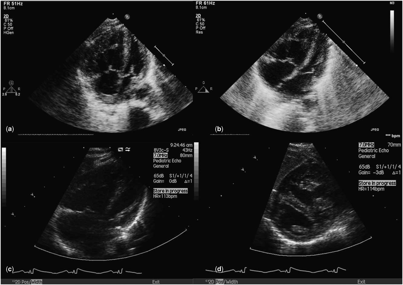
Infant S presented to us at 4 weeks of age with severe cyanosis and shock requiring high ventilatory and inotropic support. Echocardiography showed transposition of great arteries with intact ventricular septum, restrictive foramen ovale, and no ductus arteriosus. The interventricular septal and left ventricular posterior wall thickness in diastole measured 4.8 and 3.4 mm, respectively. Balloon atrial septostomy was immediately performed, creating an unrestrictive interatrial communication; however, his saturations did not improve beyond 50%. Inhaled nitric oxide was included to treat pulmonary hypertension, whereas prostaglandin infusion was initiated and titrated to re-open the closed ductus arteriosus. He also had septicaemia and continued to have poor saturations. In view of the unstable haemodynamics and septicaemia, he was deemed unfit for primary repair. Thus, the child was palliated via stenting of ductus arteriosus to improve oxygenation and haemodynamics while waiting for the infection to clear.
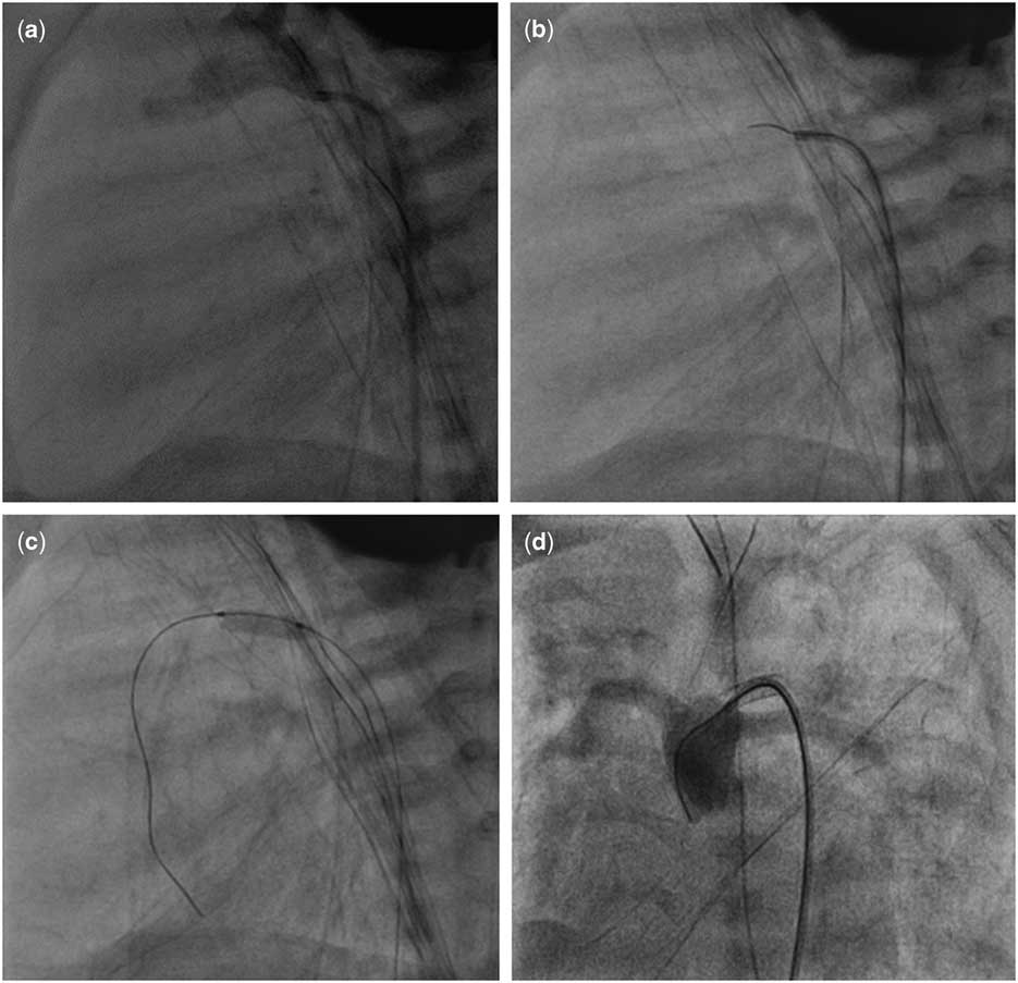
Check aortogram revealed a closed ductus arteriosus but the ampulla was present. Through a 4-French Mullins long sheath (Cooks Medical, Inc., Bloomington, Indiana, United States of America) via the femoral artery, a 4-French Judkins Right catheter (Cordis Corporation, Bridgewater, New Jersey, United States of America) was used to engage the ampulla of the ductus arteriosus. A 0.014" ChoICE® PT Floppy Guide Wire (Boston Scientific, Marlborough, Massachusetts, United States of America) was then used to probe the closed ductus arteriosus. This successfully re-canalised the closed ductus arteriosus. The position of the guide wire through the ductus arteriosus was checked using anteroposterior and lateral projections to ensure that the guide wire had moved into the pulmonary artery. The guide wire was then placed deep into the left ventricle for better anchoring. Over the wire, a coronary stent 3×9 mm was positioned at the ductus arteriosus. The position was checked via hand shots of contrast while adjusting the position of the stent. Once satisfied, the stent was inflated. Repeat angiography revealed a partially stented ductus arteriosus with an uncovered pulmonary end. A second coronary stent, 3×12 mm, was placed inside the first stent to extend the stented area in order to cover the whole ductus arteriosus. Subsequent angiography revealed adequately stented ductus arteriosus with no stent protrusion. Next, intravenous heparin was initiated, keeping activated partial thromboplastin time two times the normal value. Aspirin was overlapped with heparin infusion for at least 2 days before heparin was weaned off.
His saturations improved beyond 80%. He was weaned off ventilation and inotropic support. The post-procedural course was uneventful apart from a transient contrast-induced nephropathy, which resolved with supportive treatment.
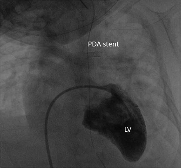
Unfortunately, he developed respiratory syncytial virus pneumonia 3 weeks after discharge. He became oxygen-dependent and was not fit for general anaesthesia for 3 months. He was otherwise haemodynamically stable. The condition of his lungs improved after supportive care. In view of a prolonged period of left ventricular training after stenting, a cardiac catheterisation was performed. It showed a left-to-right ventricular systolic ratio of 3:4. On echocardiography, interventricular septal and left ventricle posterior wall thickness in diastole remained at 4.0 and 4.4 mm, respectively. He subsequently underwent a successful arterial switch operation. After surgery, the chest was left open for 4 days and inhaled nitric oxide was administered for pulmonary hypertension totalling 5 days. He developed low cardiac output syndrome, which required the standard inotropic support but did not require extracorporeal membrane oxygenation. He was discharged after 21 days.
Discussion
Although there are reports of successful primary repairs in patients with late arterial switch operation with acceptable morbidities and mortality,Reference Kang, de Leval and Eliott 2 – Reference Bisoi, Ahmed and Malankar 5 they are associated with longer hospital stay and increased utilisation of resources in the ICU.Reference Cain, Cao and Ghanayem 6 – Reference Kothari, Ramakrishnan and Senguttuvan 8 Therefore, primary repair in patients with involuted left ventricle is not adopted by all centres.
Ductal stenting is an attractive alternative in late presenters of transposition of the great arteries.Reference Sivakumar, Francis and Krishnan 7 It provides volume loading and to a lesser extent pressure loading to the involuted left ventricle, leading to left ventricular hypertrophy. Presence of the stented duct allows for better systemic–pulmonary blood mixing, eliminating the need for aortopulmonary shunt. It has been adopted by other centres as a modality for left ventricular training before arterial switch operation, even after closure of the ductus arteriosus.Reference Kothari, Ramakrishnan and Senguttuvan 8 This method of left ventricular training is an attractive alternative, given its non-invasive nature and reduced post-procedural morbidity.Reference Sivakumar, Francis and Krishnan 7 , Reference Kothari, Ramakrishnan and Senguttuvan 8 In addition, it does not cause aortic and pulmonary annular discrepancy, as seen in patients staged via pulmonary arterial banding.Reference Kramer, Scheewe and Fischer 9
Initially, ductal stenting was performed on the patient to improve oxygenation. He was discharged home as the arterial switch operation was scheduled 4 weeks after the initial stenting. Unfortunately, he contracted respiratory syncytial virus infection and the surgery was delayed for a few months due to severe bronchiolitis. In this instance, ductal stenting managed to keep the left ventricle trained for 4 months. This is, arguably, the longest left ventricular training via ductal stenting thus far. Ductal stenting can be an option for patients with co-morbidities, which obviates early primary arterial switch operation. Nevertheless, a larger scale study is required before any definite conclusion can be made.
Conclusion
Ductus arteriosus stenting provides a practical option in late presenters of transposition of great arteries with unstable haemodynamics; however, it has its limitations and will need careful consideration before embarking on it .
( a ) Check aortogram showed a closed ductus arteriosus. ( b ) A 4-French Judkins Right catheter is positioned at the ampulla of the closed ductus arteriosus and the closed ductus arteriosus was probed using a ChoICE® PT Floppy Tip Guide Wire. ( c ) After successful re-canalisation of the ductus, the guide wire was positioned deep into the right ventricle. The ductus was stented using a coronary stent. ( d ) After placement of two coronary stents, check angiography showed good flow into both lung fields with no jailing of either branch pulmonary arteries.

Echocardiographic views of the left ventricle showing comparable left ventricular wall thickness and volume before ductal stenting ( a and b ) and after 4 months ( c and d ).

Angiography showing satisfactory left ventricular volume even 4 months after the initial ductal stenting. LV=left ventricle; PDA=patent ductus arteriosus.

Acknowledgement
We express our heart-felt gratitude to Dr Hasri Samion, Head of the Paediatric and Congenital Heart Centre at the National Heart Institute of Kuala Lumpur, for his contributions.
Financial Support
This research received no specific grant from any funding agency, commercial, or not-for-profit sectors.
Conflicts of Interest
None.
Ethical Standards
The authors assert that all procedures contributing to this work comply with the ethical standards of the National Heart Institute of Kuala Lumpur.
Supplementary Material
To view supplementary material for this article, please visit http://dx.doi.org/10.1017/S1047951115001055