Introduction
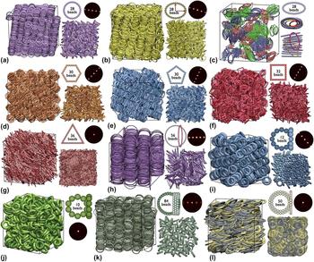
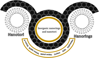
Toroidal-like nanosize structures, called as nanorings or nanotori, are not frequent among other less-common nanostructures [Reference Kharisov, Kharissova and Ortiz-Mendez1], in particular among closely related ball-type nanostructures as nano-onions, nanoballs, and nanospheres. Ring- and torus-like nanostructures are indeed “two sides of the same medal,” whose properties have been studied more theoretically using density functional theory (DFT) calculations than experimentally. However, as it will be shown below, they can possess intriguing properties, e.g., being, in some cases, ferromagnetic [Reference Vojkovic, Nunez, Altbir and Carvalho-Santos2], which could lead to novel applications. Under self-assembling, the nanorings/nanotori can form a variety of geometries (Fig. 1) on the basis of liquid-crystalline coloidal phases, very useful as storage and adsorption materials [Reference Avendaño, Jackson, Müller and Escobedo3]. Another practically undeveloped area is their use as dopants for classic oil lubricants, allowing much lesser metal–metal contact and, as a consequence, considerably higher duration of oil service in distinct mechanisms before changing the maintenance oil. As it is known, one of the main losses in metallurgical industries is related with periodic stopping of processes because of bearing wear and not because of the cost of lubricants. On the basis of only these two examples, one could propose that further development and expansion of number of toroidal nanostructures could lead to a variety of novel materials possessing unusual properties. In this review, we show a current overview of toroidal inorganic nanostructures, their properties, and applications. Some related circular organic molecules are omitted here.
Particle models and stable microstructures observed in colloidal nanorings of various geometries. Reprinted with permission from Ref. Reference Avendaño, Jackson, Müller and Escobedo3, PNAS. (See Supplementary Material for a complete caption.)

Carbon nanorings and nanotori
When compared with nanoring-like structures of other elements and compounds, the nanorings or nanotori of pure elemental carbon are rare, although they are known from long ago [Reference Liu, Jayanthi and Wu4]. These allotropic forms of carbon [Reference Liu, Zhao and Suzuki5] are generally studied by molecular dynamics (MD) simulations [Reference Sarapat and Baowan6]. It is still difficult to obtain them in laboratory experimental conditions and specially to scale up their formation. The nanoring/nanotori can possess a classic structure or that of Mobius type (one turn, see below). These nanostructures are closely related to edges in nanotubes, nanochains, graphene, and nanocones, and they can be “constructed” from carbon nanotubes [Reference Chen, Lusk, van Duin and Goddard7] (we also note the existence of V-phenylenic nanotori [Reference Alamian, Bahrami and Edalatzade8, Reference Chel Kwun, Munir, Nazeer, Rafique and Min Kang9]). Theoretically, these stable (under large tensile deformation) torus structures could be applied for networks fabrication, resulting in extremely flexible constructions with a high strength-to-density ratio. Just like graphene, the nanotori have no defects and no boundary, i.e., no any-size rings can be present other than six. In this case, such nanorings can be considered as cyclic graphene in two dimensions or elemental benzenoids (without H atoms) [Reference Balaban and Klein10]. It was reported long ago [Reference Liu, Dai, Hafner, Colbert, Smalley, Tans and Dekker11] that single-wall carbon nanotubes (SWCNTs) (∼1.4 nm in diameter) can form toroidal structure (rings made of nanotube or “crop circles”). Here, the toroidal continuous single nanotubes without end or beginning are formed, where an SWCNT is closed around onto itself. Two open tube ends fuse together, being stabilized with the aid of van der Waals forces only, resulting in a perfect toroidal SWCNT [Reference Hilder and Hill12]. The overall diameters of 600–800 nm are typical for such a toroidal structure.
MD simulation studies of carbon nanotori
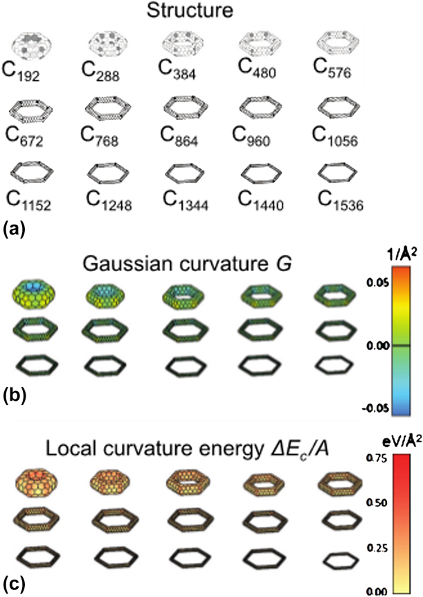
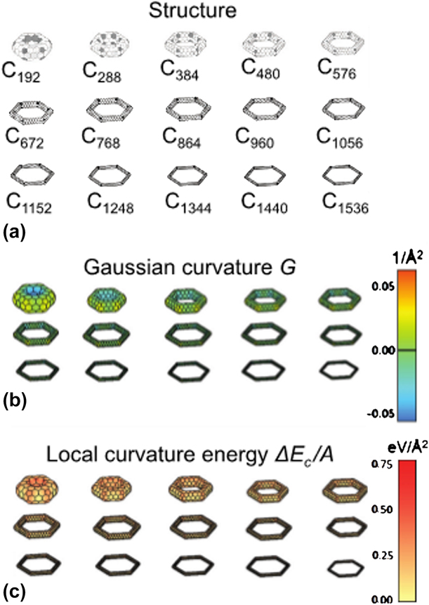
The nanotori properties, being an exotic carbon-based nanomatter [Reference Koorepazan-Moftakhar, RezaAshrafi, Ori, Putz, Putz and Ori13] had been considerably studied mainly by ab initio DFT calculations [Reference Madani and Ashrafi14]. On this basis, in addition to the approach of the continuum elasticity theory, the nanotori, formed from carbon nanotubes as precursors, were classified in accordance with their height, length, and rotational symmetry (Fig. S1; see Supplementary material) [Reference Chuang, Guan, Witalka, Zhu, Jin and Tomanek15]. Thus, Fig. 2 shows the nanotori with changing side length L. In the L → ∞ limit, the contribution from the elbow joints to the excess energy is negligible. A simple sum of energies of six constituent carbon nanotubes gives the total elastic energy. A series of polygonal nanorings resembling a segment of height H of a double-wall carbon nanotube is shown in Fig. S2 (see Supplementary material). The outer and inner tubes are connected at the bottom and the top by interactions of a lip–lip type, consisting of non-hexagonal and hexagonal rings. In the point of a H → ∞ limit, the contribution of elastic energy from the two ends is a constant value.
(a) Structural models, (b) local Gaussian curvature G, and (c) local curvature energy E c/A across the surface of torus isomers with different lengths of the nanotube segments L. The nonhexagonal rings in (a) are shaded. The values of G and E c/A have been interpolated from their values at the atomic sites. Reprinted with permission from Ref. Reference Chuang, Guan, Witalka, Zhu, Jin and Tomanek15, APS Physics.
Polygonal nanotori, having n (changing rotational symmetry number), are shown in Fig. S3 (see Supplementary material). It is focused on two nanotori families, in which the nonhexagonal ring relative positions are the same when the number n (rotational symmetry) moves from n = 4 to 13. For the two nanotori families, the strain energy depends on n in a very different manner.
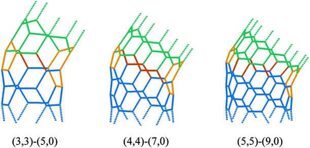
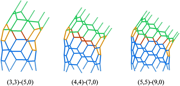
A large number of cage-like toroidal carbon moieties, made of elbows (Figs. 3 and 4), can be constructed. These structures are formed from zigzag and armchair carbon nanotubes, which are connected using a heptagonal and pentagonal defect at every bend site [Reference Cox and Hill16]. In case of elbows, made of two carbon nanotubes, it was proposed that the atoms continue to be in their preferred positions in the sections of nanotubes and can join when the bond lengths between neighboring atoms in two distinct sections are, in a least-squares sense, closest possible to the C–C bond length. For twenty possible nanotori, their geometric parameters were determined, being useful for the area of nanomechanical systems (NEMS) by incorporating there such nanotori.
Elbows formed from two nanotube sections. Reprinted with permission from Ref. Reference Cox and Hill16, American Chemical Society.
C240 molecule with structure 5(3,3)185(5,0)30. Reprinted with permission from Ref. Reference Cox and Hill16, American Chemical Society.

Investigating the electronic structure of nanorings/nanotori (the roles of curvature, hybridization, and disorder) in the rotational symmetry terms [Reference Liu and Ding17], it was established that the σ*–π* hybridization effects, being curvature induced, have an important role in determination of electronic structure of the nanotori. When the torus size increases, the electronic structures, being of a characteristic 0D system, passes to the quasi-1D system, in which the transition “semiconductor–metal phase” is revealed. Among other important results, when magnetic field perpendicular to the plane of torus, the Zeeman effect on electronic structure of carbon nanorings was observed [Reference Liu18]. Detailed studies revealed that this effect influences in a distinct manner on nanotori electronic structures, e.g., it may lead to the disappearance of periodical AB oscillation. Under variations of magnetic flux, the tori of the armchair type could be changed, resulting in gapless metals from narrow-gap semiconductors, or, on the contrary, while the nonarmchair (including chiral and zigzag types) tori reflect the behavior of a semiconductor. In addition, applying the Landauer–Büttiker formula, several calculations on the electric conductance of carbon nanotori when being contacted with SWCNTs showed the evidence that the conductance of these combinations of SWCNTs-carbon nanotorus is highly sensitive to the contacts transparency (chemistry) [Reference Chou and Guo19].
Magnetic response of carbon (being normally a nonmagnetic element) nanotori is also a derivative of curvature and disorder [Reference Liu, Chen and Ding20]. Thus, the investigations of magnetism in corrugated carbon nanotori showed the formation of ferromagnetic nanocarbons and revealed the importance of defects, symmetry, negative curvature, and defects [Reference Rodriguez-Manzo, Lopez-Urias, Terrones and Terrones21]. Among other results, the carbon nanotori made as a result of either by joining the Haeckelite tube ends (containing pentagons, heptagons, and hexagons) or coalescing C60 molecules (incorporating octagons and pentagons) along the 5-fold axis possess enhanced magnetic moments under application of an external magnetic field. It was revealed that the combination of positive and negative Gaussian curvature (which is caused by pentagonal, heptagonal, or octagonal carbon tori) has an important role in their magnetic properties. Three tori produced by coalescing C60 molecules along the axes of 2-, 3-, and 5-fold symmetry, respectively, are shown in Fig. S4 (see Supplementary material). If the structure does not contain defects, a paramagnetic response of corrugated nanotori (possessing hexagons, heptagons, pentagons, and octagons) could be got for good arrangements of atoms of nonmagnetic elements (i.e., carbon) at low temperature. At the same time, the paramagnetism and ferromagnetism in carbon nanostructures are known to be highly sensitive to the rupture of symmetry and presence of defects. Carbon nanorings possessing unusually big (giant) paramagnetic moments were also discovered theoretically [Reference Liu, Guo, Jayanthi and Wu22]. In these nanotori, such huge paramagnetic moment can exist because of the interplay between the ballistic motion of the π electrons in the metallic nanotube and the toroidal geometry.
Sizes, structural varieties, and stability of carbon nanotori
The structural stability of different-size carbon nanorings [C170 (Fig. S5, see Supplementary material), C250, C360, C520, and C750 tori] in the heating conditions, studied by MD simulations [Reference Taşci, Yazgan, Malcıoğlu and Erkoç23], established their high temperature stability: C170 nanotorus (close to the circular geometry, deformed significantly above 3700 K), C250 (pentagon-type geometry, stable up to 3000 K (deformation temperature), at a knee-region deformed again), C360 (pentagon-type geometry too, but it is more rounded when compared with C250, the strongest molecule, persisting up to 4500 K), C520 (circle-like geometry, subjected to deformation at about 4200 K, the nanotorus is also deformed at a knee-region), C750 (biggest in that report, pentagon-type inner geometry, it is deformed at a knee region at about 4100 K). Under unfolding of a carbon nanoring, deformation waves appear propagating along the carbon atomic network [Reference Glukhova and Slepchenkov24]. For a chiral torus (20 nm in diameter), a complete unfolding process time is ∼91 ps.
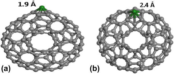
The thermal stability of the nanotori depends on a complex form on geometrical parameters; temperatures of their deformation are like those of carbon nanorods and nanotubes having comparable cross-section radius. The maximum stability was revealed for (13, 0) carbon nanotori, obtained from a zigzag carbon nanotube by its folding. These nanotori possess the lowest formation enthalpy (101 eV) [Reference Glukhova, Kolesnikova, Slepchenkov and Savostyanov25]. The nanotorus C340 possesses the most stable structure being compared with other studied objects; its values [the energy per atom (−43.02 eV) and reaction enthalpy of structure formation (10.22 kcal mol/atom)] are practically equal with parameters of the C60 fullerene. It is well known that the fullerene is one of the most stable carbon nanoclusters. Another carbon nanotorus (C460) has the greatest elasticity modulus (0.65 TPa). Indeed, the structural stability of a perfect nanotorus depends first on the nanoring diameter and nanotube diameter, meanwhile its chirality has a small effect [Reference Chang and Chou26]. Thus, by increasing the SWCNT radius (r), the radius of its corresponding ideal nanotorus (R) also grows [Reference Ajori, Ansari, Hassani and Ghighi27]. In addition, for SWCNTs having different lengths, it was established that the strain and buckling force of their related carbon nanotori increase by enhancing the R/r magnitude. Also, the DFT studies of a C120 carbon nanotorus (Fig. S6, see Supplementary material) [Reference López-Chávez, Crúz-Torres, Castillo-Alvarado, Ortíz-López, Peña-Castañeda and Martínez-Magadán28] proposed that C60 fullerene and C120 nanotorus possess a very similar chemical reactivity. The C120 Ti-decorated derivative (Fig. 5) was calculated to be a good material for hydrogen absorbance (about 5.6 wt%) [Reference López-Chávez, Peña-Castañeda, García-Quiroz, Castillo-Alvarado, Díaz-Gongora and Jimenez-Gonzalez29].
A Ti metallic atom trapped on (a) a pentagonal ring where the CTi bond length was of 1.9 Å and (b) to a hexagonal ring, where the CTi bond length was of 2.4 Å, of a C120 + 1Ti-nanotorus. Reprinted with permission from Ref. Reference López-Chávez, Peña-Castañeda, García-Quiroz, Castillo-Alvarado, Díaz-Gongora and Jimenez-Gonzalez29, Springer.

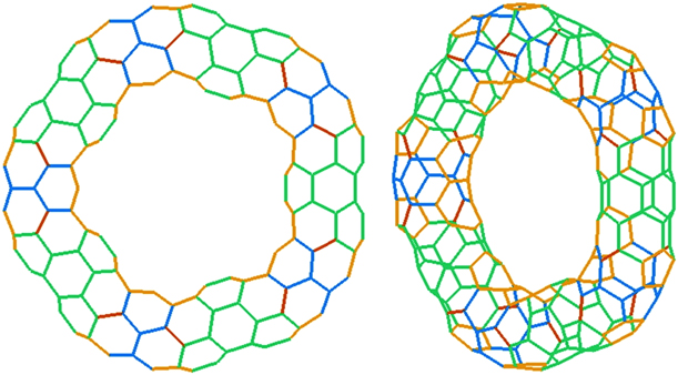
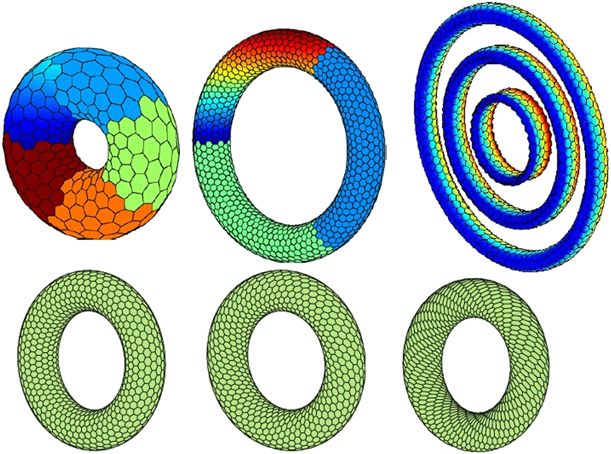
By minimizing the total ring Brenner–Tersoff potential, the equilibrium structure of nanotori was also calculated using a robust minimization method. As a result, an untwisted nanoring nice family with armchair, zig-zag, and other chiralities (Fig. 6) was discovered [Reference Pozrikidis30]. The rings having small diameters possess such cross-sectional shapes as wedding band, elliptical, and dog-bone types, meanwhile the rings having large diameters are almost circular. Also, the laser spectroscopy, time-of-flight (TOF) mass spectroscopy, and first principle ab initio calculations were used to establish the electronic structures of nanorings of C10 to C22, among others [Reference Ding and Maier31]. In case of the ring structure with alternative bond length (Peierls’ dimerization), this nanotorus was found to be unstable when having size up to C22 ring.
Selected examples of predicted types of nanorings. Reprinted with permission from Ref. Reference Pozrikidis30, Elsevier Science.

Classical MD simulations were applied to study the axial sliding in the normal nanorings on the carbon basis (NCNs, Fig. S7(a) and S7(b), see Supplementary material) and one-turn Mobius nanorings [MCNs, Fig. S7(c)] along coaxial inner carbon nanotubes [Reference dos Santos, Mendes Filho, Freire, Caetano and Albuquerque32]. It was shown that the NCN:CNT system revealed an oscillatory shift having an initial frequency of about 10 GHz reducing slowly with time during the transfer of the vibrational energy to torsional and radial modes of the nanotorus. It was established that the sliding of normal and Mobius carbon nanorings along a set of armchair nanotubes depend strongly on the topology of nanotori. The evaluation of the response of a quasi 1D ballistic carbon nanotorus to the field of an electromagnetic wave, passing to the ring plane along the normal plane, when a constant electric field is collinear to the wave field, was made [Reference Fedorov, Yanyushkina and Belonenko33]. In such conditions, the carbon nanotorus may work as a terahertz radiation source. It is possible to control effectively the dynamics of the electron in the ring by varying the incident radiation parameters.
Carbon nanotori containing distinct carbon structural units and doping elements
The MD simulations were applied to study the thermal transport across a hybrid structure formed by carbon nanorings (CNRs; Fig. S8, see Supplementary material) and two graphene nanoribbons [Reference Shi, Zhang, He, Ju and Jiang34]. It was revealed that with two covalent-bonded CNRs, the thermal conductivity of overlapped sheets of graphene considerably increases and has a top at 68.8 W/(m K), representing 28% of that of single-layer graphene sheet and being 11 times higher than that of overlapped graphene sheets without CNRs. The CNR density and diameter were emphasized as two most important parameters, influencing on the thermal transport in the CNR–graphene composite nanostructures. In another classic example of carbon–carbon nanocomposite, a carbon nanotorus containing fullerene, the van der Waals energy, is responsible for the main interaction between the two structure counterparts [Reference Sarapat, Baowan and Hill35], and it is calculated as a surface integral over the toroidal and the spherical surfaces. The most stable arrangement corresponds to the torus with the minor radius greater than 6.31 Å and major radius around 20–22 Å. Another example is a carbon nanotori containing a single-carbon atomic chain, which is symmetrically located inside the nanotorus [Reference Sumetpipat, Lee, Cox, Hill and Baowan36]. It was predicted that when the nanotorus cross-sectional radius r is higher than 3.17 Å, this carbon chain can fit inside the carbon nanotorus. A relative object is a nanotube–nanotorus hybrid, where the van der Waals forces are responsible for its formation [Reference Hilder and Hill37]. This nanotorus oscillates along the CNT axis due to the opposite and equal pulse-like forces acting at any nanotube end. Oscillation behavior and optimal configuration of the system, when atoms interact with a carbon nanotorus, are described using the Lennard-Jones potential [Reference Sarapat, Hill and Baowan38]. It was revealed that when the atom is along the torus axis in a symmetrical form, there is a linear relationship between the minor radius of the nanotorus and its optimal major radius. When the atom shifts away from this axis, the location of minimum energy also moves from the center when the ratio “major/minor radii” is higher than 0.90. In case of the presence of alien atoms in the nanotori, the doping B(N) atoms into the pentagonal (heptagonal) sites in the nanorings is thermodynamically favorable; in this case, the electronic properties of these hybrids can be influenced by B/N doping [Reference Liu, Zhang, Gao and Zhao39]. In addition to pure and doped carbon nanorings, we note well-studied sp [Reference Vojkovic, Nunez, Altbir and Carvalho-Santos2]-carbon–containing organic hydrocarbon nanorings of syn-tetrabenzoheptafulvalene [Reference Yin Cheung, Yang and Miao40], related cycloparaphenylenes (Fig. S9, see Supplementary material) [Reference Omachi, Segawa and Itami41, Reference Franklin-Mergarejo, Ondarse Alvarez, Tretiak and Fernandez-Alberti42], and their fullerene adducts [Reference Kawase and Oda43, Reference Miki, Matsushita, Inoue, Senda, Kowada and Ohe44, Reference Kawase, Tanaka, Seirai, Shiono and Oda45].
Experimental preparation of carbon nanotori
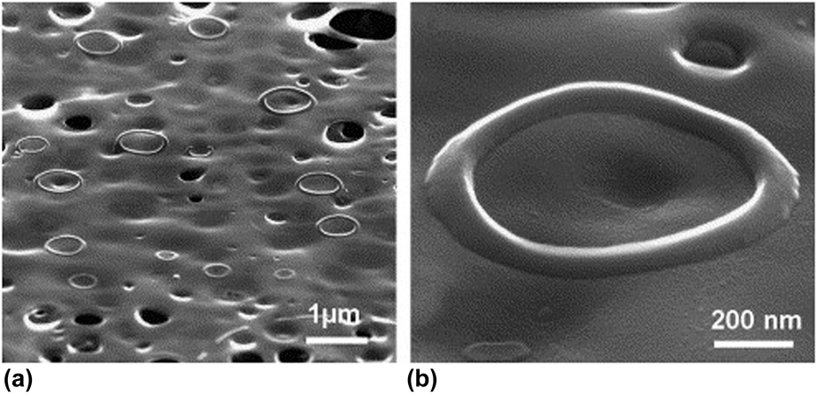
Amorphous carbon micro- and nano-sized rings (diameter of 2–10 μm and rim thickness of ∼50 nm, Fig. 7) were obtained from carbon nanostructures prepared by the carbonization of a composite of mesoporous SBA-15 rods containing Pluronic P123 polymer inside them; the alumina membrane was used as a porous medium [Reference Sai Krishna and Eswaramoorthy46]. The presence of the alumina and silica walls can efficiently prevent the oxygen diffusion during the process of calcination and contribute to an easier carbonization stage. The obtained carbon-containing nanostructures were revealed to be self-assembled around the H2O droplets in the presence of carbon tetrachloride and yield stable ring-like structures (Fig. S10, see Supplementary material).
(a) Field emission scanning electron microscopy (FESEM) image of carbon nanorings obtained after 2 h HCl treatment. (b) High magnification image of a single carbon nanoring tilted at 60° showing a 3-D ring structure. Reprinted with permission from Ref. Reference Sai Krishna and Eswaramoorthy46, Elsevier Science.

Potential applications of carbon nanotori
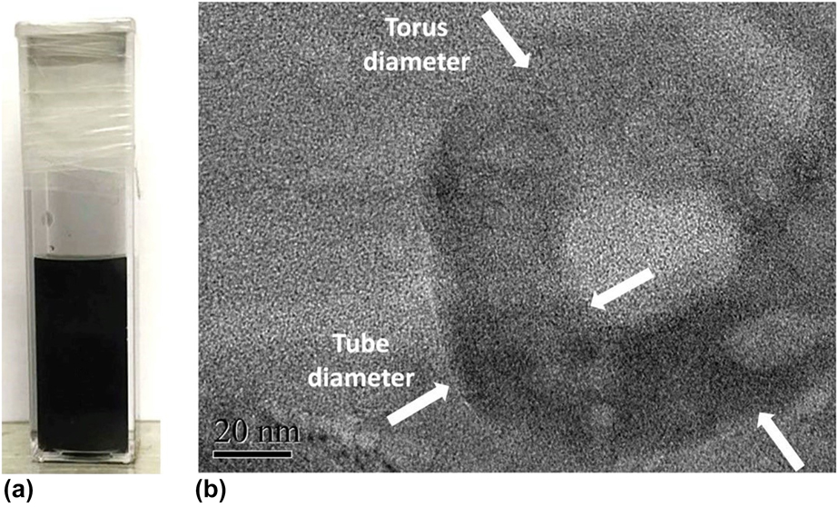
Carbon nanotori could be used as traps for atoms and ions [Reference Chan, Cox and Hill47]. According to their structure, similarly to nano-onions and fullerenes, and resistance to mechanical stresses and deformations, they also can be used as effective additives for oils. Thus, lubricants were prepared on the basis of carbon nanotori (Fig. 8) dispersed in distilled water from MWCNTs (30–50 nm in diameter) as precursors by their sequential acid and oxidative treatment steps [Reference Peña-Parás, Maldonado-Cortés, Kharissova, Itzel Saldívar, Contreras, Arquieta and Castaños48], demonstrating significant improvements in load-carrying capacity (up to 950% with 2.0 wt%), and excellent anti-wear (96%) and friction-reducing (76%) properties. These improvements can be attributed (Fig. S11, see Supplementary material) to their hollow multiwalled toroidal shape and high elastic modulus that enabled them to withstand the loads of the tests, thus limiting the metal–metal contact. We note that these carbon nanotori obviously contain a host of oxygen-containing groups, taking into account their preparation method in highly oxidative media.
(a) Carbon nanotori suspensions in DW, (b) TEM image of carbon nanotori suspended in DW. Reprinted with permission from Ref. Reference Peña-Parás, Maldonado-Cortés, Kharissova, Itzel Saldívar, Contreras, Arquieta and Castaños48, Elsevier Science.

Nanotori of metals, metal oxides, and salts
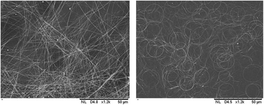
Ring-type nanostructures of noble metals are relatively common, especially, Ag and Au [Reference Gong, Liu, Yu, Wu and Yin49, Reference Shamraiz, Raza, Hussain, Badshah, Green, Ahmad Kiani and Al-Harrasi50]. Thus, reducing Ag+ ions in trisodium citrate buffers using NaBH4, coloidal nanorings were prepared, whose shape was found to be dependent on the reaction temperature, NaBH4/AgNO3 ratio, and pH [Reference Drogat, Granet, Sol and Krausz51]. Silver nanorings with almost ideal circular shape (Fig. S12, see Supplementary material) can also be produced by a solvothermal route (pressure 150 kPa) with high yields [Reference Azani and Azin Hassanpour52]. These nanorings can be easily dispersed in EtOH and can form conductive films (Fig. 9) on the polyethylene terephthalate (PET) substrate via spray coating, thus could be potentially used as optoelectronic materials.
Scanning electron microscopy (SEM) images of sprayed films of silver nanowires (left) and nanorings (right) on PET substrate. Reprinted with permission from Ref. Reference Azani and Azin Hassanpour52, Wiley.

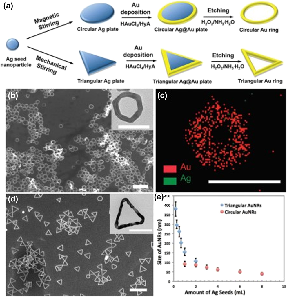
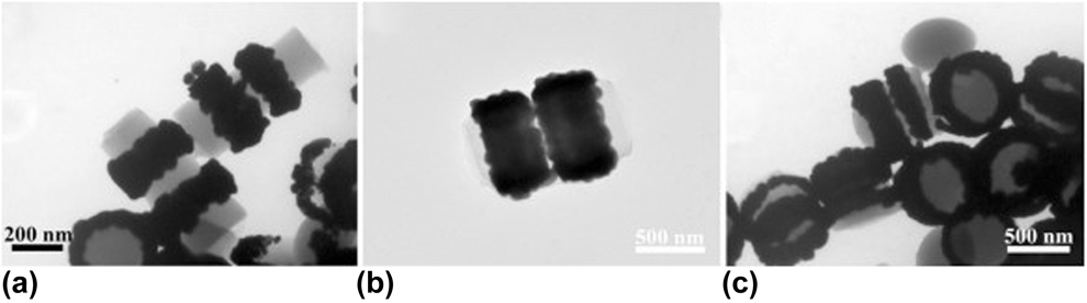
Gold nanorings are most common circle-type structures and have got various applications in the catalysis, electronic devices, bioimaging, and biosensors. There are several techniques for their fabrication. Thus, monodispersed triangular and circular gold nanorings with smooth surface were recently produced using HAuCl4 as a precursor by a scalable method (Fig. 10), including several steps, such as Au deposition on Ag nanoplates, silver elimination by mild chemical etching with H2O2–NH3 mixture, and further deposition of gold to reach necessary thickness (5–30 nm) and diameter (50–400 nm) of toroidal structures [Reference Lin, Liu, Lin, Zhang and Nie53]. The parameters of metal deposition process and template size directly influence on the size and shape of final products. Also, Pd and Au nanorings and their arrays were prepared from H2PdCl4 and HAuCl4 as raw materials and hydroxylamine hydrochloride and L-ascorbic acid solution as reducing agents, respectively, by a route on the basis of colloidal lithography (where polymer arrays of nanorods were first formed and further used as templates for Au adsorption) and nanoparticle polymer-assisted self-assembly (Fig. S13, see Supplementary material) [Reference Chen, Mu, Fang, Shen, Zhang and Yang54]. Thickness of polymer spacing layer influences on the distance between neighboring Au nanorings. This method is characterized by high yields of dispersible nanorings and their arrays (Fig. 11). Well-ordered arrays of Au nanocrescents and nanorings were fabricated by a nanosphere lithographic approach [Reference Lewicka, Li, Bohloul, Yu and Colvin55]. Comparing disk- and ring-like Au nanoparticles, it was established that the excitation wavelengths of Au nanoparticles depend much on the particle geometry, exhibiting near-IR resonance in case of Au torus, whereas similar disk-type Au nanoparticles do not reveal this effect [Reference Aizpurua, Blanco, Hanarp, Sutherland, Kall, Bryant and Garcıa de Abajo56].
(a) Schematic illustration for the synthesis of circular and triangular gold nanorings (AuNRs). (b and c) SEM and transmission electron microscopy (TEM) (inset) images of circular AuNRs with a diameter of 83 ± 12 nm (b) and triangular AuNRs with an edge length of 115 ± 22 nm (c). (d) EDX mapping of Au and Ag in a circular AuNR. (e) The average diameter/edge length of AuNRs as a function of the amount of Ag seeds added in the step of template synthesis. Scale bars are 300 nm in (b and c), and 100 nm in (d) and all insets. Reprinted with permission from Ref. Reference Lin, Liu, Lin, Zhang and Nie53, Royal Society of Chemistry.

TEM images of gold nanorings with different diameters. The inner diameters are 200 nm (a), 420 nm (b), and 540 nm (c). Reprinted with permission from Ref. Reference Chen, Mu, Fang, Shen, Zhang and Yang54, Springer.

In addition to individual gold nanorings above, mixed-metal Au/Co/Au magnetoplasmonic nanorings are known [Reference Feng, Luo, Kekesi, Granados, Meneses‐Rodríguez, García, García‐Martín, Armelles and Cebollada57], exhibiting multiresonant properties and magneto–optical activity, and they can be applied in isolators, optical communications, and sensing. Non-noble nanorings are represented by aluminum nanorings (90 nm in diameter), prepared by electron beam lithography [Reference Lehr, Dietrich, Helgert, Käsebier, Fuchs, Tünnermann and Kley58].
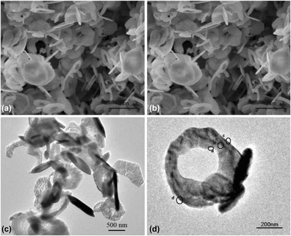
A few metal oxides have been found in circular form, as, e.g., α-Fe2O3 nanorings [Reference Cao, Cao, Sun and He59], hydrothermally prepared at 180 °C for 96 h using sulfate and phosphate ions as additives. Their size can vary depending on ratios of these additives to ferric ions. A classic nano-object, zinc oxide, can exist in a large variety of nanostructures, in particular in those with toroidal form. For obtaining zinc oxide nanorings, solid-vapor processes are generally applied. Thus, flexible ZnO nanobowls and nanorings, possessing superior mechanical toughness, can be produced at high purity and yielded in large quantities by physical evaporation of ZnO powder [Reference Hughes and Wang60]. Their morphology can be tuned by pressure variations during the synthesis process. Alternatively, during the growth of polar nanobelts by a solid-vapor process using ZnO–In2O3 and Li2CO3 as raw materials, ZnO freestanding single-crystal toroidal structures were formed [Reference Kong, Ding, Yang and Wang61]. In this process, a circular folding of a nanobelt led to the nanoring formation because of electrostatic interactions and nanobelt loop-by-loop winding. On the contrary, S-doped MoO2 ring-in-ring architectures (0.5 nm in size, which means 2–3 atomic layers only), resembling carbon nano-onions, were fabricated by scalable solvothermal route (200 °C, 6 h) from thiourea and ammonium heptamolybdate [(NH4)6Mo7O24·4H2O] as starting materials, oleyl alcohol as solvent and oleyamine as a surfactant [Reference Yang, Yang, Chen, Lu, Song, Wei and Wang62]. The ligand–surface interactions and sulfur doping were established to be the main contributors in the formation of the ring structure. In case of SiO2 nanotori, no real experimental data are available, except a patent [Reference Narducci, Cerofolini and Romano63] and theoretic calculations of its distance-based topological indexes [Reference Arockiaraj, Klavžar, Mushtaq and Balasubramanian64]. Also, Co(OH)2 nanorings (Fig. 12) and nanodisks were hydrothermally prepared from Ni and Co acetates as precursors at different molar ratios and hydrazine monohydrate as the reducing agent without the use of surfactants [Reference Chen, Wang and Guo65]. The Ni(OH)2 redissolution under extreme hydrothermal conditions (high pressure and temperature) was possibly the principal factor of formation of Co(OH)2 nanorings.
Morphology of the hexagonal Co(OH)2 nanoring: (a, b) SEM images of the hexagonal ring; (c, d) TEM images of the hexagonal Co(OH)2 nanoring (side view). Reprinted with permission from Ref. Reference Chen, Wang and Guo65, Springer.

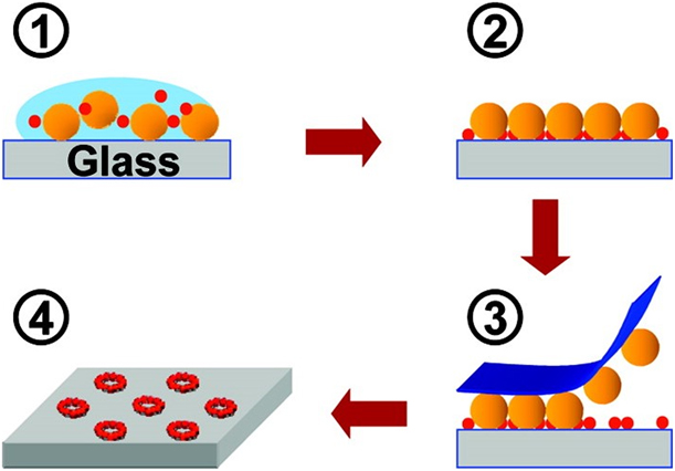
As well as in case of ZnO (see above), thermal evaporation methods also serve for zinc chalcogenides. Thus, evaporating ZnSe powder, its free-standing nanowires and nanorings (wurtzite phase; thickness 50–100 nm) were obtained on Si substrates coated with gold [Reference Leung and Choy66]. ZnSe shape (Fig. S14, see Supplementary material) can be controlled during the growth process by varying only the pressure and maintaining other conditions the same. It was proposed that the favorable nucleation of hexagonal phase leads to the appearance of metastable wurtzite phase. In addition, 2D arrays of 4-nm CdSe nanorings, made from quantum dots (QDs), were prepared by an evaporative method using hexagonal arrays of polystyrene microspheres as templates (Fig. 13) [Reference Chen, Liao, Chen, Yang, Wark, Son, Batteas and Cremer67]. Atomic force microscopy studies (Fig. S15, see Supplementary material) showed that the nanostructures containing at least six layers (each layer of 5 nm) of nanoparticles are formed in the ratio of 104:1 QDs:polymer spheres and the number of layers decreases to three in the ratio of 4000:1 and 1–2 at 1000:1, sometimes containing individual QDs in a single-layer nanoring.
Schematic diagram of the evaporation templating procedure employed for forming CdSe nanorings (red particles) on planar substrates using microsphere templates (orange particles). The drawing is not to scale. Reprinted with permission from Ref. Reference Chen, Liao, Chen, Yang, Wark, Son, Batteas and Cremer67, American Chemical Society.

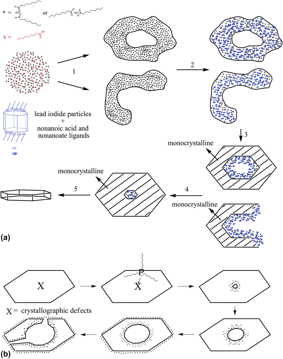
PbI2 nanorings were prepared by several steps (Fig. 14), including the formation of 2D structures (nanosheets) in nonanoic acid and their subsequent etching in trioctylphosphine presence, which acts in the weakest central area of this intermediate structure [Reference Klein, Heymann, Hungri, Lesyuk and Klinke68]. This last step is responsible for the hole size in the formed rings (thickness 20–85 nm and outer diameter 2–10 μm). Studies of the formation mechanism revealed that the first prepared nanosheets in nonanoic acid micelles at an early stage of the reaction were further transformed to rings. The formation of the clouds occurs due to polarity. These nanorings can be potentially applied in photodetectors, showing high gain values, fast response, and other important characteristics. Nitride derivatives are also known in the nanotorus form. Thus, Si3N4-core SiO2-shell nanotori with average diameter of 2–10 μm, obtained by the chemical vapor deposition (CVD) method at 1350 °C in a horizontal quartz tube from NH3, a mixture of Si (previously pulverized for 24 h by high-energy ball milling) and SiO2 powders, can emit a peak of 3.0 eV at 412 nm [Reference Du, Zhang and Li69]. In addition, boron nitride (BN) nanotori were studied by MD simulations, showing thermal stability at more than 3400 K [Reference Loh and Baillargeat70]. Simulations were used also for the elucidation of BaTiO3 nanotori properties [Reference Thorner, Kiat, Bogicevic and Kornev71].
Proposed mechanism for the formation of PbI2 nanorings. Reprinted with permission from Ref. Reference Klein, Heymann, Hungri, Lesyuk and Klinke68, Royal Society of Chemistry. (See complete caption in Supplementary material.)

Conclusions and further outlook
The field of nanorings/nanotori is currently not well-developed. Main contributions in this field correspond to the carbon nanotori; several metals (Ag, Au, and Al), mixed-metal nanorings (Au/Co/Au), metal (ZnO, MoO2, and Fe2O3) and nonmetal (SiO2) oxides, hydroxides [Co(OH)2], and salts (PbI2 and metal selenides) are also known, possessing distinct sizes, structural varieties, and stability. The carbon nanorings are closely related to edges in nanotubes, nanochains, graphene, and nanocones, and they can be “constructed” from carbon nanotubes. Nanorings/nanotori possess several useful properties (studied mainly by ab initio DFT calculations), in particular multiresonant properties and magneto-optical activity, paramagnetism, and ferromagnetism (even for carbon, which is normally a nonmagnetic element, whose nanotori possess magnetic response as a derivative of curvature and disorder). These nanostructures can be applied in isolators, optical communications, sensing, as traps for atoms and ions, counterparts in lubricants, etc., showing considerable improvement in material properties. Because of the insufficient number of reports on toroidal nanostructures of other inorganics in addition to the above noted, one could suppose that a considerable higher number of inorganic systems need to be prepared in this form, looking for their unusual properties. First of all, more transition metals in elemental form, as well as their oxides, could exist in this form and exhibit new properties and corresponding applications. Upon certain efforts, more variety of metal and nonmetal oxides, as classic objects in nanotechnology, could also be got in this unusual form, providing other applications.
Supplementary material
To view supplementary material for this article, please visit https://doi.org/10.1557/jmr.2019.370.
Acknowledgment
The authors are very grateful to César Máximo Oliva González, M.Sc. (UANL), for technical assistance in the preparation of this manuscript.