Introduction
The 129 inbred mouse strain is useful as an animal model of testicular teratoma (Stevens, Reference Stevens1967; Matin, Reference Matin2007), and it greatly contributes to production of genetically engineered mice as a supplier of embryonic stem (ES) cells (Evans & Kaufman, Reference Evans and Kaufman1981; Martin, Reference Martin1981). Apart from these useful traits for genetic research, it has been reported that the litter size of 129 mice is considerably smaller than that of other popular inbred strains such as C3H and C57BL/6 (Verley et al., Reference Verley, Grahn, Leslie and Hamilton1967; Nagasawa et al., Reference Nagasawa, Miyamoto and Fujimoto1973; Festing, Reference Festing1979). The low reproductive performance of 129 mice may be attributable to failure in fertilization rather than embryonic death because the in vitro fertilization (IVF) rates are usually low (Sztein et al., Reference Sztein, Farley and Mobraaten2000; Byers et al., Reference Byers, Payson and Taft2006; Kawai et al., Reference Kawai, Hata, Suzuki and Matsuda2006). Our previous study found that the in vivo fertilization rate was less than 50% even when 129 females were mated with C57BL/6J males with normal fertility (Hino et al., Reference Hino, Oda, Nakamura, Toyoda and Yokoyama2009). These findings strongly suggest that the low fertilization rate of 129 mice arises from oocytes. One of the possible causative factors is chemical alteration of the zona pellucida enclosing the oocyte; thus, spermatozoa cannot pass through the structure. Another is that the capability of the oolemma to fuse with the sperm plasma membrane is low in this strain.
To determine the exact causative factor(s) of the low fertilization rates of strain 129 mice, the sensitivity of the zona pellucida to protease and the fertilizability of oocytes were examined at different times after ovulation. The capability of the oolemma to fuse with the sperm plasma membrane was evaluated by IVF assay using zona-free oocytes. In addition, development of fertilized eggs was followed up to full term.
Material and methods
Animals
The 129 inbred mouse strain used in the present study consisted of two 129 substrains; 129+Ter/Sv mice were purchased from CLEA Japan and 129/SvEv mice were purchased from Biological Research Laboratories. Inbred C57BL/6J mice with normal reproductive performance were purchased from CLEA Japan and used as a standard of comparison. Females from 2 to 4 months of age and males from 3 to 4 months of age (strain 129 and C57BL/6J mice) were used in the experiments. A mature MCH (ICR) hybrid mouse strain from CLEA Japan served as recipients of embryo transfer. The 129+Ter/Sv, 129/SvEv, and C57BL/6J were referred to as 129T, 129S and B6/J, respectively. All mice were kept under specific pathogen-free conditions for at least 1 week before use. They were fed ad libitum under controlled lighting conditions (light: 08:00 h to 20:00 h) at a temperature of 23 ± 1 °C and humidity of 55 ± 10%. All experimental procedures were approved by the Animal Care and Use Committee of the Mitsubishi Kagaku Institute of Life Sciences.
Media
Organic and inorganic reagents were purchased from Wako Pure Chemical Industries, Ltd, unless specifically stated. The medium used for oocyte collection and in vitro fertilization (IVF) was TYH medium (Toyoda et al., Reference Toyoda, Yokoyama and Hosi1971a). The culture medium for embryos was modified Whitten medium (mWM) (Nomura & Katsuki, Reference Nomura and Katsuki1987), which consists of 109.51 mM NaCl, 4.78 mM KCl, 1.19 mM KH2PO4, 1.19 mM MgSO4·7H2O, 22.62 mM NaHCO3, 5.56 mM glucose, 0.23 mM sodium pyruvate (Nacalai Tesque), 1.49 mM calcium lactate·5H2O, 75 mg/l penicillin G potassium (Meiji Seika), 50 mg/l streptomycin sulphate (Meiji Seika), 0.01 mM 2-mercaptoethanol (Nacalai Tesque), 0.05 mM EDTA·2Na (Nacalai Tesque), 1 mg/l phenol red, and 3 g/l bovine serum albumin (BSA) (Yagai Co. Ltd).
Time of ovulation after hCG injection
Females were intraperitoneally injected with 7.5 IU eCG followed 48 h later with 7.5 IU hCG. They were sacrificed by cervical dislocation at 1-h intervals from 10 to 15 h after hCG injection and their oviducts were removed. When cumulus-enclosed oocytes were detected in the ampullary region of the oviducts, they were collected and put in a droplet (50 μl) of TYH medium containing 0.01% hyaluronidase (Sigma-Aldrich) under paraffin oil (Fisher Scientific). Five to 10 min later, cumulus-free oocytes were washed twice with TYH medium. The number of oocytes was recorded.
Dissolution of the zona pellucida by chymotrypsin
Cumulus-enclosed oocytes were obtained from females at 14, 17, and 20 h after hCG injection and 1-cell embryos from females 1 day after mating (24 h after hCG injection). After removal of the cumulus cells by treatment with 0.01% hyaluronidase, they were washed thoroughly with TYH medium and transferred to droplets (50 μl; two to four oocytes/embryos per droplet) of TYH medium containing 1.5 IU α-chymotrypsin (Sigma-Aldrich) under paraffin oil at 37 °C under 5% CO2 in air. They were observed for the absence of the zona pellucida at 10, 30, 60, 120, 180, and 280 min. For each case, 46 to 73 oocytes and 26 to 33 1-cell embryos were examined.
IVF assay with zona-intact oocytes
To examine passage of spermatozoa through the zona pellucida, IVF was performed according to the procedure by Toyoda et al. (Reference Toyoda, Yokoyama and Hosi1971a,Reference Toyoda, Yokoyama and Hosib). Spermatozoa obtained from the cauda epididymis of 129T, 129S, and B6/J males were introduced into a droplet (300 μl) of TYH medium under paraffin oil and were incubated for 1.5 to 2 h at 37 °C under 5% CO2 in air to induce capacitation.
Cumulus-enclosed oocytes were obtained at 14 and 17 h after hCG injection. They were transferred to a droplet (300 μl) of TYH medium, and inseminated by adding the preincubated sperm suspension. The final concentration of spermatozoa at the time of insemination was 150–200 cells/μl. In every IVF experiment, two to three females were used.
Five to 6 h after insemination, the eggs were washed thoroughly with mWM, and the formation of pronuclei and extrusion of a second polar body were microscopically examined. Ova with both male and female pronuclei and a second polar body were recorded as monospermic ova, those with more than two pronuclei and a second polar body as polyspermic ova, and those with one pronucleus as parthenogenetic ova.
IVF assay with zona-free oocytes
The cumulus-enclosed oocytes were obtained from 129T and B6/J females at 16 h after hCG injection. Cumulus cells were dispersed by hyaluronidase treatment, and the zona pellucidae were completely dissolved in acidic Tyrode's solution (pH 2.5) followed by three rapid washes in TYH medium. The zona-free eggs were directly transferred to a droplet (300 μl) of TYH medium containing 129T spermatozoa following preincubation for 1.5 to 2 h at a concentration of 3 and 10 cells/μl. Insemination was completed 17 h after hCG injection. Nine hours after insemination, the number of pronuclei was scored.
Cortical granule staining and quantification
Zona-free eggs of 129T, 129S, and B6/J females were obtained 14 and 17 h after hCG injection as mentioned above. To examine whether the spontaneous release of cortical granules occurs in vitro, some of cumulus-enclosed oocytes recovered 14 h after hCG injection were cultured in TYH medium for 3 h. The zona-free oocytes were fixed in 3.7% paraformaldehyde in Dulbecco's PBS (D-PBS) for 30 min and blocked in D-PBS containing 0.3% BSA (blocking solution). They were washed three times in blocking solution, and then permeabilized in D-PBS containing 0.1% Triton X-100 for 5 min. After washing three times in blocking solution, they were incubated for 30 min in D-PBS containing 100 μg/ml FITC conjugate-lens culinaris agglutinin LCA (Sigma-Aldrich) which specifically attaches to the cortical granules (Ducibella et al., Reference Ducibella, Anderson, Albertini, Aalberg and Rangarajan1988). They were washed three times in blocking solution and mounted with Vectashield containing DAPI (Vector, RL-1000). All procedures were conducted at room temperature.
The cortical granule density in 100 μm2 area of the cortex was determined under fluorescence microscope by counting LCA-labeled cortical granules. For each case, 26 to 39 oocytes from three to four females were examined.
Embryo transfer
Some of monospermic zygotes produced by IVF assay with 129T and 129S zona-intact oocytes recovered at 14 and 17 h after hCG injection were cultured in mWM medium under 5% CO2 in air at 37 °C. On the next day, resultant 2-cell embryos were transferred into the oviduct of pseudopregnant ICR females. Two to four females were used as recipients, and 20 embryos were transferred into each recipient. Pregnant females were either sacrificed 19 days after the transfer or allowed to deliver in order to count live pups.
Statistical analysis
Fertilization rates of zona-intact oocytes were analyzed by one-way ANOVA after transformation into arcsine values. The average number of ovulated oocytes and density of cortical granules were compared by Student's t-test. Results of IVF assay with zona-free oocytes and embryo development were analyzed by chi-squared test. Differences were considered to be significant with p < 0.05.
Results
Time of ovulation after hCG injection
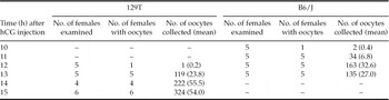
Table 1 presents the number of females with oocytes in the oviducts and the number of oocytes recovered at different times after hCG injection in 129T and B6/J strains. In 129T, oocytes were recovered from only one female at 12 h after hCG injection and from all females at and after 13 h after hCG injection. The number of oocytes recovered reached the maximum level at 14 h after hCG injection, indicating that ovulation was completed between 12 and 14 h after hCG injection in this strain. By contrast, all females had ovulated by 12 h after hCG injection in B6/J. Thus, it appears that ovulation after hCG injection in 129T females occurs approximately 2 h later compared to B6/J females. Interestingly, the mean number of oocytes recovered on completion of ovulation was obviously larger in 129T than in B6/J (p < 0.01).
Ovulation in 129T and B6/J females at different intervals after hCG injection.

Change in sensitivity of the zona pellucida to chymotrypsin
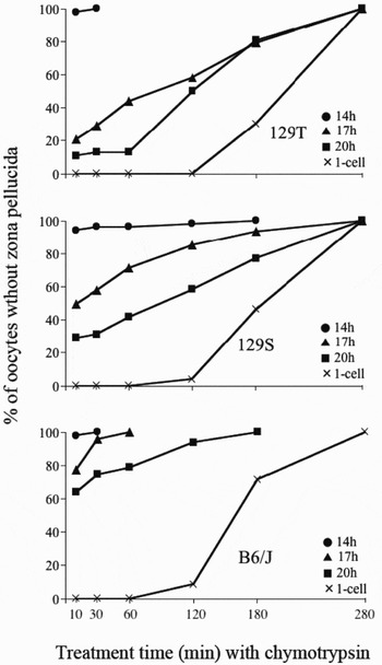
Sensitivity of the zona pellucida of oocytes to chymotrypsin was markedly dependent upon both the time after ovulation and the mouse strain (Figure 1). When oocytes were recovered at 14 h after hCG injection (shortly after ovulation) in 129T and 129S, the zona pellucida was usually digested by the enzyme within 10 min. However, the structure became considerably resistant to the enzyme when oocytes were recovered at 17 h (approximately 3 h after ovulation) and 20 h (approximately 6 h after ovulation) after hCG injection. In B6/J oocytes, a high sensitivity of the zona pellucida to chymotrypsin persisted until 17 h after hCG injection (approximately 5 h after ovulation). Even in oocytes recovered at 20 h after hCG injection (approximately 8 h after ovulation), digestion of the zona pellucida was seen in more than 90% of oocytes by 120 min. The zona pellucida of 1-cell embryos of all strains was highly resistant to the enzyme until 120 min.
Difference in dissolution time of the zona pellucida by chymotrypsin among oocytes recovered at 14, 17, and 20 h after hCG injection in 129T, 129S and B6/J mice.

IVF assay with zona-intact oocytes
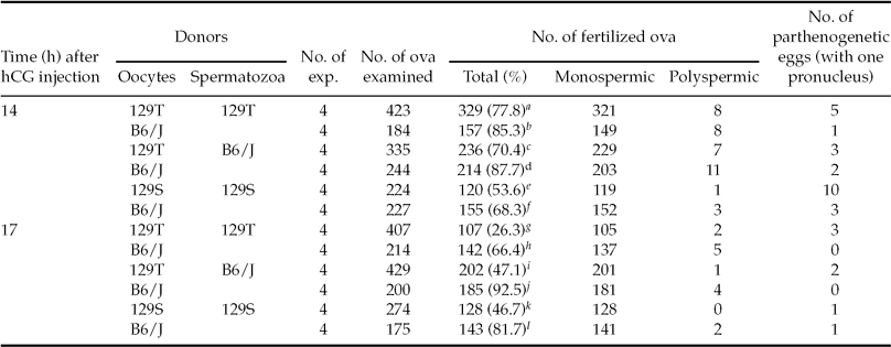
Results of the IVF assay are presented in Table 2. In the IVF assay with 129T oocytes, B6/J spermatozoa were also used for insemination to check on the results obtained by 129T spermatozoa. When 129T oocytes recovered 14 h after hCG injection were used, fertilization rates were similar to those of B6/J oocytes regardless of donors of spermatozoa. A similar result was found when 129S oocytes were inseminated 14 h after hCG injection. When 129T oocytes were inseminated 17 h after hCG injection, however, the fertilization rates were significantly reduced and the low fertilization rate was inadequately improved by use of B6/J spermatozoa. Consequently, the fertilization rates of 129T oocytes recovered 17 h after hCG injection were much lower than that of B6/J oocytes recovered at the same time. When 129S and B6/J oocytes were inseminated 17 h after hCG injection, the fertilization rate was significantly lower in 129S oocytes than in B6/J oocytes. In this study, monospermy was seen in more than 90% of fertilized eggs regardless of the time of oocyte recovery after hCG injection and origin of gametes.
IVF assay with 129T, 129S and B6/J zona-intact oocytes recovered at 14 and 17 h after hCG injection.
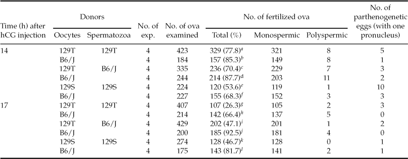
Statistical significance of B6/J oocytes: g vs. h (p < 0.05), i vs. j (p < 0.01), and k vs. l (p < 0.01).
Statistical significance between 14 and 17 h post-hCG: a vs g (p < 0.05).
IVF assay with zona-free oocytes
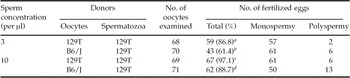
Immediately after the zona-free oocytes were placed in a droplet of sperm suspension, they were quickly attached by some spermatozoa. In both low and high sperm concentrations, fertilization rates were significantly higher in 129T oocytes than in B6/J oocytes (Table 3), indicating that the capability of oolemma of 129 mouse oocytes to fuse with sperm plasma membrane never deteriorated up to at least 3 h after ovulation.
IVF rate of 129T and B6/J zona-free oocytes inseminated 17 h after hCG injection.

Statistical significance of B6/J oocytes: a vs. b (p < 0.01), c vs. d (p < 0.05).
Observation of cortical granules
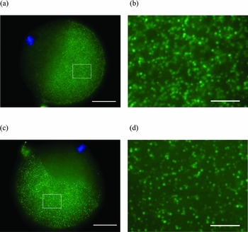
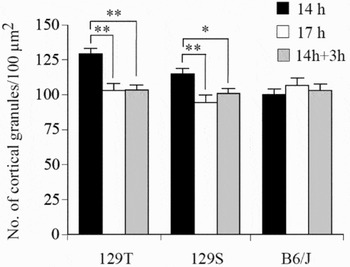
All of 129T and 129S oocytes recovered at 14 and 17 h after hCG injection showed typical metaphase II configuration and cortical granule domain (Figures 2a and 2c). However, the density of cortical granules in 129T and 129S oocyte recovered 17 h after hCG injection significantly decreased (Figures 2b, 2d and 3).This reduction occurred when oocytes recovered 14 h after hCG injection were cultured in vitro for further 3 h. Although the density of cortical granules in B6/J oocytes recovered 14 h after hCG injection was low, there was no such reduction in density of cortical granules. Thus, partial release of cortical granules occurred in a time-dependent manner with 129T and 129S oocytes even when the oocytes were cultured in vitro.
Fluorescence micrographs of cortical granules in oocytes of 129T mice. (a) Oocyte recovered 14 h after hCG injection; (b) higher magnification of the frame in (a); (c) oocyte recovered 17 h after hCG injection; (d) higher magnification of the frame in (c). Scale bar represents 30 μm in (a) and (c), and 5 μm in (b) and (d), respectively.

Comparison of cortical granule density in oocytes recovered 14 and 17 h after hCG injection in 129T, 129S and B6/J mice. White and black bars represent the cortical granule density when oocytes were recovered 14 and 17 h after hCG injection, respectively. Grey bars represent the cortical granule density when oocytes recovered 14 h after hCG injection was cultured in vitro for 3 h. *p < 0.05; **p < 0.01.

Embryo development
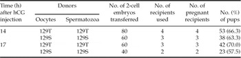
In an IVF assay with oocytes recovered 14 and 17 h after hCG injection, almost all (98–100%) of monospermic zygotes in 129T and 129S developed to 2-cell embryos. After embryo transfer, all of the recipient females became pregnant (Table 4). The embryos of both substrains well developed to term and there was no significant difference in percentage of live pups delivered between both oocyte recovery times after hCG injection. These percentages in 129T and 129S were comparable with that obtained in our previous study (Suzuki-Migishima et al., Reference Suzuki-Migishima, Hino, Takabe, Oda, Migishima, Morimoto and Yokoyama2009).
Offspring from 129T and 129S embryos produced by IVF assay with oocytes recovered 14 and 17 h after hCG injection.

Discussion
The present study found that the zona pellucida of 129 mouse oocytes became resistant to chymotrypsin approximately 3 h after ovulation (17 h after hCG injection), and concomitantly rates of successful IVF in the 129 mouse oocytes significantly decreased; however, their oolemma maintained the capability to fuse with the sperm plasma membrane. The decrease of IVF rates persisted even when B6/J spermatozoa were used. These findings indicate that the zona pellucida of 129 mouse oocytes primarily hampers fertilization. Because there was a significant reduction in the density of cortical granules, the partial release of cortical granules might have caused the zona pellucida to become resistant to the enzyme.
Usually, the zona pellucida acquires resistance to proteases after penetration of the spermatozoa into the oocyte cytoplasm (Smithberg, Reference Smithberg1953; Krzanowska, Reference Krzanowska1972; Mintz & Gearhart, Reference Mintz and Gearhart1973; Schmell & Gulyas, Reference Schmell and Gulyas1980; Gulyas & Yuan, Reference Gulyas and Yuan1985), ensuring an oocyte monospermy (Barros & Yanagimachi, Reference Barros and Yanagimachi1971; Sato, Reference Sato1979). Xu et al. (Reference Xu, Abbott, Kopf, Schultz and Ducibella1997) reported that spontaneous activation, which is accompanied by release of some cortical granules, modification of the zona pellucida, and transition of metaphase to anaphase, occurs in post-ovulatory aged oocytes of CF-1 mice. Compared with freshly ovulated oocytes, the aged mouse oocytes were reportedly susceptible to artificial parthenogenetic stimuli (Fulton & Whittingham, Reference Fulton and Whittingham1978; Collas et al., Reference Collas, Balise, Hofmann and Robl1989; Kubiak, Reference Kubiak1989; Xu et al., Reference Xu, Abbott, Kopf, Schultz and Ducibella1997). Therefore, partial release of cortical granules found in 129 mouse oocytes approximately 3 h after ovulation might have resulted from spontaneous activation of the oocytes. However, it appeared that the activation was too weak to induce the transition of metaphase to anaphase because there were no oocytes exhibiting the spindle of anaphase configuration.
In most mammalian species, the epithelial cells of the oviducts secrete glycoproteins (OGPs), which can interact with the zona pellucida of ovulated oocytes (Buhi, Reference Buhi2002). Coy et al. (Reference Coy, Cánovas, Mondéjar, Saavedra, Romar, Grullón, Matás and Avilés2008) reported that the OGPs made the zona pellucida of bovine and swine oocytes resistant to proteases and thereby contributed to prevent polyspermy. More recently, it has been found that OGPs function as adhesive ligands for mouse spermatozoa (Lyng & Shur, Reference Lyng and Shur2009). However, it remains unknown whether OGPs can chemically alter the zona pellucida to block polyspermy.
Our previous study demonstrated that 129 female mice showed reduction in litter size after natural mating (Hino et al., Reference Hino, Oda, Nakamura, Toyoda and Yokoyama2009). The time interval between ovulation and sperm penetration in vivo has been estimated to be 3 to 5 h in spontaneously ovulated females and 1 to 3 h in superovulated females in common mouse strains (Braden & Austin, Reference Braden and Austin1954; Edwards & Gates, Reference Edwards and Gates1959; Braden, Reference Braden1962). If this is the case in 129 mice, the small litter size following natural mating could be due to poor fertilization via chemical alteration of the zona pellucida before penetration of spermatozoa.
Sztein et al. (Reference Sztein, Farley and Mobraaten2000) and Byers et al. (Reference Byers, Payson and Taft2006) reported that 129 female mice had relatively low IVF rates (53% and 24%, respectively) even when oocytes were recovered 13 to 14.5 h after hCG injection; however, in our study, IVF rates with oocytes recovered 14 h after hCG injection were 78% in 129T mice and 54% in 129S mice. According to our results, ovulation would be either in progress or finished at this time; therefore, the oocytes should maintain normal fertilizability. Although it is difficult to directly compare our results with previous results because of different IVF methodologies, the time lag of fertilization following insemination may cause a discrepancy in the IVF rates between studies. In the present study, spermatozoa were adequately capacitated by preincubation for 90 to 120 min; however, the sperm preincubation was short (about 10 min) in the previous studies (Sztein et al., Reference Sztein, Farley and Mobraaten2000; Byers et al., Reference Byers, Payson and Taft2006). Toyoda et al. (Reference Toyoda, Yokoyama and Hosi1971b) reported that the passage of spermatozoa through the zona pellucida was delayed following the preincubation for less than 15 min compared with the preincubation for 60 to 120 min because of inadequate sperm capacitation. It is therefore possible that the low IVF rates reported in the previous studies may be explained by the alteration of the zona pellucida before the penetration of spermatozoa through it.
In humans, even though production of spermatozoa is normal in number and motility, fertilization failure (0%) and/or low fertilization (<25%) occurs in 4–20% of the couples undergoing IVF (Barlow et al., Reference Barlow, Englert, Puissant, Lejeune, Delvigne, Van Rysselberge and Leroy1990; Molloy et al., Reference Molloy, Harrison, Breen and Hennessey1991; Roest et al., Reference Roest, Van Heusden, Zeilmaker and Verhoeff1998). Although the causes remain unclear, the possibility exists that spermatozoa fail to pass through the zona pellucida. Olds-Clarke (Reference Olds-Clarke1996) reported that not only the sperm velocity but also the quality of the zona pellucida influences the success of IVF. Männikkö et al. (Reference Männikkö, Törmälä, Tuuri, Haltia, Martikainen, Ala-Kokko, Tapanainen and Lakkakorpi2005) reported that gene mutation affecting the structure of the zona pellucida is associated with the IVF failure. If an adverse alteration of the zona pellucida actually occurs in the oocytes of the infertile women, the 129 mice may be useful as a relevant animal model of unexplained fertilization failure.
In conclusion, 129 mouse oocytes exhibit a short fertilizable life span. This is due to accelerated alteration of the zona pellucida, which is probably caused by the spontaneous release of cortical granules. The use of oocytes immediately after ovulation can improve the IVF rate and enhance the reproductive efficiency of 129 inbred mouse strains.
Acknowledgements
We thank Dr R. Yanagimachi, University of Hawaii, for invaluable discussion and comments. We also wish to acknowledge to Ms Y. Motegi and Ms C. Kaneko for their technical assistance, and Dr. S. Kamijo for caring for the animals. This study was supported by Special Coordination Funds of Ministry of Education, Culture, Sports, Science, and Technology in Japan (M.Y.).