Introduction
Body dysmorphic disorder (BDD) is a mental disorder characterized by a preoccupation with an imagined defect in physical appearance, or if a slight abnormality is present, the concern about it is excessive. Individuals with BDD often engage in repetitive and ritualistic behaviours, including skin picking, camouflaging their supposed defect, and checking their appearance in the mirror. The prevalence of BDD is approximately 1.8% in community self-report samples (Buhlmann et al. Reference Buhlmann, Glaesmer, Mewes, Fama, Wilhelm, Brähler and Rief2010), with slightly higher prevalence among females. Individuals with BDD have impaired psychosocial functioning and quality of life (Phillips et al. Reference Phillips, Menard, Fay and Weisberg2005b), they may experience prolonged unemployment, severe social isolation and suicidal ideation, with approximately 25% of individuals with BDD attempting suicide (Phillips et al. Reference Phillips, Coles, Menard, Yen, Fay and Weisberg2005a; Buhlmann et al. Reference Buhlmann, Glaesmer, Mewes, Fama, Wilhelm, Brähler and Rief2010). Many individuals with primary BDD also fulfil the criteria for other mental disorders, including social phobia (Coles et al. Reference Coles, Phillips, Menard, Pagano, Fay, Weisberg and Stout2006), major depression (Phillips et al. Reference Phillips, Didie and Menard2007a) and obsessive–compulsive disorder (OCD) (Stewart et al. Reference Stewart, Stack and Wilhelm2008). The high level of co-morbidity may represent conceptual similarities between BDD and other disorders or aetiological links.
The severe body image distortions that individuals with BDD experience suggest that fundamental cognitive and perceptual abnormalities are involved. Indeed, neuropsychological research has revealed a range of deficits in BDD, including in executive function (Hanes, Reference Hanes1998; Dunai et al. Reference Dunai, Labuschagne, Castle, Kyrios and Rossell2009; Labuschagne et al. Reference Labuschagne, Castle and Rossell2011), selective attention (Buhlmann et al. Reference Buhlmann, McNally, Wilhelm and Florin2002), information processing, verbal and non-verbal memory (Deckersbach et al. Reference Deckersbach, Savage, Phillips, Wilhelm, Buhlmann, Rauch, Baer and Jenike2000; Dunai et al. Reference Dunai, Labuschagne, Castle, Kyrios and Rossell2009), recognition of emotion in others (Buhlmann et al. Reference Buhlmann, McNally, Etcoff, Tuschen-Caffier and Wilhelm2004) and visual processing (Feusner et al. Reference Feusner, Townsend, Bystritsky and Bookheimer2007, Reference Feusner, Moller, Altstein, Suger, Bookheimer, Woon and Hembacher2010a). The combination of deficits in response inhibition, combined with heightened attention to threatening stimuli compared with controls, has suggested that frontal–striatal circuits may be important in BDD (Buchanan et al. Reference Buchanan, Rossell and Castle2011).
Several neuroimaging studies have directly investigated structural and functional brain abnormalities in BDD. To date, these investigations have focused primarily on grey matter structures. The right amygdala has been shown to have increased activation in response to visual stimuli in a functional magnetic resonance imaging (fMRI) study (Feusner et al. Reference Feusner, Townsend, Bystritsky and Bookheimer2007), while structural MRI techniques have shown that right amygdala volume correlates to symptom severity (Rauch et al. Reference Rauch, Phillips, Segal, Makris, Shin, Whalen, Jenike, Caviness and Kennedy2003; Feusner et al. Reference Feusner, Townsend, Bystritsky, McKinley, Moller, Bookheimer, Feusner, Townsend, Bystritsky, McKinley, Moller and Bookheimer2009). Other abnormalities in areas related to emotion processing, such as the cingulate cortex (Atmaca et al. Reference Atmaca, Bingol, Aydin, Yildirim, Okur, Yildirim, Mermi and Gurok2010), suggest that there are deficits in the limbic system. The orbitofrontal cortex has been shown to be reduced in volume compared with controls using structural MRI (Atmaca et al. Reference Atmaca, Bingol, Aydin, Yildirim, Okur, Yildirim, Mermi and Gurok2010), and an fMRI study found hyperactive activation in the same area (Feusner et al. Reference Feusner, Moody, Hembacher, Townsend, McKinley, Moller and Bookheimer2010b). Furthermore, neuropsychological (Dunai et al. Reference Dunai, Labuschagne, Castle, Kyrios and Rossell2009) research has shown deficits in cognitive functioning that may be related to the orbitofrontal cortex. Consistent with BDD symptomatology centred around visual stimuli, occipital lobe activation has been found to be different from that of controls (Feusner et al. Reference Feusner, Moody, Hembacher, Townsend, McKinley, Moller and Bookheimer2010b). Less consistent abnormalities have been shown in the caudate nucleus (Atmaca et al. Reference Atmaca, Bingol, Aydin, Yildirim, Okur, Yildirim, Mermi and Gurok2010), and temporal lobes (Carey et al. Reference Carey, Seedat, Warwick, van Heerden and Stein2004), which could possibly be associated with memory dysfunction found in BDD samples (Deckersbach et al. Reference Deckersbach, Savage, Phillips, Wilhelm, Buhlmann, Rauch, Baer and Jenike2000).
BDD and OCD are often compared as they share symptomatology (McKay et al. Reference McKay, Neziroglu and Yaryura-Tobias1997; Stewart et al. Reference Stewart, Stack and Wilhelm2008). There is a high co-morbidity rate (Stewart et al. Reference Stewart, Stack and Wilhelm2008) between these disorders, they have comparable demographic characteristics (Frare et al. Reference Frare, Perugi, Ruffolo and Toni2004), and have a genetic overlap (Monzani et al. Reference Monzani, Rijsdijk, Iervolino, Anson, Cherkas and Mataix-Cols2012). In terms of neurobiology, abnormalities of the caudate nucleus (Rauch, Reference Rauch2000; Whiteside et al. Reference Whiteside, Port and Abramowitz2004) and orbitofrontal cortices (Atmaca et al. Reference Atmaca, Bingol, Aydin, Yildirim, Okur, Yildirim, Mermi and Gurok2010; Rotge et al. Reference Rotge, Langbour, Guehl, Bioulac, Jaafari, Allard, Aouizerate and Burbaud2010) are common to both disorders. However, neuroimaging data are well replicated in OCD but scarce in BDD, and no study has directly compared the two disorders using neuroimaging techniques.
Diffusion tensor imaging (DTI) is a neuroimaging technique developed to examine the integrity of white matter tracts. Recent methodological advances in DTI have motivated a growing interest in disconnection models proposing that white matter structural connectivity modulates symptoms in various mental disorders. In OCD samples there are at least nine separate studies that have reported DTI data, whereas this technique has not yet been reported on in a BDD sample. Within OCD samples DTI has found abnormalities in the fronto-striatal neural pathway, the corpus callosum, superior longitudinal fasciculus as well as a generalized disorganization among neural tracts (Garibotto et al. Reference Garibotto, Scifo, Gorini, Alonso, Brambati, Bellodi and Perani2010; Bora et al. Reference Bora, Harrison, Fornito, Cocchi, Pujol, Fontenelle, Velakoulis, Pantelis and Ycel2011).
Within DTI analysis, fractional anisotropy (FA) is the most widely used measurement, and represents the normalized standard deviation of three tensor eigenvalues. In white matter the movements of water molecules are restricted by various tissue components (e.g. myelin sheath or membranes), so that diffusion is reported to be anisotropic (Basser et al. Reference Basser, Mattiello and LeBihan1994; Basser & Pierpaoli, Reference Basser and Pierpaoli1996). FA reflects the degree of directionality (anisotropy) within a voxel. High FA suggests that there is highly directional diffusion such as that seen in white matter fibre tracts, and low FA values are associated disorganized white matter (Mori et al. Reference Mori, Wakana, Nagae-Poetscher and Van Zijl2005). This technique, therefore, can allow investigators to examine the neural organization of white matter, which reflects the efficiency of how different parts of the brain communicate with each other.
This study has two main aims: (1) to investigate whether white matter abnormalities exist within a BDD cohort by comparing them with healthy controls; and (2) to characterize the biological abnormalities underlying the disorder, using FA. Given that OCD shares nosological links with BDD, our hypotheses were partly based on the reported OCD white matter abnormalities (although it is acknowledged that there is some inconsistency in the OCD literature). In addition the neuropsychological deficits and grey matter differences reported within BDD samples were used to inform our hypothesis that white matter integrity may be compromised in BDD. Specifically, it was hypothesized that the BDD sample would have widespread significantly reduced FA, including in the corpus callosum, superior longitudinal fasciculus, and inferior fronto-occipital fasciculus. Past neuroimaging studies (Feusner et al. Reference Feusner, Townsend, Bystritsky, McKinley, Moller, Bookheimer, Feusner, Townsend, Bystritsky, McKinley, Moller and Bookheimer2009, Reference Feusner, Moody, Hembacher, Townsend, McKinley, Moller and Bookheimer2010b) have suggested a relationship between structural and functional abnormalities and BDD symptom severity. Thus, the relationship between symptoms severity and FA was also investigated in this study. In addition, FA was correlated with current anxiety and depression scores to help investigate the influence of these co-occurring symptoms on white matter integrity.
Method
Participants
Two cohorts were recruited comprising 20 individuals with BDD and 20 (after one exclusion) healthy controls, aged between 19 and 64 years. The BDD participants were recruited from the St Vincent's Hospital Body Image Clinic, Melbourne, Australia. Recruitment was conducted via referrals from this clinic, where clients were identified as having BDD and introduced to the research project. Participants gave their informed consent and diagnosis was then confirmed using the Body Dysmorphic Disorder Diagnostic Module (BDD-DM) and symptom severity was recorded using the Yale–Brown Obsessive Compulsive Scale Modified for Body Dysmorphic Disorder (BDD-YBOCS; Phillips et al. Reference Phillips, Hollander, Rasmussen, Aronowitz, DeCaria and Goodman1997). BDD patients were excluded if they had a past or current psychotic disorder, OCD, bulimia nervosa, anorexia nervosa, alcohol or substance abuse history, intellectual/cognitive impairment, metal implants or neurological disturbance. Furthermore, BDD participants were excluded if they had a co-morbid mental disorder that was considered to be their primary diagnosis, ensuring that all individuals in the patient sample had BDD as their primary concern. Because primary diagnosis can be difficult to delineate when there is co-morbid OCD, individuals with OCD were excluded, whereas individuals who also fulfilled the criteria of social phobia or major depression were allowed if BDD was clearly assessed as their primary diagnosis. As BDD shows very high rates of psychiatric co-morbidity (Phillips et al. Reference Phillips, Pinto, Menard, Eisen, Mancebo and Rasmussen2007b) our exclusion criterion was calibrated to obtain a highly representative sample.
Healthy control participants were sourced from a voluntary healthy research database, comprising members of the public. The control group had no personal or family history of a mental disorder and met the same exclusion criteria outlines for the BDD group. All participants had a Wechsler Test of Adult Reading pre-morbid intelligence quotient (IQ) score of >80, and all participants had English as their preferred language. Of the participants, one control was excluded after MRI acquisition due to incidental brain pathology and is not presented in any of the data tables.
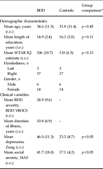
Participants were assessed with the Mini-International Neuropsychiatric Interview (MINI; Sheehan et al. Reference Sheehan, Lecrubier, Sheehan, Amorim, Janavs, Weiller, Hergueta, Baker and Dunbar1998) to evaluate the presence or absence of other mental disorders. Handedness was assessed with the Edinburgh Inventory, and social anxiety and depression were measured by the Social Interaction Anxiety Scale (SIAS) (Brown et al. Reference Brown, Turovsky, Heimberg, Juster, Brown and Barlow1997) and the Zung Self-rating Depression Scale (Zung, Reference Zung1965), which has been shown to be sensitive to the clinical severity of depression in psychiatric samples (Biggs et al. Reference Biggs, Wylie and Ziegler1978). Group comparisons were computed using independent-samples t tests at p = 0.05. Demographic and clinical data are displayed in Table 1.
Demographic and clinical variables

BDD, Body dysmorphic disorder; s.d., standard deviation; WTAR, Wechsler Test of Adult Reading; IQ, intelligence quotient; BDD-YBOCS, Yale–Brown Obsessive Compulsive Scale Modified for Body Dysmorphic Disorder; Zung, Zung Self-rating Depression Scale; SIAS, Social Interaction Anxiety Scale.
a Degrees of freedom = 38.
The two participant groups were well matched on age, gender, education, estimated IQ and handedness. The clinical data indicate that mean BDD severity was in the ‘moderate’ range which is defined as scores between 16 and 30 on the BDD-YBOCS (Phillips et al. Reference Phillips, Hollander, Rasmussen, Aronowitz, DeCaria and Goodman1997). Scores below 15 indicate ‘mild’ BDD symptoms and scores from 31 to 48 are defined as ‘severe’ symptoms. At the time of MRI acquisition our sample comprised two participants in the mild range, 13 with moderate symptoms and five in the severe range.
The depression scores as measured by the Zung Self-rating Depression Scale indicated that the BDD sample had a higher level of depressive symptoms compared with controls. On average the BDD group fell in the subclinical range of depressive symptoms, defined as a score below 50 (Zung, Reference Zung1965). A score between 50 and 59 on the scale represents mild depression, between 60 and 69 indicates moderate symptoms and above 70 indicates severe symptoms (Zung, Reference Zung1965). The SIAS scores showed that the BDD sample had a moderate level of social anxiety symptoms; less than people diagnosed with social phobia but higher than other anxiety disorders (Peters, Reference Peters2000), which is typical of BDD samples (Coles et al. Reference Coles, Phillips, Menard, Pagano, Fay, Weisberg and Stout2006). Assessment with the MINI showed that four BDD participants fulfilled the diagnostic criteria for current major depressive disorder or dysthymia, and five had current agoraphobia or social phobia, showing that our sample was representative of a typical BDD profile (Coles et al. Reference Coles, Phillips, Menard, Pagano, Fay, Weisberg and Stout2006). Areas of aesthetic concern for our sample were generally the face, skin and hair. All but two members of the BDD sample were taking psychoactive medication: five where taking seroquel, four escitalopram, two duloxetine, two desvenlafaxine, two diazepam, and one each paroxetine, mirtazapine, lorazepam, methylphenidate, sodium valproate or clomipramine.
MRI acquisition
Participants were scanned using a 3 T scanner (Siemens Magnetom TrioTim, Germany) at the Murdoch Childrens Research Institute (Royal Children's Hospital, Melbourne, Australia). The DTI scanning sequence was conducted as a component of a 1 h long scan. The scanner acquired an isotropic DTI sequence for FA estimations (number of directions = 60, b value = 2000s/m2, slice thickness = 2.5 mm). Data were transferred to a Linux workstation for image processing and analyses.
FA analysis
Tract-based spatial statistics (TBSS) developed by Smith et al. (Reference Smith, Jenkinson, Johansen-Berg, Rueckert, Nichols, Mackay, Watkins, Ciccarelli, Cader, Matthews and Behrens2006) was used to create a mean FA skeleton that was representative of our 40 participants. TBSS uses non-linear registration to create a template for FA comparisons that allows voxelwise analysis of multi-subject diffusion data. The TBSS module in the FSL package (http://www.fmrib.ox.ac.uk/fsl/) was used to compute statistics. A two-sample t test was conducted using the randomize tool, which tests the t value at each voxel against a null distribution generated from 5000 random permutations of group membership. The output contained statistical maps corrected for multiple comparisons (p < 0.05) using threshold-free cluster enhancement. This method has a high level of sensitivity to true differences while minimizing false positives by avoiding the specification of a subjective cluster-forming threshold (Smith & Nichols, Reference Smith and Nichols2009). Specific white matter areas were defined using integrated white matter atlases within FSL: the ICBM-DTI-81 White-Matter Labels Atlas and the JHU White-Matter Tractography Atlas, both developed in the Laboratory of Brain Anatomical MRI at Johns Hopkins University (USA).
In addition, a Pearson product-moment correlation analysis at p < 0.05 was performed between voxelwise FA and symptom severity as measured by the BDD-YBOCS, Zung Self-rating Depression Scale and SIAS.
Results
FA
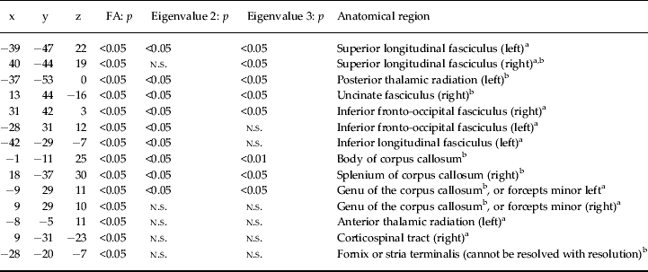
The BDD patients exhibited widespread significant reductions in corrected FA values compared with controls. Table 2 shows decreased FA values in BDD divided into anatomical regions, and shows areas of significant increase in eigenvalues 2 and 3. There were no significant differences between BDD and controls on eigenvalue 1.
Areas of decreased FA and increased eigenvalue 2 and 3 values in BDD
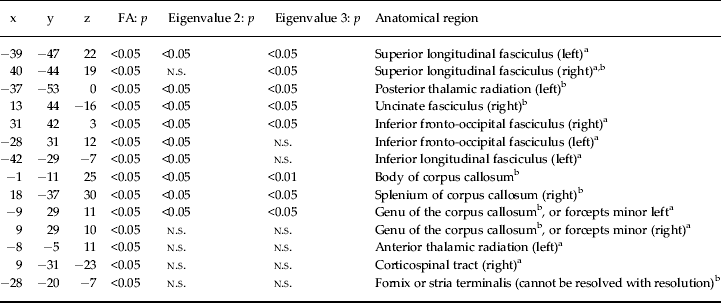
FA, Fractional anisotropy; BDD, body dysmorphic disorder; n.s., not significant.
a Area identified using the JHU White-Matter Tractography Atlas.
b Area identified using the ICBM-DTI-81 White-Matter Labels Atlas.
Table 2 shows that there are widespread white matter differences in BDD. Since FA is a composite of eigenvalues 1, 2 and 3 and there was no differences on eigenvalue 1, the majority of FA reductions can be accounted for by an increase of radial diffusivity as represented by eigenvalues 2 and 3.
Correlation analysis
The correlation analysis found that there was no significant correlation between BDD symptom severity scores and corrected FA in any voxel within the BDD sample. Likewise, there was no significant correlation between depression scores and FA in the BDD group. In the BDD group, social anxiety scores were found to correlate negatively with corrected FA values on the left superior longitudinal fasciculus (x = –38, y = 2, z = 22) at p < 0.05. There was no correlation between depression and anxiety in any voxel in the control group.
Discussion
As far as we are aware, this is the first study to examine white matter differences within a BDD sample using DTI. Additionally, this study had the largest BDD sample in a neuroimaging study to date, and our sample was thoroughly screened to ensure that BDD was the primary condition and that it was representative of the patient population (Phillips et al. Reference Phillips, Didie and Menard2007a). Importantly, this study provides preliminary evidence of neural abnormalities in BDD; specifically, evidence for a widespread loss of integrity in white matter connectivity.
Consistent with the hypothesis that we would find similar reductions in FA in BDD as has been shown in OCD (Szeszko et al. Reference Szeszko, Ardekani, Ashtari, Malhotra, Robinson, Bilder and Lim2005; Garibotto et al. Reference Garibotto, Scifo, Gorini, Alonso, Brambati, Bellodi and Perani2010), we found reduced FA in the corpus callosum, superior longitudinal fasciculus, and inferior fronto-occipital fasciculus, bilaterally. The widespread FA reductions in our BDD sample can be accounted for through increases in radial diffusivity rather than reduced eigenvalue 1. Of the 12 areas identified in Table 2 as having significantly reduced FA, nine can be accounted for by eigenvalues 2 and 3. Indeed a comparison between Figs. 1 and 2 shows that mean diffusivity was more widespread than FA differences. This suggests loss of integrity in the white matter whereby attenuated white matter pathways allow water diffusion in directions that are not consistent with overall white matter directionality. Reduced FA has been shown in other work to be driven by myelin abnormalities which is largely under genetic control (Menzies et al. Reference Menzies, Williams, Chamberlain, Ooi, Fineberg, Suckling, Sahakian, Robbins and Bullmore2008). Therefore, neurodevelopment irregularities leading to abnormal myelination may be important to the predisposition to BDD. Reduced FA due to increased radial diffusivity has also been found in OCD (Bora et al. Reference Bora, Harrison, Fornito, Cocchi, Pujol, Fontenelle, Velakoulis, Pantelis and Ycel2011).
Statistically significant fractional anisotropy reductions in body dysmorphic disorder sample compared with controls. Numbers in red represent distance (mm) from the anterior commissure.
Statistically significant mean diffusivity increases in body dysmorphic disorder sample compared with controls, coronal view. Numbers in red represent distance (mm) from the anterior commissure.
While the loss of white matter integrity was widespread, it is worth considering the impact of specific neural pathways on BDD cognition. For example, reduced FA in the corpus callosum indicates attenuated interhemispheric communication. This may explain the neuropsychological research in BDD that has shown difficulties with integration of detailed and global information processing (Deckersbach et al. Reference Deckersbach, Savage, Phillips, Wilhelm, Buhlmann, Rauch, Baer and Jenike2000; Feusner et al. Reference Feusner, Moller, Altstein, Suger, Bookheimer, Woon and Hembacher2010a). Moreover, white matter operating inefficiently may explain the Stroop task difficulties associated with BDD (Hanes, Reference Hanes1998), as reduced FA and other neurobiological correlates in frontostriatal–limbic regions have been shown to relate to performance on the Stroop and planning tasks in geriatric depression and OCD (van den Heuvel et al. Reference van den Heuvel, Veltman, Groenewegen, Witter, Merkelbach, Cath, van Balkom, van Oppen and van Dyck2005; Murphy et al. Reference Murphy, Gunning Dixon, Hoptman, Lim, Ardekani, Shields, Hrabe, Kanellopoulos, Shanmugham and Alexopoulos2007). Furthermore, reduced FA in the bilateral superior longitudinal fasciculi suggests that connectivity between the prefrontal, parietal, occipital and temporal regions (Makris et al. Reference Makris, Kennedy, McInerney, Sorensen, Wang, Caviness and Pandya2005) may be compromised.
The reduced FA in the uncinate fasciculus is of particular interest because it is a major white matter fibre tract that connects the inferofrontal and anterotemporal cortices, and it travels over the lateral nuclei of the amygdala. Thus, lower FA within this tract suggests compromised fronto-amygdala structural connectivity in BDD. In the context of BDD, this is interesting when considering that frontal regions such as the orbitofrontal cortex serve important roles in top-down regulation of amygdala reactivity to control negative affect and mediate threat perception (Ochsner et al. Reference Ochsner, Bunge, Gross and Gabrieli2002; Barrett et al. Reference Barrett, Bliss-Moreau, Dunca, Rauch and Wright2007).
It is clear from past research that there are grey matter differences in BDD patients compared with non-psychiatric controls, but how these interact with the white matter abnormalities reported here remains speculative. Volumes of the right amygdala and the inferior frontal gyrus have been shown to correlate with current BDD symptom severity (Feusner et al. Reference Feusner, Townsend, Bystritsky, McKinley, Moller, Bookheimer, Feusner, Townsend, Bystritsky, McKinley, Moller and Bookheimer2009), suggesting that differences in these grey matter structures are the most proximal contributor to symptoms, while white matter differences can be considered as a more distal, or perhaps predisposing, factor in BDD.
Our examination of whether BDD symptom severity correlated with FA was not significant, despite such a correlation being found in OCD samples (Garibotto et al. Reference Garibotto, Scifo, Gorini, Alonso, Brambati, Bellodi and Perani2010). Given that we had a large range of symptom severity and a sample size that was able to provide robust correlation results, our non-significant results suggest that BDD has a distinct neurobiological makeup that is state-independent. Such a finding suggests important trait-related white matter abnormalities in BDD. This result would need to be replicated; further research could establish if these brain differences are present in an individual whether or not illness is active, and provide preliminary evidence for a BDD phenotype that is perhaps related to the emerging phenotype considered important in OCD (Menzies et al. Reference Menzies, Williams, Chamberlain, Ooi, Fineberg, Suckling, Sahakian, Robbins and Bullmore2008).
The results showing a significant negative correlation between social anxiety scores and FA in the left superior longitudinal fasciculus are interesting, considering that the main measure of BDD symptom severity (the BDD-YBOCS) was not significantly correlated. It is possible that our social anxiety measure was sensitive to an important feature of BDD that our main symptom severity rating scale was not. Indeed, social anxiety can be seen as a important factor within BDD symptomatology and is best conceptualized as an expression of higher BDD symptoms rather than a separate problem of social phobia (Coles et al. Reference Coles, Phillips, Menard, Pagano, Fay, Weisberg and Stout2006). The finding that there was no correlation between depression scores and FA in any voxel indicated that the FA differences among the two groups can be accounted for by the diagnosis of BDD independent from depression ratings. Indeed, this finding taken together with the non-significant contribution of BDD symptoms to FA suggests that state-based symptoms scores bear less importance to white matter than state-independent diagnosis.
The parallels that have been drawn between OCD and BDD in terms of clinical features (Stewart et al. Reference Stewart, Stack and Wilhelm2008) are yet to be robustly supported using DTI imaging techniques. In fact, to date, the nine DTI studies in OCD have yielded inconsistent results, with both local FA increases and decreases reported (e.g. Menzies et al. Reference Menzies, Williams, Chamberlain, Ooi, Fineberg, Suckling, Sahakian, Robbins and Bullmore2008; Bora et al. Reference Bora, Harrison, Fornito, Cocchi, Pujol, Fontenelle, Velakoulis, Pantelis and Ycel2011, Lochner et al. Reference Lochner, Fouché, du Plessis, Fineberg, Chamberlain and Stein2012). The inconsistent data in OCD and the preliminary evidence of widespread FA decreases provided by the current study in BDD should be supplemented by future studies to help determine biological similarities and differences between the two disorders.
A limitation of this study is that nearly all of our BDD participants were taking medication at the time of MRI acquisition. This may be problematic as treatment is likely to have reduced the severity of BDD symptoms, thereby possibly influencing FA. However, as white matter integrity is generally considered state independent, current BDD symptomatology is unlikely to have significantly influenced the results. At the time of MRI acquisition two of the BDD participants were rated as having mild BDD symptoms on the BDD-YBOCS, although an active diagnosis of BDD was still confirmed using the BDD-DM.
In conclusion, we believe this to be the first study to examine white matter integrity in a BDD sample. The main contribution of our data is that they provide evidence that individuals with BDD have compromised white matter fibres, reflected by changes in fibre directionality as indicated by eigenvalues 2 and 3. This reduced connectivity among different regions of the brain is widespread in nature and is not related to symptom severity.
Acknowledgements
This study was funded by a Monash Strategic Grant.
Declaration of Interest
None.