Introduction
Fish and metazoan parasites that inhabit deep water (400–1000 m mesopelagic; >1000 m bathypelagic) require various adaptations to cope with the adverse conditions of this environment (i.e. high pressure, low temperatures, lack of light, and extremely scarce food supply). The fish behaviour, including extensive daytime vertical migration and characteristic feeding ecology, affects the oceanic food web and the pathways of metazoan parasite transmission in the deep sea. Most parasitological studies of midwater fish have revealed larval or juvenile stages of helminths that mature in large pelagic fish and marine mammals rather than adult helminths (e.g. Gartner & Zwerner, Reference Gartner and Zwerner1989; Mateu et al., Reference Mateu, Nardi, Fraija-Fernández, Mattiucci, de Sola L, Raga, Fernández and Aznar2015). However, in most cases the number of parasite species and individuals has been rather low (e.g. Klimpel et al., Reference Klimpel, Busch, Kellermanns, Kleinertz and Palm2009, Reference Klimpel, Busch, Sutton and Palm2010; Woodstock, Reference Woodstock2018). This suggests that most of these mesopelagic infections represent sporadic accidental events that most likely occur in the photic zone.
Although the study of the parasites of deep-sea fish has a long history (e.g. von Linstow, Reference von Linstow1888; Manter, Reference Manter1934; Collard, Reference Collard and Snieszko1970; Noble & Collard, Reference Noble, Collard and Snieszko1970; Polyanskii, Reference Polyanskii, Dogiel, Petrushevski and Polyanskii1970; Noble, Reference Noble1973; Campbell et al., Reference Campbell, Haedrich and Munroe1980; Ñacari & Oliva, Reference Ñacari and Oliva2016), our knowledge of the species composition and geographic distribution of these parasites is far from complete. There are about 3800–4200 different deep-sea fish species (~10–12% of the known fish fauna) (Froese & Pauly, Reference Froese and Pauly2019), and according to Klimpel et al. (Reference Klimpel, Busch, Sutton and Palm2010), only a fraction of deep-water fish (~511 fish species) have been studied for parasites. For the Gulf of Mexico (GoM), published information for parasites of these fish comes exclusively from the northern GoM (i.e. Andres et al., Reference Andres, Peterson and Overstreet2016; Woodstock, Reference Woodstock2018).
In 2015, the Mexican government offered investment opportunities to international private oil companies for the exploration and extraction of deep-sea hydrocarbons (500–3650 m depth) from the Perdido region in north-west GoM (CNH, 2015). For this, a major research project was carried out with the aim of establishing the baseline for environmental and biological statuses of the pelagic and benthic zone in the Perdido region prior to oil-extraction activities. As part of this effort, we had the opportunity to undertake a survey of the fish parasites from a subsample of the fish caught. Therefore, the objective of the present study was to taxonomically describe the metazoan parasite fauna from a subsample of mesopelagic and bathypelagic fish hosts recovered during oceanographic surveys in the Perdido region, north-west GoM.
Materials and methods
Study area and sampling techniques
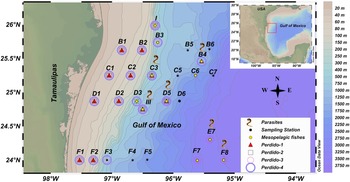
The study area encompassed 27 sampling stations, of which 15 stations were between 468 and 3680 m depth in the Perdido region, western GoM (Figure 1). Fish were collected from the sampling stations during four cruises (Perdido 1: May 2016, Perdido 2: September–October 2016, Perdido 3: June 2017, Perdido 4: September–October 2017), using a benthic sled with a 25.4 mm mesh size, on the oceanographic vessel (OV) ‘Justo Sierra’ (UNAM) (Supplementary Table S1). For each planned sampling site, a Kongsberg EM 302 multibeam echo sounder was used to determine both the flatness of the sea bottom and the depth at which the benthic sled was deployed. Once the benthic sled was detected as touching the oceanic bottom, the trawls were carried out over 0.5 h or one nautical mile. It should be clarified that the benthic sled did not have automatic opening and closing systems; therefore, it was not possible to validate the specific depth at which the fish got trapped in the mesh. However, the depth at which the benthic sled was deployed, as indicated by the echo sounder is presented in Supplementary Table S1. Most of the fish specimens were kept by an ichthyologist for taxonomic purposes and vouchers were deposited at the ichthyological collection (code: YUC.PEC.084.0999) of the Fish Taxonomy and Ecology Laboratory of the Centro de Investigación y de Estudios Avanzados del Instituto Politécnico Nacional, Unidad Mérida (CINVESTAV) as reference material. However, a subsample from the different sampling points for each of the four cruises in Perdido was kept for the parasitological study. All sampling stations are shown in Figure 1, and those in which fish were collected for parasitological study are marked with a yellow dot.
Sampling stations visited during the four oceanographic cruises carried out in the Perdido region (north-western Gulf of Mexico). Sampling stations where mesopelagic and bathypelagic fish were collected are marked with a yellow dot. Those sampling stations where fish were infected are marked with a symbol for parasites. The black dots correspond to sampling stations of Perdido cruises where fish were not found.

Parasitological examination
On board, fish were collected by hand from the net attached to the benthic sled, maintained in seawater at 4 °C in a refrigerator, and examined within 24 h. These fish were preliminarily identified and later confirmed in the laboratory of Fish Taxonomy and Ecology of CINVESTAV based on their morphometric characteristics, using specialized references (McEachran & Fechhelm, Reference McEachran and Fechhelm1998, Reference McEachran and Fechhelm2005; Carpenter, Reference Carpenter2002a, Reference Carpenter2002b, Reference Carpenter2002c; among others). For each individual fish, the total length, standard length, maximum height (±0.1 cm) and weight (±0.1 g) were recorded. Parasitological examinations were conducted on board. The body surface, cavities and all internal organs were individually examined for metazoan parasites using a dissection microscope and marine water during the search of ectoparasites and 0.7% saline solution for endoparasites. All metazoan parasites from each individual host were counted in situ and preserved in 96% alcohol (for digeneans, cestodes, nematodes and parasitic crustaceans) in labelled vials for subsequent morphological or molecular taxonomic identification. The parasite shape, movement, colour, site of infection and number were recorded. Small pieces of cardboard were used along opposite sides of the coverslip to avoid deformation of the helminths and crustaceans by pressure. Digeneans and nematodes previously fixed in 96% OH were cleared in glycerine jelly for 1–2 h and their dimensions measured under a light microscope (Olympus BX50). All other helminthological methods were those recommended by Vidal-Martínez et al. (Reference Vidal-Martínez, Aguirre-Macedo, Scholz, González-Solís and Mendoza-Franco2001). Sketches or high-quality digital photographs from a camera attached to the light microscope were used. Images of the metazoan parasites were used to produce drawings on a digitizing tablet with CorelDRAW 12 software. All measurements are in micrometres (μm) unless otherwise specified. Both prevalence and mean intensity values were calculated following Bush et al. (Reference Bush, Lafferty, Lotz and and Shostak1997). Synonyms and additional information for each host species were obtained from FishBase (Froese & Pauly, Reference Froese and Pauly2019). Voucher specimens of each species were deposited in the Helminthological Collection of the Laboratory of Parasitology, Centre for Research and Advanced Studies, National Polytechnic Institute, Mérida, Yucatán, México (CHCM).
DNA extraction, polymerase chain reaction (PCR) amplification and sequencing
DNA was extracted from both metazoan parasites and fish using the DNeasy Blood and Tissue kit (Qiagen, Hilden, Germany) according to the manufacturer's instructions. A partial 18S rDNA and internal transcribed spacer (ITS) gene for nematodes, a partial 28S rDNA and the mitochondrial cytochrome oxidase subunit 1 (COI) gene for cestodes, and a COI gene for copepods were amplified (García-Varela & Nadler, Reference García-Varela and Nadler2005; Van Steenkiste et al., Reference Van Steenkiste, Locke, Castelin, Marcogliese and Abbott2015). Additionally, a partial COI was amplified and sequenced for molecular identification of fish (Folmer et al., Reference Folmer, Black, Hoeh, Lutz and Vrijenhoek1994). All PCR tests were carried out in a 25 μl reaction volume using 12.5 μl Green GoTaq Master Mix (Promega, Madison, WI, USA), 8.5 μl ultrapure water, 1 μl of each primer (10 μM) and 2 μl genomic DNA. The primers and cycling conditions used for identification of fish and metazoan parasites are presented in Supplementary Table S2. The PCR products were analysed by electrophoresis on 1% agarose gel in the presence of Redgel then visualized under ultraviolet light. The purification and sequencing of the PCR products were carried out by Genewiz (South Plainfield, NJ, USA; https://www.genewiz.com/).
Results
Fish species examined
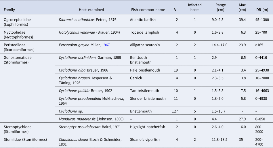
A total of 187 individual fish from the oceanographic cruises were provided by the ichthyologists for parasitological examination. Twelve fish species of six families (Gonostomatidae, Sternoptychidae, Stomiidae, Myctophidae, Peristediidae and Ogcocephalidae) from an amplified fragment of 650 bp barcode region of Cytochrome C oxidase Subunit I (COI) were identified. All fish species identified for this study are presented in Table 1.
Morphological measurements and number of infected individuals of the mesopelagic and bathypelagic fish species collected in the Perdido region (north-western Gulf of Mexico)

N, number of fish examined; Range, standard length range of the fish examined; Max, maximum length (standard length) reported for that fish species in FishBase (http://www.fishbase.se); DR, depth range reported for that fish species in FishBase (http://www.fishbase.se).
Metazoan parasite species of some meso- and bathypelagic fish from the Perdido region
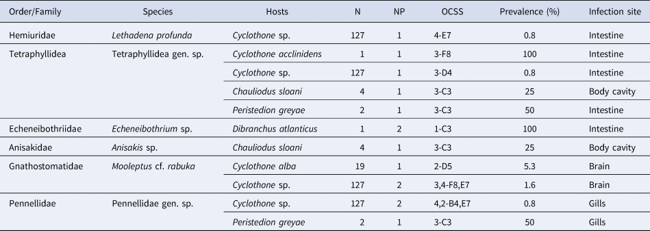
Six parasite species were found infecting six of the 12 fish species examined (Table 2). Cyclothone sp. was the host species with the highest number of parasites species (four), followed by Chauliodus sloani Bloch and Schneider, 1801 and Peristedion greyae Miller, Reference Miller1967 with two each. Cyclothone acclinidens Garman, 1899; Cyclothone alba Brauer, 1906; and Dibranchus atlanticus Peters, 1876 each harboured one parasite species. Cyclothone braueri Jespersen and Tåning, 1926; Cyclothone pallida Brauer, 1902; Cyclothone pseudopallida Mukhacheva, 1964; Manducus maderensis (Johnson, 1890); Notolychnus valdiviae (Brauer, 1904); and Sternoptyx pseudobscura Baird, 1971 were examined, but no parasites were found.
Metazoan parasites recovered from mesopelagic and benthopelagic fishes in the Perdido fold belt, north-western Gulf of Mexico between May 2016 and October 2017

N, Total number of fishes examined; NP, Number of parasites; OCSS, Oceanographic cruise-sampling station.
SYSTEMATICS
Class TREMATODA Rudolphi, 1808
Subclass DIGENEA Carus, 1863
Order PLAGIORCHIIDA La Rue, 1957
Suborder HEMIURATA Skrjabin & Guschanskaja, 1954
Family HEMIURIDAE Looss, 1899
Genus Lethadena Manter, 1947
Lethadena profunda (as Sterrhurus profundus) (Manter, Reference Manter1934) Manter, Reference Manter1947
(Figure 2A)
MATERIAL EXAMINED
One adult individual collected from Cyclothone sp., fixed in 96% OH and mounted in glycerine jelly, during the oceanographic cruise Perdido 4 (Perdido region, western GoM) at sampling station E7 (25 September 2017) (see Table 2). One specimen deposited as a voucher specimen CHCM No. 661 (voucher). V. Vidal-Martínez collector, 25 September 2017.
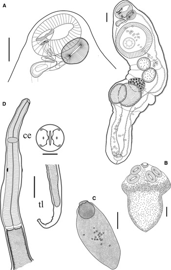
Metazoan parasite species of mesopelagic and bathypelagic fishes from the Perdido fold belt, north-western Gulf of Mexico. (A) Adult digenean Lethadena profunda (Manter, Reference Manter1934) Manter, Reference Manter1947 recovered from the intestine of Cyclothone sp. (B) Larval cestode Tetraphyllidea gen. sp. recovered from the intestine of Cyclothone acclinidens. (C) Larval Echeneibothrium recovered from the intestine of Dibranchus atlanticus. (D) Larval nematode Mooleptus rabuka recovered from the brain of Cyclothone alba; (ce) Cephalic end, (tl) tail of larva. All scale bars in the figure are 100 μm.

MORPHOLOGY
Based on one specimen fixed in 96% ethanol and cleared in glycerine jelly (Figure 2A). Body elongated, cylindrical, smooth, widest at the level of the ventral sucker; 2230 long × 570 wide. Ecsoma 720 long with a fold anterior to the vitellaria. Oral sucker at the anterior end of the body; preoral lobe absent; mouth almost terminal. Ventral sucker circular, at level of first 1/3 of body length, 420 long × 481 wide. Ratio of ventral sucker length to oral sucker length 1:2.9. Prepharynx absent; pharynx large (101 long × 145 wide); oesophagus inconspicuous; narrow ceca extending to the extreme posterior tip of the ecsoma. Genital pore median, overlapping the oral sucker. Genital atrium present. Seminal vesicle undivided, sac-shaped (45 long × 121 wide), thick-walled, intercaecal, slightly overlapping ventral sucker posteriorly. Prostate gland not visible. Testes oblique, posterior to ventral sucker, smooth, longer than wide; anterior testis 178 long × 171 wide, posterior testis 201 long × 171 wide. Ovary median, large, smooth, immediately posterior to testis, 194 long × 224 wide. Two vitellaria, larger than ovary and testis, unlobed, slightly elongated, immediately posterior to the ovary, within ecsoma, left 281 long × 217 wide, right 315 long × 315 wide. Uterus coils to the posterior end of the tail, soon becoming very swollen, extending anteriorly as almost straight tube; eggs tiny, thin-shelled, colourless, about 21–26 long × 16–19 wide. Excretory vesicle subterminal, excretory pore terminal. Since only one specimen was recovered, we decided to keep it complete for deposition, so molecular identification was not attempted.
REMARKS
We identified this digenean as L. profunda (originally described as Sterrhurus profundus) because most of the morphology and measurements lie within the range of the original description (Manter, Reference Manter1934). The dorsal papillae anterior to the oral sucker described by Manter (Reference Manter1934) were not observed. This species was originally described from a species of Xenodermichthys (Günther) (as Aleposomus sp. Gill) (Alepocephalidae) and Pronotogrammus sp. Gill (Serranidae) from Dry Tortugas (Florida) (Manter, Reference Manter1934). Another record of the species was provided by Gibson (Reference Gibson, Margolis and Kabata1996) stating that L. profunda was found parasitizing the stomach of Urophycis chesteri (currently Phycis chesteri) Goode and Bean (Phycidae) in the Scotian shelf. Lethadena sp. has also been described parasitizing Myctophum punctatum Rafinesque (Myctophidae) in the Mid-Atlantic Ridge (Klimpel et al., Reference Klimpel, Busch, Sutton and Palm2010). In this study, L. profunda (as Sterrhurus profundus) was found infecting Cyclothone sp., which represents a new host record for this digenean.
Class CESTODA Rudolphi, 1808
Subclass EUCESTODA
Order TETRAPHYLLIDEA (Carus, 1863)
(Figure 2B)
MATERIAL EXAMINED
Four specimens collected, all in larval stage, were preserved in 96% alcohol: one from Cyclothone acclinidens (16 June 2017); one from Cyclothone sp. (16 June 2017); one from Chauliodus sloani (5 June 2017); and one from Peristedion greyae (5 June 2017) (see Table 2), all from oceanographic cruise Perdido 3, Perdido region, western GoM. The morphological description was based on one individual fixed with ammonium picrate because all other larvae were used for molecular analysis. One specimen deposited as a voucher specimen CHCM No. 662 (voucher) (larval stage). V. Vidal-Martínez collector, 25 September 2017.
MORPHOLOGY
Body elongated, tapered posteriorly, 1925 long × 950 wide. Scolex oval, 1350 wide, with four lateral suckers and a small apical sucker. The remaining three specimens were used for molecular identification, but no DNA was obtained.
REMARKS
Specific identification was not possible because of the absence of genital organs and incomplete knowledge of the life cycle. Tetraphyllidean larvae possessing four lateral suckers and a muscular apical sucker have been designated as Scolex pleuronectis Muller, 1780. This taxon undoubtedly includes a variety of tetraphyllidean larvae belonging to different genera, including members of the families Phyllobothriidae and Prosobothriidae among others (Euzet, Reference Euzet, Khalil, Jones and Bray1994). Usually, adults of tetraphyllideans are found parasitizing elasmobranchs; therefore, the host species from which these larvae were recovered probably act as secondary intermediate or paratenic hosts (Euzet, Reference Euzet, Khalil, Jones and Bray1994; Vidal-Martínez et al., Reference Vidal-Martínez, Aguirre-Macedo, Scholz, González-Solís and Mendoza-Franco2001). For the northern Gulf of Mexico Andres et al. (Reference Andres, Peterson and Overstreet2016) and Woodstock (Reference Woodstock2018) have reported the presence of larval tetraphyllideans in fish. Andres et al. (Reference Andres, Peterson and Overstreet2016) reported larval tetraphyllideans in three mesopelagic fish species: Pollichthys mauli, Maurolicus weitzmani and Polyipnus clarus. Additionally, Woodstock (Reference Woodstock2018) reported larval tetraphyllideans parasitizing the fish Benthosema suborbitale, Diaphus lucidus, Nealotus tripes, Sternoptyx diaphana and Sternoptyx pseudobscura. Larval tetraphyllidean cestodes parasitizing mid-water fish have also been described by Klimpel et al. (Reference Klimpel, Kellermanns, Palm and Moravec2007, Reference Klimpel, Palm, Busch and Kellermanns2008a, Reference Klimpel, Busch, Sutton and Palm2010) from the Mid-Atlantic Ridge. Cyclothone acclinidens, Cyclothone sp., Chauliodus sloani and Peristedion greyae represent new host records for Tetraphyllidea gen. sp.
Class CESTODA Rudolphi, 1808
Subclass EUCESTODA
Order RHINEBOTHRIIDEA Healy, Caira, Jensen, Webster & Littlewood, 2009
Family ECHENEIBOTHRIIDAE de Beauchamp, 1905
Genus Echeneibothrium Van Beneden, 1849
(Figure 2C)
MATERIAL EXAMINED
Two specimens in the larval stage (preserved in 96% alcohol) from one Dibranchus atlanticus at sampling station C3 during oceanographic cruise Perdido 1 (see Figure 1), Perdido region, western GoM (Table 2). One larva deposited as voucher specimen CHCM No. 663 (voucher) (larval stage). V. Vidal-Martínez collector, 20 May 2016.
MORPHOLOGY
Body elongated, tapered posteriorly; its length is approximately three times its width, 398–448 long × 142–190 wide. Scolex not defined; with a snout-like prominent organ at the apical end (the myzorhynchus), 104–112 long × 96–111 wide. Multiple calcareous bodies. No structures such as bothridia were observed (Figure 2C). Additionally, a fragment of 974 bp of 28S rRNA was obtained and sequenced from each of two processed individuals. The Basic Local Alignment Search Tool (BLAST) analysis (Zhang et al., Reference Zhang, Schwartz, Wagner and Miller2000) revealed 97% homology with Echeneibothrium multiloculatum (MH688748.1). However, this per cent similarity is not enough to determine the identity of these cestodes; therefore, we decided to keep the identification as species of Echeneibothrium.
REMARKS
Camacho (Reference Camacho1958) described the larvae of species of Echeneibothrium with anterior holdfast organs that comprised outgrowths of four ear-like structures adhering closely to the body and the apical end as a snout-like organ (myzorhynchus). However, in our specimens, only the snout-like structure is present in the apical end. Apparently, our specimens appear to still be in the procercoid stage; therefore, these ear-like structures are not yet evident. Larvae of species of Echeneibothrium have been reported in several fish species: Cynoscion regalis (Bloch & Schneider, 1801) (Sciaenidae); Limanda ferruginea (Storer, 1839) (Pleuronectidae); Lophius piscatorius Linnaeus, 1758 (Lophiidae); Paralichthys dentatus (Linnaeus, 1766) (Paralichthyidae); Stenotomus chrysops (Linnaeus, 1766) (Sparidae) and Fistularia petimba (Lacepède, 1803) (Fistulariidae) from Massachusetts, north-eastern USA (Linton, Reference Linton1897); Lotella phycis (Temminck & Schlegel, 1846) (Moridae), Uranoscopus japonicus Houttuyn, 1782 (Uranoscopidae) and Rhinoplagusia japonica (accepted as Paraplagusia japonica) (Temminck & Schlegel, 1846) (Cynoglossidae) from Tomaya Bay, Japan (Yamaguti, Reference Yamaguti1934). In the GoM, species of Echeneibothrium larvae have been recorded in Thunnus albacares (Bonnaterre, 1788) (Scombridae) (Jensen, Reference Jensen, Felder and Camp2009). Adult-stage Echeneibothrium species have also been described from bathypelagic and bathydemersal fish, such as the deep-sea skate Raja bathyphila (accepted as Rajella bathyphila) (Holt & Byrne, 1908) (Rajidae) (Campbell, Reference Campbell1977) and Atlantic horse mackerel Trachurus trachurus (Linnaeus, 1758) (Carangidae) (Young, Reference Young1956). Dibranchus atlanticus represent a new host record for species of Echeneibothrium.
Phylum NEMATODA Rudolphi, 1808
Class CHROMADOREA Inglis, 1983
Order RHABDITIDA Chitwood, 1933
Family GNATHOSTOMATIDAE Railliet, 1895
Genus Mooleptus Özdikmen, 2010
Mooleptus cf. rabuka (Machida et al., Reference Machida, Ogawa and Okiyama1982) Özdikmen, Reference Özdikmen2010
(Figure 2D)
MATERIAL EXAMINED
Three individuals fixed in 96% OH and mounted in glycerine jelly, all larval stage: one from the brain of Cyclothone alba collected during the oceanographic cruise Perdido 2 at sampling station D5 (6 October 2016), one from the brain of Cyclothone sp. during the oceanographic cruise Perdido 3 at sampling station F8 (16 June 2017), and one from the brain of Cyclothone sp. during the oceanographic cruise Perdido 4 at sampling station E7 (25 September 2017) (Figure 2D; Table 2). One larva was deposited as voucher specimen CHCM No. 664 (voucher). V. Vidal-Martínez collector, 6 October 2016.
MORPHOLOGY
Body 1277–2778 (N = 3), long 56–96 (N = 3) at the widest region (junction of oesophagus and intestine). Mouth bounded by two lateral labia, each with three lobes. Nerve ring at 216–467 (N = 3) from anterior end. Cervical papillae at 354 (N = 1) from anterior end. Oesophagus 341–623 (N = 3) long, 22–26% of body length with anterior end slightly swollen and 27–56 (N = 3) wide near its posterior end; undivided and connected to intestine by a well-developed transverse ventriculus-like formation 8–26 (N = 3) long, 33–70 (N = 3) wide. Ventriculus was a two-layered structure with oesophagus layer slightly broader than the intestinal layer. Excretory pore slightly above cervical papilla at 339 (N = 1) from anterior end. Tail 58 (N = 1) long; gradually tapering with a blunt tip.
REMARKS
This species was originally described by Machida et al. (Reference Machida, Ogawa and Okiyama1982) as Metaleptus rabuka from the stomach and intestine of Chlamydoselachus anguineus Garman 1884 (Chlamydoselachidae), a deep-sea shark off Choshi, Pacific coast of central Honshu, main island of Japan. In 2000, Moravec and Nagasawa described an adult individual from the intestine of deep-sea catsharks Apristurus fedorovi Dolganov 1983 (Scyliorhinidae) (1290–1300 m depth), captured off Iwate and Aomori Prefectures, northern Honshu, Japan. Based on morphological characteristics, such as trilobed pseudolabia, undivided oesophagus, and structures of the male caudal end, these authors transferred this species from Physalopteroidea to Gnathostomatoidea where they created the new subfamily Metaleptinae to accommodate it. Later, Özdikmen (Reference Özdikmen2010) noted that the generic name Metaleptus was already used for parasitic nematodes of insects and proposed the genus Mooleptus for these parasitic nematodes of deep-sea sharks. Finally, the most recent record of M. rabuka comes from the large-nose shark Apristurus nasutus deBuen 1959 (Scyliorhinidae), caught off Juan Fernández Archipelago (400–950 m deep), south-eastern Pacific Ocean, Chile (Rodríguez et al., Reference Rodríguez, D'Elía and George-Nascimento2012). The morphology and measurements of our specimens from C. alba and Cyclothone sp. are very similar to those reported for this species by Machida et al. (Reference Machida, Ogawa and Okiyama1982). The larva of M. rabuka was also recovered from the brain of a deep-sea bristlemouth Cyclothone atraria Gilbert 1905 by Machida et al. (Reference Machida, Ogawa and Okiyama1982). We identified this nematode as the larval stage of M. cf. rabuka based on the presence of two lateral labia, an oesophagus connected with the intestine by a well-developed transverse ventriculus-like two-layered structure, the top of the brain as a habitat, and the host genus (Cyclothone). However, there were differences in size between our material and the adult worms described by Machida et al. (Reference Machida, Ogawa and Okiyama1982), Moravec & Nagasawa (Reference Moravec and Nagasawa2000) and Rodríguez et al. (Reference Rodríguez, D'Elía and George-Nascimento2012), because we recovered only the larval stages of this species. We have no adult specimens with all the characteristics for a proper identification at species level, and for this reason we decided to leave it as M. cf. rabuka. The finding of M. rabuka in Cyclothone alba and Cyclothone sp. from Perdido region, western GoM, represent a new host and new geographic record for this species.
Phylum NEMATODA Rudolphi, 1808
Class CHROMADOREA Inglis, 1983
Order RHABDITIDA Chitwood, 1933
Family ANISAKIDAE Railliet & Henry, 1912
Genus Anisakis Dujardin, 1845
MATERIAL EXAMINED
One larval specimen (preserved in 96% alcohol) from Chauliodus sloani at sampling station C3 during oceanographic cruise Perdido 1 (see Figure 1), Perdido region, western GoM (see Table 2). V. Vidal-Martínez collector, 6 October 2016.
MORPHOLOGY AND MOLECULAR IDENTIFICATION
The only specimen collected was processed by error of the technician for molecular biology; consequently, we do not have morphological measurements. However, a fragment of 1920 bp of 18S rRNA was obtained and sequenced from the nematode larvae. No amplification was obtained using 28S or ITS primers (Table 1). The BLAST analysis (Zhang et al., Reference Zhang, Schwartz, Wagner and Miller2000) revealed 96% homology with species of Anisakis (U94365.1).
REMARKS
Klimpel et al. (Reference Klimpel, Busch, Sutton and Palm2010) reported the presence of species of Anisakis infecting Poromitra crassiceps (Günther) (Melamphaidae) and C. sloani from the Mid-Atlantic Ridge. For the continental platform of the Yucatan Peninsula, González-Solís et al. (Reference González-Solís, Vidal-Martínez, Antochiw-Alonso and Ortega-Argueta2006) reported Anisakis simplex (Rudolphi, Reference Rudolphi1809); Anisakis brevispiculata Dollfus, Reference Dollfus1966; and Pseudoterranova ceticola (Deardorff & Overstreet, Reference Deardorff and Overstreet1981) from pygmy whales Kogia breviceps de Blainville 1838 (Kogiidae) based on morphology. Later, Cavallero et al. (Reference Cavallero, Nadler, Paggi, Barros and D'Amelio2011) found seven genetically distinct species of Anisakis infecting cetaceans: Anisakis simplex (sensu stricto); Anisakis pegreffii Campana-Rouget & Biocca, Reference Campana-Rouget and Biocca1955; Anisakis typica (Diesing, Reference Diesing1860); Anisakis ziphidarum Paggi et al., Reference Paggi, Nascetti, Webb, Mattiucci, Cianchi and Bullini1998; Anisakis brevispiculata Dollfus, 1966; Anisakis paggiae Mattiucci et al., Reference Mattiucci, Nascetti, Dailey, Webb, Barros, Cianchi and Bullini2005; and Anisakis physeteris (Baylis, Reference Baylis1923). These species were reported from the coasts of North Carolina, South Carolina, Florida, Belize and Curaçao. From the northern Gulf of Mexico Andres et al. (Reference Andres, Peterson and Overstreet2016) described larval A. brevispiculata infecting the mid-water fishes Pollichthys mauli, Argyropelecus aculeatus and Polyipnus clarus. This author also described the presence of A. typica in Polymetme corythaeola, and species of Anisakis in Echiostoma barbatum. Finally, Woodstock (Reference Woodstock2018) reported the presence of species of Anisakis in Diaphus lucidus. At this moment, it is not possible to ascribe our larval nematode to one of these species. It will be necessary to obtain additional material to further investigate the specific identity of the larva of species of Anisakis recovered from C. sloani.
Phylum ARTHROPODA von Siebold, 1848
Class HEXANAUPLIA Oakley, Wolfe, Lindgren & Zaharof, 2013
Subclass COPEPODA Milne Edwards, 1840
Order SIPHONOSTOMATOIDA Burmeister, 1835
Family PENNELLIDAE Burmeister, 1835
MATERIAL EXAMINED
Three adult specimens (preserved in 96% alcohol): one from Peristedion greyae at sampling station C3 during oceanographic cruise Perdido 3 (5 June 2017), and two from Cyclothone sp. at sampling stations B4 and E7 during oceanographic cruise Perdido 4 (25 September 2017), Perdido region, western GoM (Figure 1, Table 2). V. Vidal-Martínez collector, 25 September, 2017.
MORPHOLOGY AND MOLECULAR IDENTIFICATION
The three adult specimens showed basic morphological characteristics, such as an elongated body, highly modified long trunk, and unsegmented abdomen, which allowed us to allocate them within the family Penellidae, as defined by Boxshall (Reference Boxshall1986). Unfortunately, the three specimens were in very bad condition, and it was not possible to carry out a more detailed description or take their morphometric measurements. Therefore, these were processed by molecular biology. A fragment of 672 bp of Cytochrome C oxidase Subunit I (COI) gene was obtained and sequenced. The BLAST analysis (Zhang et al., Reference Zhang, Schwartz, Wagner and Miller2000) revealed 84% homology with Peniculus cf. fistula (KU557420.1). Clearly, this per cent similarity is insufficient to determine the genus or species of these copepods; therefore, we decided to keep the identification at the family level.
REMARKS
Within the family Pennellidae, 140 species of 20 genera have been described (e.g. Boxshall & Halsey, Reference Boxshall and Halsey2004; Ho et al., Reference Ho, Nagasawa and Kim2007; Uyeno & Nagasawa, Reference Uyeno and Nagasawa2010a, Reference Uyeno and Nagasawa2010b; Uyeno et al., Reference Uyeno, Wakabayashi and Nagasawa2012; Ismail et al., Reference Ismail, Ohtsuka, Venmathi Maran, Tasumi, Zaleha and Yamashita2013; Uyeno, Reference Uyeno2013), which are ectoparasites of marine fish and cetaceans (Kabata, Reference Kabata1979, Reference Kabata1992). Several species of the Pennellidae family have been found on mesopelagic and bathypelagic fish (Boxshall, Reference Boxshall1998; Boxshall & Halsey, Reference Boxshall and Halsey2004; Cherel & Boxshall, Reference Cherel and Boxshall2004; Ho et al., Reference Ho, Nagasawa and Kim2007; Uyeno et al., Reference Uyeno, Wakabayashi and Nagasawa2012). Additionally, species such as Sarcotretes scopeli Jungersen, Reference Jungersen1911 have been recorded in at least 27 species of deep-sea fish (belonging to eight families) (Ohtsuka et al., Reference Ohtsuka, Lindsay and Izawa2018). Likewise, copepods, such as Sarcotretes eristaliformis (Brian, Reference Brian1908); Sarcotretes longirostris (Ho et al., Reference Ho, Nagasawa and Kim2007); Sarcotretes umitake (Uyeno et al., Reference Uyeno, Wakabayashi and Nagasawa2012); and Lernaeenicus gonostonae (Kensley & Grindley, Reference Kensley and Grindley1973), have been reported infecting deep-sea fish, including species of the genus Cyclothone (Jungersen, Reference Jungersen1911; Wilson, Reference Wilson1917; Leigh-Sharpe, Reference Leigh-Sharpe1934; Yamaguti & Utinomi, Reference Yamaguti and Utinomi1953; Shiino, Reference Shiino1958; Lewis, Reference Lewis1959; Boxshall, Reference Boxshall1986, Reference Boxshall2000; Hogans, Reference Hogans1988; Cherel & Boxshall, Reference Cherel and Boxshall2004; Ho et al., Reference Ho, Nagasawa and Kim2007; Klimpel et al., Reference Klimpel, Busch, Sutton and Palm2010; Boxshall & Walter, Reference Boxshall and Walter2012; Uyeno et al., Reference Uyeno, Wakabayashi and Nagasawa2012; Ohtsuka et al., Reference Ohtsuka, Lindsay and Izawa2018). Therefore, it is possible that our specimens belong to one of these species. However, it is not possible to be more specific from the available information. Consequently, more material will be needed to clarify the taxonomic identification of these parasitic copepods from the Perdido region. The copepods were recovered from gills of both hosts (Table 2). This is a microhabitat that has been reported for other members of the family Pennellidae such as Haemobaphes diceraus on gills of Theragra chalcogramma (Grabda, Reference Grabda1975) and Lernaeocera branchialis on gills of Gadus morhua (Smith et al., Reference Smith, Wootten and Sommerville2007). The finding of copepods of the family Pennellidae from the Perdido region, western GoM, represents a new geographic record. Peristedion greyae represent a new host record for this family.
Discussion
With respect to the parasite species composition, four of the six parasite species reported for the fish of the Perdido region have been recorded before from other localities of the northern GoM by Manter (Reference Manter1934), Henson (Reference Henson1975), Andres et al. (Reference Andres, Peterson and Overstreet2016) and Woodstock (Reference Woodstock2018). This is the case of species of Echeneibothrium, Tetraphyllidea gen. sp., species of Anisakis and Lethadena profunda. This means that 66% of the parasite fauna was similar between Perdido and the northern GoM, even when all the fish species reported by the studies in the northern GoM were different from those reported here (with exception of Sternoptyx pseudobscura). However, we do not consider that our fish sample is representative of the fish fauna of Perdido. In fact, high similarity on the fish species composition of the northern (e.g. McEachran, Reference McEachran, Felder and Camp2009; Ross et al., Reference Ross, Quattrini, Roa-Varon and McClain2010; Woodstock, Reference Woodstock2018) and southern GoM (San Vicente-Añorve et al., Reference San Vicente-Añorve, Flores-Coto and Sanchez-Velasco1998; Ramírez et al., Reference Ramírez, Vázquez-Bader and Gracia2019) has been previously suggested. Thus, if we are speaking of a similar fish fauna, it is highly probable that the parasite fauna would also be similar. The remaining 44% of the parasite fauna of the fishes from Perdido, were two parasites that have not been recorded before for mesopelagic and bathypelagic fish of the northern or southern GoM. This was the case for Mooleptus cf. rabuka from Cyclothone alba and Cyclothone sp. and copepods of the Penellidae family from Cyclothone sp. and Peristedion greyae.
With respect to the number of parasite species recovered, six parasite species were found infecting five species of bathypelagic fish and one demersal fish (P. greyae) of the Perdido region during our opportunistic sampling (Table 2). The number of parasite species recovered by this study was low compared with the report of Woodstock (Reference Woodstock2018) for the northern GoM. The most likely reason for such a difference in the number of parasite species compared with Woodstock (Reference Woodstock2018) was the small sampling size for our study (187 individuals of 12 fish species) compared with the large sample size (699 individuals of 26 fish species) used by this author. In contrast, Andres et al. (Reference Andres, Peterson and Overstreet2016) studied 296 individuals of five fish species for the northern GoM and only found six parasite species. It is possible that even when the fish species composition between our study and that of Andres et al. (Reference Andres, Peterson and Overstreet2016) were different, the number of the most frequent and abundant larval parasite species in the meso- and bathypelagic zones of the GoM is limited and that was the reason why the number of parasite species recovered in both studies was the same. However, Woodstock (Reference Woodstock2018) reported 29 taxa in his study with only 17% identified at species level. Thus, on one hand further taxonomic identification of the material of Woodstock is necessary, and on the other hand further sampling in the Perdido region is needed to know whether the species found by Woodstock (Reference Woodstock2018) are also present there. An additional factor possibly contributing to the low number of parasite species from the Perdido region is that the host fish of the most abundant genus examined (Cyclothone) (147 out of 187 individuals) are not strong vertical migrants (Thompson & Kenchington, Reference Thompson and Kenchington2016). Instead, they wait at specific depths, depending on the species, for copepods to migrate into deeper waters.
Parasites common to Perdido and the Northern Gulf of Mexico
Several records have been made for Lethadena profunda (as Sterrhurus profundus) and Lethadena sp. in the GoM and other regions (Manter, Reference Manter1934; Gibson, Reference Gibson, Margolis and Kabata1996; Overstreet et al., Reference Overstreet, Cook, Heard, Felder and Camp2009; Klimpel et al., Reference Klimpel, Busch, Sutton and Palm2010). The knowledge on the life cycle of L. profunda is limited as Klimpel et al. (Reference Klimpel, Busch, Sutton and Palm2010) discussed. Gibson (Reference Gibson, Gibson, Jones and Bray2002) and Klimpel et al. (Reference Klimpel, Busch, Sutton and Palm2010) have suggested that the life cycle of hemiurids includes molluscs as primary intermediate hosts and pelagic invertebrates (e.g. copepods, euphausiids, chaetognaths) as secondary intermediate hosts. Remarkably, L. profunda found in Cyclothone sp. was an adult, which means that the life cycle of the parasite occurs entirely within the water column. In this sense, Køie (Reference Køie, Last, White, de Carvalho, Séret, Stehmann and Naylor1995) mentioned that hemiurids use pelagic gastropods, such as pteropods and heteropods, as primary intermediate hosts. Support for this claim comes from the fact that there are at least two records of cystophorous cercariae (typical of hemiurids) in these kind of gastropods in the literature (Bonnevie, Reference Bonnevie1916; Vande Vusse, Reference Vande Vusse1980). Furthermore, Køie (Reference Køie, Last, White, de Carvalho, Séret, Stehmann and Naylor1995) also pointed out that cystophorous cercariae have been found attached to the body of free-swimming copepods, which could be acting as secondary intermediate hosts, as demonstrated by Ching (Reference Ching1960). Therefore, it is possible that both the primary and secondary intermediate hosts of L. profunda are these types of zooplanktonic gastropods and copepods in the Perdido region and in the entire GoM.
The larval tetraphyllidean cestodes were the parasites infecting the largest number, by far, of fish species in the present study (Cyclothone acclinidens, Cyclothone sp., Chauliodus sloani and Peristedion greyae) (Table 2). These larval cestodes have been reported previously from other fish hosts in the continental shelf of the northern GoM (e.g. Jensen, Reference Jensen, Felder and Camp2009). For mesopelagic fish of the northern GoM, Woodstock (Reference Woodstock2018) found the following fish species infected with tetraphyllidean larval cestodes: Benthosema suborbital (Gilbert, 1913), Diaphus lucidus (Goode & Bean, 1896) (both Myctophidae), Nealotus tripes Johnson, 1865 (Gempylidae), Scombrolabrax heterolepis Roule, 1921 (Scombrolabracidae), Sternoptyx diaphana Hermann, 1781 (Sternoptychidae) and Sternoptyx pseudobscura Baird, 1971 (Sternoptychidae). The few partial life cycles of tetraphillidean cestodes known involve copepods as primary intermediate hosts (Riser, Reference Riser1956; Mudry & Dailey, Reference Mudry and Dailey1971). The definitive hosts of these larval cestodes are elasmobranchs, such as sharks or rays (Jensen & Bullard, Reference Jensen and Bullard2010). However, at this point, it is extremely difficult to link these larval stages to a specific genus or species without good quality molecular evidence.
The records of species of Echeneibothrium in the northern GoM include the presence of these larval cestodes in the bivalves Argopecten irradians concentricus (Say, 1822), Macrocallista nimbosa ([Lightfoot], 1786) and Spisula solidissima similis (Say, 1822) (Cake, 1973), and in yellowfin tuna Thunnus albacares (Nikolaeva & Paruhin, Reference Nikolaeva and Paruhin1968; Jensen, Reference Jensen, Felder and Camp2009). Larval cestodes of this genus have also been reported for Argopecten gibus (Linnaeus, 1758) in North Carolina (Singhas et al., Reference Singhas, West and Ambrose1993) and by Katkansky & Warner (Reference Katkansky and Warner1969) in Protothaca laciniata (Carpenter, 1864) in California. Except for T. albacares, all reports of larval Echeneibothrium in the GoM and elsewhere have been in bivalves. Henson (Reference Henson1975) is the only published report of an adult form of species of Echeneibothrium parasitizing Dasyatis sabina (Lesueur, 1824) (from Texas). Apparently, D. atlanticus acts as a secondary intermediate host in the life cycle of species of Echeneibothrium, whereas the primary intermediate hosts are likely to include some of the bivalves mentioned above and definitive host rays such as D. sabina.
Larvae of Anisakis sp. have been previously reported from the GoM by several authors (e.g. Cavallero et al., Reference Cavallero, Ligas, Bruschic and D'Amelio2012), in both shallow-water fish (<130 m depth) and mesopelagic fish (Woodstock, Reference Woodstock2018). Woodstock (Reference Woodstock2018) found larvae of Anisakis in Diaphus lucidus and Echiostoma barbatum (Stomiidae). The most likely explanation for the presence of Anisakis sp. in C. sloani is related to the feeding habits of this fish species, which include many fish species of the Myctophidae family and crustaceans such as peneids, euphausiids and copepods (Sutton & Hopkins, Reference Sutton and Hopkins1996; Klimpel et al., Reference Klimpel, Kellermanns and Palm2008b). Most likely, C. sloani searches the water column and acquires prey during their daily vertical migration, and it is difficult to determine whether this fish species acts as an intermediate or paratenic host (or both). A high possibility is that the life cycle of Anisakis sp. is completed in the GoM at mesopelagic depths where pygmy whales and other cetaceans are infected with adult stages of Anisakis (González-Solís et al., Reference González-Solís, Vidal-Martínez, Antochiw-Alonso and Ortega-Argueta2006; Cavallero et al., Reference Cavallero, Ligas, Bruschic and D'Amelio2012).
Parasites considered new findings for the Gulf of Mexico
Mooleptus cf. rabuka in Cyclothone sp. represents the first record of this nematode in the GoM. With respect to its geographic distribution, M. rabuka has been previously reported only from the Pacific Ocean off California, Japan and Chile (Machida et al., Reference Machida, Ogawa and Okiyama1982; Moravec & Nagasawa, Reference Moravec and Nagasawa2000; Rodríguez et al., Reference Rodríguez, D'Elía and George-Nascimento2012). The discovery of this nematode in Cyclothone sp. suggests an epipelagic and mesopelagic distribution. This suggestion is based on the fact that Cyclothone species are not strong vertical migrants and normally can be found in depths between 138–1149 m along the northern GoM (Ross et al., Reference Ross, Quattrini, Roa-Varon and McClain2010). With respect to the life cycle of M. rabuka, Machida et al. (Reference Machida, Ogawa and Okiyama1982) found copepods, chaetognaths, amphipods and ostracods in the intestine of Cyclothone sp. and suggested that some of these invertebrates may act as intermediate hosts for the nematode. The definitive hosts reported for M. rabuka are deep-water sharks, such as the frill shark Chlamydoselachus anguineus (Machida et al., Reference Machida, Ogawa and Okiyama1982), catsharks Apristurus fedorovi (Moravec & Nagasawa, Reference Moravec and Nagasawa2000) and Apristurus nasutus (Rodríguez et al., Reference Rodríguez, D'Elía and George-Nascimento2012). Therefore, we would expect to find the adult stages of M. cf. rabuka in deep-sea sharks in the Perdido region. Finally, this nematode was found in the brain of Cyclothone sp. in the Perdido region (Table 2). A similar microhabitat was reported by Machida et al. (Reference Machida, Ogawa and Okiyama1982) for larval M. rabuka in Cyclothone atraria from Japan and for Cyclothone spp. in California, according to larvae sent by Prof. Elmer Noble to the Japanese authors for identification.
The copepods of the family Pennellidae are ectoparasites of marine fish and cetaceans (see Remarks section). These parasitic copepods have not been previously described from the GoM; therefore, their presence in Cyclothone sp. and P. greyae represent new geographic records for the entire GoM. Copepods of the family Pennellidae have been reported to infect Cyclothone sp. in the Mid-Atlantic Ridge (Klimpel et al., Reference Klimpel, Busch, Sutton and Palm2010). For this reason, we did not consider Cyclothone sp. as a new host finding for these parasites. However, in the case of P. greyae, we did not find any previous record for the GoM or elsewhere. Therefore, we consider this fish species to be a new host for members of the Pennellidae. As mentioned above, the copepods recovered were, unfortunately, too poorly degraded for morphological identification. However, the molecular identification provided clear results and the sequence is available in GenBank for further taxonomic work.
In conclusion, the parasite species we have reported for the Perdido region were very similar to those reported for the northern GoM. This is not surprising since it has been previously suggested that the mesopelagic fish species composition in the GoM varies little over long distances (e.g. Ross et al., Reference Ross, Quattrini, Roa-Varon and McClain2010). Therefore, it is only a matter of time before the discovery of parasites of mesopelagic fish, such as M. rabuka and copepods of the family Pennellidae, in the northern GoM. Due to the similarities of the fish and parasite fauna of the Perdido region and those of the northern GoM, we concur with previous authors (e.g. Ross et al., Reference Ross, Quattrini, Roa-Varon and McClain2010) that we are likely describing the same meso- or bathypelagic fish and the same parasite fauna for the entire northern GoM.
Supplementary material
The supplementary material for this article can be found at https://doi.org/10.1017/S0025315422000649.
Acknowledgements
We are indebted to Jovita Mirella Hernández de Santillana, Giuliana Ivette Cruz Trejo, Daniel Aguirre Ayala, Carlos Alberto Puch Hau, Juan Pablo Ek Huchim, Emmanuel Uc Sánchez, Jesus Aragón González, Cap. Alt. Leobardo Ríos Mora, officials Guillermo del Angel Román and Abraham Sasoé Vázquez, the mariners Juan Manuel Castillo Castañeda, Guillermo González Hernández, Justo Cuauhtemoc Ramírez Hernández and Luis Manuel Lorenzo Castan, and Dr Lee Couch (University of New Mexico) for her help with English language. This paper is dedicated to the friendship and memory of Robin Overstreet.
Authors' contributions
LCSL participated in the design of the study, performed the figures, and drafted the manuscript. MLAM participated in the design of the study and helped to draft the manuscript. MEVC provided the fish samples for the analysis and helped to draft the manuscript. JGGT helped to draft the manuscript. CEEO provided the oceanographic data for the analysis and helped to draft the manuscript. VMVM conceived of the study, obtained the data in the field, produced the first draft of the manuscript, and helped to draft the manuscript. All authors read and approved the final manuscript.
Financial support
This research was funded by the CONACyT-SENER (Mexican Ministry of Energy) Hydrocarbon Trust, project 201441. This is a contribution of the Gulf of Mexico Research Consortium (CIGoM). We acknowledge PEMEX's specific request to the Hydrocarbon Fund to address the environmental effects of oil spills in the Gulf of Mexico.
Conflict of interests
The authors declare no conflict of interests.