Introduction
Musculoskeletal injuries arise largely from traumatic events, disease, and birth defects. Collectively, 30–40 million musculoskeletal repair and regenerative surgeries are performed in the United States each year. In some instances of severe injury, the diseased or damaged tissues and limb are surgically removed, with 135,000 new amputations performed each year. There are approximately 1.25 million amputees living in the United States, and they are largely between 41 and 70 years of age, with 75% of the procedures performed in those over age 65. Reference Pidcoke, Aden, Mora, Borgman, Spinella, Dubick, Blackbourne and Cap1–Reference Miyajima, Shirai, Yamamoto, Okada and Matsushita7
The loss of function that results due to limb disease or its removal accounts for a significant number of clinical disorders, resulting in great social and economic cost. The successful restoration of tissue and organ function, especially for musculoskeletal tissues, is quantified by the generation and transmission of mechanical forces that facilitate simple and complex limb movement. Tissues such as bone, muscle, ligament, tendon, and cartilage are highly specialized both in composition and structure, providing a unique functionality that requires the selection of an optimal biomaterial and implant design. The characteristics of musculoskeletal tissues such as mechanical loading, viscoelastic properties, maintenance and differentiation of stem cells, sequestration of multiple chemical factors, and vasculature, are parameters that must be taken into consideration when developing engineered tissue substrates to augment and regenerate multiscale tissues.
The challenge moving forward is to understand the developmental complexities found in nature and translate this deep understanding into clinically successful strategies. For instance, we must learn from organisms such as newts and salamanders, for which a chopped limb is completely regenerated and functional within a period of 10 weeks. Reference Reichert, Ratner, Anderson, Coury, Hoffman, Laurencin and Tirrell8 The limb is a complex organ consisting of various specialized tissues, and the challenge is to regenerate multiple tissues (bone, muscle, tendon, blood vessels, nerves, skin) simultaneously into a single integrated and functioning organ. As we pursue these challenges, we must pay attention to the fact that the regenerative capacity is inversely related to complexity of the organism.
Human beings possess some limited regenerative capabilities, and surgeons are able to surgically reattach severed appendages that heal almost completely under specific conditions. A key strategy is to utilize biomaterials and engineered substrates as guides that can influence phenotype maturation of undifferentiated cell types through chemical and biophysical cues. This methodology known as “tissue engineering” (TE) seeks to restore the function of diseased or damaged tissues and organs using cues to promote the proliferation and phenotype development of donor cells. It is an interdisciplinary field that applies the principles of engineering and life sciences toward the development of biological substitutes that restore, maintain, or improve tissue function. Reference Langer and Vacanti9 To date, few engineered substrates have been clinically proven, with the root cause being poor implant integration. This leads to failure under physiological loading and in poor cell survival due to nutrient transport limitations.
As the field of TE was being developed, the available knowledge and toolkits were not mature and extensive, and in some instances, were yet to be discovered. Materials science has been applied to TE, but major advances were only realized by a shift in focus from micro- to nanosubstrate architectures. We gained insight that beyond the microarchitecture, it is the nanoscale features and topography mimicking the native extracellular matrix (ECM) that are the critical influencers of localized cellular behavior, where they support a host of cell–ECM, cell–cell, and cell–soluble factor interactions. Reference Taipale and Keski-Oja10 For example, with our understanding that the stem cell niche is essentially a natural web of hierarchically organized nanofibers composed of cell adhesive and structural proteins, the therapeutic strategies have pivoted to realize mechanisms that can ensure maintenance of local stem cell populations within individual tissues. The stem cell niche is a unique microenvironment in specific anatomical locations where stem cells reside, which interacts with stem cells to regulate cell fate. This provides cell–cell and cell–ECM signaling and feedback communications to promote tissue repair, regeneration, and homeostasis. These new advances are redefining how we approach regeneration, and the shifting paradigm of TE is more accurately described as “regenerative engineering” (RE). Reference Laurencin and Khan11
Advanced processing and fabrication toolboxes have enabled us to manipulate and exert control at molecular-, nano-, and microlength scales, thereby introducing dynamic and complex behavior into biomaterials and engineered scaffolds. Reference Pashuck and Stevens12 Developmental biology provides an acute understanding of the bottom-up approaches through which native tissues are self-assembled from highly proliferative and differentiated cell layers. The field of RE puts forward a convergence approach, moving beyond individual tissue repair to the regeneration of multi-tissue systems. This new field has been defined as the integration of TE with advanced materials science, stem cell science, physics, developmental biology, and clinical translation toward the goal of multicellular tissue regeneration. Reference Reichert, Ratner, Anderson, Coury, Hoffman, Laurencin and Tirrell8,Reference Deng, James, Laurencin and Kumbar13
Advanced materials
War and conflict events in the last two decades have resulted in a significant increase in younger individuals undergoing extremity amputations, increasing the need for a more functional prosthetic limb system and regenerative solutions. In the United States, there is increased funding and several active national programs to support this initiative. Reference Laurencin and Khan3,Reference Zlotolow and Kozin14–Reference Kumbar, James, Nukavarapu and Laurencin23 Amputation results in significant functional and cosmetic changes that cause an individual to be affected psychologically. They undergo extensive occupational therapy to relearn how to perform basic tasks and psychotherapy to overcome emotional problems, depression, and helplessness.
Electrical stimulations (ESs) have been investigated over the last several decades to promote bone healing, demonstrating advantages such as fast fracture consolidation, increased fracture callus (the mass of heterogeneous tissue that fills the fracture gap), fracture resistance, bone proliferation, and reduced disuse osteoporosis. Reference Shayesteh, Eslami, Dehghan, Vaziri, Alikhassi, Mangoli and Khojasteh24–Reference James and Laurencin30 At the cellular level, ES has been shown to significantly enhance cell proliferation, mineralized nodule formation by the deposition of calcium hydroxyapatite crystals indicating conversion into bone, and ECM protein synthesis. Reference Hartig, Joos and Wiesmann31,Reference Wiesmann, Hartig, Stratmann, Meyer and Joos32 Normal fracture healing is driven by a piezoelectric effect in response to mechanical stimulation, whereas failure of the fractured ends to join over time (known as nonunions) are typically categorized as electrically silent. Reference Bassett33 This phenomenon can be described by Wolff’s Law of reorganization, which states that mechanical strain is largely responsible for the balance between formation of new bone tissue and the breakdown of existing bone tissue. Reference Rubin and Hausman34 For instance, when a long bone is compressed, bone formation occurs at the outer surface of the compressed side, whereas bone resorption occurs on the tension side. A negative charge occurs on the compressed side, and likewise, an external application of current achieves a similar response ( Figure 1 ). Reference Trock35 The electric transduction of mechanical deformation promotes bone differentiation. Reference Friedenberg, Harlow and Brighton36
(a) A compressive force on a femur. (b) The piezoelectric effect occurs after compression. (c) Wolff’s Law is the transduction of mechanical stress into bone growth and remodeling, guided by electricity. Reprinted with permission from Reference Reference Otterness, Eskra, Bliven, Shay, Pelletier and Milici38. © 2000 Elsevier.

Hydroxyapatite and collagen are natural piezoelectric biomaterials and their deformation creates an electric potential. The deformation of crystalline structures (e.g., bone) causes electrons to migrate to the compressed side, creating a negative potential that quickly disappears if the compression is maintained. For this reason, cyclic loading especially under weight-bearing activities, has been demonstrated to have beneficial effects in preserving bone density and, similarly, in dense connective tissues such as cartilage. Reference Palmoski and Brandt37,Reference Otterness, Eskra, Bliven, Shay, Pelletier and Milici38 Furthermore, exogenous electric fields induce currents through ionic solutions and affect cell behavior. Reference Trock35 In response to compression or mechanical deformation, a “streaming potential” is generated by the movement of mobile ions within the fluid stream past the fixed negative charges of the sulfated proteoglycans present in bone tissue. An electric current is thus generated upon each compressive cycle.
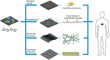
Osteoblasts are responsible for new bone formation, and upregulating of their activity is necessary to accelerate bone regeneration. The first successful use of electric treatment for a human nonunion fracture was demonstrated in 1971 when a 10 mA dc current was applied using a stainless-steel electrode placed directly in the bone marrow of the fracture void. Reference Friedenberg, Harlow and Brighton39 The ES therapeutic approach is minimally invasive and free of complications, with success rates upward of 80% in comparison to surgical procedures. Reference Scott and King40 Electrical stimulation thus clearly represents a promising and powerful tool in RE to manipulate or modulate cellular activity. Meng et al. fabricated an electrically conductive substrate for bone regeneration made of 5% polypyrrole, a conductive polymer and 95% poly-L-lactic acid, a biodegradable polyester. Reference Meng, Zhang and Rouabhia41,Reference Shi, Rouabhia, Wang, Dao and Zhang42 Electrical stimulation was found to promote osteoblast adhesion and growth, resulting in significantly higher calcium and phosphate content in the surface-deposited minerals on the conductive membranes. Furthermore, the gene expression of osteoblast-specific markers was significantly upregulated, suggesting that ES applied through a synthetic conductive substrate could be a viable option to regenerate bone. Figure 2 Reference Hardy, Lee and Schmidt43 represents a schematic of the aspects (e.g., conductivity properties under hydration, polymer biological activity, biodegradation mechanism of the engineered substrate, mechanical properties, surface chemistry and topographical properties) to be considered when designing biomimetic conductive polymer-based regenerative substrates.
Schematic illustrating the most important aspects considered when designing biomimetic conductive polymer-based materials. Reprinted with permission from Reference Reference Hardy, Lee and Schmidt43. © 2013 Elsevier.

Musculoskeletal soft tissues such as muscle and tendon function as a single unit to generate force, transmitting it to the bone to facilitate limb movement. Most clinical tissue-repair strategies are based on strengthening the site of injury via autograft (tissue harvested from the same patient) or allograft (tissue harvested from cadavers and processed prior to use) augmentation. Reference Longo, Lamberti, Maffulli and Denaro44 The regeneration of critical defects such as those occurring at the tendon-muscle unit remains a large and significant clinical problem. The rotator cuff (RC) of the shoulder, for example, is susceptible to muscle atrophy and fatty expansion in the tendon tissue post-trauma, and patients experience pain, weakness, a limited range of shoulder motion and an inability to reach above their head. Reference Gerber, Fuchs and Hodler45–Reference Safran, Derwin, Powell and Iannotti50 Biocompatible materials and biomimetic substrates are critical to harnessing the full range of regenerative abilities in an individual. Nanoscale fibrous morphology mimicking collagen fiber bundles are one of the defining structural features of the ECM, and skeletal muscle presents a hierarchical ECM fiber organization and myoblast alignment, which is a key step in musculoskeletal myogenesis. Reference McCullen, Ramaswamy, Clarke and Gorga51
Musculoskeletal tissues supplied with nerves, such as muscle and muscle-tendon junctions respond to ES, especially for the attenuation of muscle atrophy, and ES therapy has been investigated to restore mobilization and recover from muscle disuse and spinal cord injury. Reference Ku, Lee and Park52 The effectiveness of ES treatment is influenced by stimulation parameters, particularly the applied frequency and duration. Ku et al. investigated random and aligned poly(ε-caprolactone) (PCL) nanofiber matrices containing polyaniline (PANi), a conductive polymer, to determine the combinatorial effects of topographical cues and electroactivity on muscle fiber (known as mytoubes) formation by C2C12 mouse myoblasts seeded onto these nanofiber substrates. The skeletal myoblasts grew well on PCL/PANi nanofibers irrespective of PANi concentration or fiber alignment, but their morphology was highly dependent on fiber alignment. Myosin heavy chain (MHC) and gene expression levels of myotube phenotype markers were positively influenced by conductive polymer concentration and nanofiber alignment, leading to enhanced myoblast attachment, proliferation, and differentiation into muscle tissue phenotype (myogenic).
Laurencin et al. developed a smart biointerface consisting of the conducting polymer poly(3,4-ethylenedioxythiophene) poly(styrene sulfonate) (PEDOT:PSS) in combination with an aligned biodegradable substrate composed of PCL. Reference Tang, James and Laurencin53 This flexible conductive substrate was hierarchically fabricated by first preparing aligned electrospun nanofiber substrates of PCL onto which dopamine was surface functionalized to form a polydopamine coating ( Figure 3 ). Reference Tang, James and Laurencin53 The deposited layer of polydopamine enhanced affinity for the deposition of PEDOT:PSS solutions with varying concentration (1%, 10%, 33%, and 100% w/v). Electroconductive nanofiber substrates with controlled conductivity were successfully achieved by modifying the concentration of PEDOT:PSS, with the 100% group having an average sheet resistance of ∼5 × 10⁴ Ω cm, while the conductivities of the 33% and 10% groups were 13 × 105 Ω cm and 17 × 106 Ω cm, respectively. An optimal PEDOT:PSS coating concentration of 10% was found to be effective in promoting myoblast differentiation due to its intrinsic conductivity. Mechanistically, differentiation proceeded via an upregulation of myogenic regulatory factors, such as Myf5, MyoD, myogenin (MyoG), and troponin T type 1. Reference Tang, James and Laurencin53
Schematic illustration of novel conducting polymer-coated matrices and myotube formation on electroactive matrices. Intrinsic conductivity facilitated by 10% poly(3,4-ethylenedioxythiophene) poly(styrene sulfonate) (PEDOT:PSS) solution was found to be effective in promoting myoblast differentiation into fused myotubes or muscle fibers. Reference Tang, James and Laurencin53

Ionically conducting bioelectronics as previously described have demonstrated the ability to independently electrically stimulate muscle cells and regulate cellular response. Such a feature has important implications for the design of functional muscle constructs with inbuilt self-control features. The capabilities of this technology are extremely promising as it impacts muscle regeneration via muscle stem cell differentiation, and could have a significant impact in the clinical setting, where muscle atrophy and fatty infiltration prevents the healing of RC disease and injury. The signaling cascades involved in differential phenotype expression upon ES have not been completely elucidated. However, preliminary studies suggest that the participation of cell-cycle-regulating intracellular signaling pathways (e.g., the PI3-AKT pathway) and the communication of signals from cell surface receptors to the cell nucleus (e.g., mitogen-activated protein kinases pathway) in response to ES act to maintain intracellular pathways at levels similar to the native tissue under mechanical loading. Reference Chin, Olson, Richardson, Yang, Humphries, Shelton, Wu, Zhu, Bassel-Duby and Williams54 Low-frequency ES elevates intracellular calcium and induces sustained, low-amplitude calcium influx, which activates the protein calcineurin phosphatase. The calcineurin-transcription factor pathway uniquely promotes skeletal muscle differentiation, fiber-type transformation, muscle regeneration, and hypertrophy, thus attenuating muscle atrophy.
There is a need for a systematic materials platform that provides tailored substrate stiffness, porosity, and hydrophilicity to modulate and study cellular response, especially of complex soft-tissue systems. Reference Park, Gong, Park, Moon, Wall, Kim, Lee and Knowles55,Reference Xu, Li, Fang, Thouas, Cook, Newgreen and Chen56 Ma et al. designed and synthesized a series of shape-memory copolymers exhibiting multifunctional characteristics, including electroactivity, super stretchability, and tunable recovery temperature, and is based on degradable copolymers of PCL and conductive amino-capped aniline trimer. These novel substrates improved the proliferation of C2C12 myoblasts in vitro compared to polyester biomaterials, indicating that they are excellent substrates to repair skeletal muscle and could be applied to regenerate other tissues. Reference Deng, Guo, Zhao, Li, Dong, Guo and Ma57 These shape-memory copolymers display a high electroactivity and elasticity at break, including recovery at body temperature, high recovery ratio, good mechanical deformation ratio, and short recovery time. The stretchable electroactive copolymers showed significant positive effect on myogenic differentiation (MHC and MyoG) of C2C12 myoblasts compared to PCL substrates for gene expression levels.
Stem cells
Stem cells are a critical component of regenerative biomedical technologies due to their two unique abilities—the ability to self-renew and the ability to differentiate into multiple lineages under appropriate stimuli, giving rise to numerous cell types that comprise tissues and organs. Stem cells are typically categorized into two distinct classes, those presenting pluripotency and multipotency. Pluripotent stem cells include embryonic stem cells (ESCs) and induced pluripotent stem cells (iPSCs), and in contrast to multipotent stem cells, they exhibit the unique property to self-renew indefinitely and differentiate into one of the three germ layers, namely endoderm, ectoderm, and mesoderm. Reference Thomson, Itskovitz-Eldor, Shapiro, Waknitz, Swiergiel, Marshall and Jones58–Reference Fox, Daley, Goldman, Huard, Kamp and Trucco60 ESCs are isolated from the blastocyst during embryological development, and iPSCs are obtained by genetically modifying somatic cells (i.e., differentiated cells). Examples of multipotent stem cells include bone marrow-derived mesenchymal stem cells (MSCs), hematopoietic stem cells, neural stem cells (NSCs), and adipose-derived stem cells. These stem cells are present in specialized niches within the differentiated tissues and can undergo self-renewal into advanced ages. Stem cells can be easily expanded in vitro, and therefore, a large number of cells can be obtained for seeding onto three-dimensional (3D) substrates of clinically relevant volume for transplantation, and give rise to committed cell types, such as osteoblasts (bone), chondrocytes (cartilage), adipocytes (fat), and neuronal cells, when they receive the necessary stimulatory cues. Reference Zhang and Kohn61
Studies have demonstrated that polymer material properties affect the activity of stem cells in vitro, and tissue regeneration in vivo, and thus, the incorporation of appropriate physical, chemical, mechanical, and biological cues to guide stem cell adhesion, proliferation, and differentiation is critical to successful translation. Reference Laurencin and Khan3,Reference Laurencin, Daley and James16 Natural polymers (e.g., collagen, fibrin, and polysaccharides) contain a variety of biological cues, allowing for cell recognition and promoting cellular adhesion. However, their manufacturing process is inherently prone to significant batch-to-batch variation, leading to engineering concerns for medical devices incorporating them. In comparison, synthetic polymers of precise molecular weight and composition are reliably synthesized, thereby minimizing engineering concerns. Synthetic polymers, including polyesters, polyglycols, polycaprolactones, and polyanhydrides have been extensively investigated and show excellent biocompatibility, controlled drug release, and support cell adhesion and cellular proliferation. Reference Zhang and Kohn61,Reference Guo and Ma62 However, in the long term, these polymers and their degradation remnants in vivo become encapsulated by a layer of fibroblasts and inflammatory cells, becoming suboptimal in function, and inhibiting the formation or regeneration of new functional tissue.
Advances in directing stem cells are inspired by cellular mechanisms involved in embryonic development, leading to the discovery of inductive cues such as growth factors and small molecules to recapitulate the embryonic mechanisms and differentiation into desired cellular phenotype and specific tissues. The behavior of stem cells can be modulated by incorporation of directional cues into engineered substrates, including chemical and insoluble factors. Reference Zhang and Kohn61 The cellular microenvironment is unique to every tissue, and its composition of matrix proteins, structure, dimensions, and interaction with cells and factors is under constant dynamic pressure responding to common issues such as injury, disease, and increasing age of the individual. Roughness, stiffness, topography, pore size, porosity, pore connectivity, fiber diameter, fiber orientation, and surface charge have been shown to exhibit controlling effects on stem cells ( Figure 4 ). Reference Coyle, Jia and Mei63–Reference Khalil, Xie and Murphy65
Stem cell interactions with chemical and physical cues. (a) Chemical interactions on materials can regulate growth factor signaling. Engineered materials may incorporate (i) covalently bound glycosaminoglycans (GAGs) or proteoglycans (PGs) or (ii) moieties that bind GAGs/PGs, which in turn, can sequester growth factors from the stem cell microenvironment. Alternatively, materials may be functionalized with (iii) moieties that bind growth factors or (iv) moieties that directly interact with growth factor receptors (GFRs), to upregulate or downregulate GFR signaling. Finally, GFRs and their associated signaling pathways may synergize with (v) integrin-mediated adhesion and signal downstream of adhesion. (b) Mechanical properties of the microenvironment. Resistance to deformation of stiff materials increases the cytoskeletal tension of human mesenchymal stem cells (hMSCs) through focal adhesion kinase and Rho-associated kinase activity, leading to differentiation (left). The introduction of topography results in rearrangement of integrins at the cell-material interface, promoting topography-dependent hMSC proliferation, self-renewal, or differentiation (right). Reprinted with permission from Reference Reference Khalil, Xie and Murphy65. © 2014 American Chemical Society.

Nanofiber-based technology for tissue healing and RE has received significant attention, leading to a comprehensive reference library of peer-reviewed literature. Reference James, Toti, Laurencin, Kumbar and Hurst66–Reference Zhang68 The attractiveness of nanofiber technology lies in the unique biological responses and cellular phenomena that are directed or guided by their small length scale. This is strongly correlated to events within the native tissue environment. Electrospun manufacturing of nanofibers provides for a wide range of fiber diameters, pore-diameter distribution, high porosity, and variable mechanical properties to recapitulate the native microenvironment. This paradigm contrasts with the well-investigated approach of modulating cell activity and function using chemical and biomacromolecular cues.
The mature technologies available today for engineered nanofiber substrates began with our work in nanofiber technology. This helped develop new areas of biomedical research. Reference Li, Laurencin, Caterson, Tuan and Ko69 Laurencin et al. reported RC healing and regeneration using a hybrid PCL nanofiber substrate coated with a hydrophilic polyphosphazene derivative. This mimics the intrinsic cues and microenvironmental influences that specify and maintain cell identity. Reference Peach, Kumbar, James, Toti, Balasubramaniam, Deng, Ulery, Mazzocca, McCarthy, Morozowich, Allcock and Laurencin70 Exogenous stem cell populations delivered using this hybrid scaffold did not integrate with the host, but affected tissue functionality and composition through paracrine (nearby cell-to-cell communication factors) influences. In addition, other groups have leveraged dynamic control of the substrate’s elastic modulus to reveal bias differentiation in stem cell populations. Reference Engler, Sen, Sweeney and Discher71,Reference Lv, Li, Sun, Zhang, Chen, Rong and Li72 We and others have derivatized mechanical, organizational, and compositional cues, encoding them into biocompatible substrates using nanofiber technology. Reference Li, Laurencin, Caterson, Tuan and Ko69,Reference Seidi, Sampathkumar, Srivastava, Ramakrishna and Ramalingam73,Reference Zhang, Reagan and Kaplan74 As a prerequisite for moving beyond single-tissue repair to complex multi-tissue regeneration, nanofiber substrates have been applied to the regeneration of different soft tissues. Reference Nelson, Khan and Laurencin75,Reference Wang, Valmikinathan, Liu, Laurencin and Yu76 Dermagraft is a cell-material hybrid substrate approved by the US Food and Drug Administration for use in the clinic for wound healing. It is composed of a polyglactin mesh onto which human neonatal dermal fibroblasts are cultured in vitro. It is then cryopreserved. Reference Pham, Greenwood, Cleland, Woodruff and Maddern77 Dermagraft has been approved as a skin substitute for wound closure of full-thickness diabetic foot ulcers presenting for longer than six weeks.
Cell molecular mechanisms respond to different topographies, translating to differences in the focal adhesion structures of cells cultured on these substrates. These differences include change in cell size, their adhesion strength, and ECM composition, which correlate to the formation of stress fibers, cell spreading, and determination of cell fate. Reference Khalil, Xie and Murphy65 MSCs differentiate along an osteogenic lineage upon RhoA activation, leading to cell spreading, whereas adipogenic phenotype is directed upon inhibition and restriction of cell spreading. A key regulator of mechanotransduction are focal adhesion kinases (FAK) which are activated upstream of RhoA and are also influenced by changes in substrate nanotopography. Differential activation of FAK triggers downstream intracellular signaling cascades that deliver information about the extracellular environment to the cell nucleus. Stem cell populations cultured on nanofiber substrates have enhanced nuclear and peroxisomal (energy production) functionality, and exhibit upregulation of differentiation capacity potentially driven by changes in the structure, positioning, and function of these organelles. Reference Tutak, Jyotsnendu, Bajcsy and Simon78 Mechanistic studies thus far have provided a brief preview, suggesting the involvement of the FAK/RhoA signaling pathway in substrate topography influenced cell fate decisions.
Patterning technology has been used to fabricate polymer microarrays containing monomer and polymer solutions melt-deposited by noncontact printing methods onto a substrate ( Figure 5 ). Reference Coyle, Jia and Mei63,Reference Oliveira and Mano79 These advanced technologies (lithography and nanoimprinting) allow for microarray fabrication of spatially defined polymers with varied chemistries and topographies. The variations in chemical composition also allow for polymeric substrates with a range of physical properties such as elasticity, hydrophilicity, and adhesiveness. These techniques have been used to robotically fabricate high-throughput material arrays to rapidly identify the influence of physical and chemical cues on stem cell behavior. For example, Mei et al. fabricated microarrays composed of light-polymerized methacrylates, evaluating their ability to adsorb serum and ECM proteins to bind cell-surface receptors. Reference Coyle, Jia and Mei63,Reference Mei, Saha, Bogatyrev, Yang, Hook, Kalcioglu, Cho, Mitalipova, Pyzocha, Rojas, Van Vliet, Davies, Alexander, Langer, Jaenisch and Anderson80
A conceptual diagram showing the steps required to perform the evaluation of the in vivo inflammatory response to biomaterials in a high-throughput manner using superhydrophobic surfaces patterned with wettable regions as chips. (a) The preparation of superhydrophobic surfaces with arrays of wettable regions, where droplets of aqueous solutions can be dispensed with different volumes in spots with different shapes and sizes. Such solutions may be polymeric and are amenable to being freeze-dried on-chip, generating biomaterial arrays of miniaturized porous scaffolds, widely used in the tissue engineering field. (b) The chips with arrays of combinatorial biomaterials are implanted subcutaneously in a Wistar rat model. (c) After a determined time of implantation (t), the chip is retrieved from the animal, and the scaffolds on the chip are analyzed by immunocytochemistry. (d) Immunocytochemistry and cell-staining images are then collected by microscopy techniques and analyzed, preferably by automated processes using software such as ImageJ (National Institutes of Health). (e) The results must be analyzed for statistical significance and with factorial analysis so that the main and combined effects of the biomaterial design and processing can be analyzed in relation to the biological phenomena that occur in vivo. Reprinted with permission from Reference Reference Oliveira and Mano79. © 2014 Elsevier.

Studies that correlated physical characteristics such as surface wettability, surface roughness, and indentation elastic modulus of each polymer helped determine the structure–function relationship between biological performance and material properties. The results showed that substrate properties affected the undifferentiated growth of human ESCs to varying degrees, with surface chemistry being the most dominant parameter. Combinatorial polymer microarrays enabled screening of culture substrates to identify new chemical moieties that promote stem cell adhesion. Reference Anderson, Putnam, Lavik, Mahmood and Langer81 Others have directly printed biodegradable polymers onto glass slides to investigate their effects on human MSCs, neural stem cells (NSCs), and primary articular chondrocytes, demonstrating the need for unbiased screening to discover suitable substrates. In further technological advancements, high-throughput methods have been applied to evaluate heterogeneous cell populations based on surface topography. Chen et al. demonstrated that the undifferentiated human ESCs prefer smooth surfaces while the differentiated human ESCs favorably attach to surfaces featuring nanoroughness. Reference Chen, Villa-Diaz, Sun, Weng, Kim, Lam, Han, Fan, Krebsbach and Fu82
Conclusion
New breakthroughs and translational developments are emerging from the convergence of varied entities and toolboxes within advanced materials science, stem cell science, and developmental biology. Reference Laurencin and Khan3,Reference Tibbitt, Rodell, Burdick and Anseth83 No single entity is sufficient by itself. Recent advances in biomaterial synthesis and fabrication tools have made it possible to guide cells into complex 3D structures by using appropriate substrate templates. Reference Dvir, Timko, Kohane and Langer84,Reference Kelleher and Vacanti85 Polymer array technology has come to the forefront as a powerful tool to rapidly evaluate the relationship between biophysical properties and polymer chemistry, and cellular responses. High-throughput materials discovery, “hit” optimization and the identification of “leading” compounds can efficiently improve discovery methodologies by predicting cellular responses using extensive combinatorial libraries. Reference Celiz, Smith, Patel, Hook, Rajamohan, George, Flatt, Patel, Epa, Singh, Langer, Anderson, Allen, Hay, Winkler, Barrett, Davies, Young, Denning and Alexander86 Scientists and engineers will be able to rapidly triage the optimal candidates that meet desired specifications, such as stem cell response and phenotype development without the need to experimentally examine each candidate. This leads the way in personalized/precision regenerative medicine and disease modeling.
While previous research efforts largely focused on identifying or developing optimal substrates to support cell culture, the delivery and maintenance of donor stem cells and feedback communication with the host tissue is the new regenerative direction. The emergence of an optimally engineered substrate to drive multipotent cells toward desired lineages solely through the material and design-based cues holds great promise, and we expect this technology to undergo significant development over the next decade to further harness the ability of the body to self-heal. Reference Metavarayuth, Sitasuwan, Zhao, Lin and Wang87 Approaches such as “organs on a chip” and 3D organ printing have emerged, and mergers of seemingly disparate technologies, such as mechanisms of signaling pathways and bioelectronics, will be distilled to specify precise parameters of regenerative mechanisms. Reference Derby88,Reference Park, Lim, Park and Lee89
Acknowledgments
We gratefully acknowledge funding from The Raymond and Beverly Sackler Center for Biomedical, Biological, Physical and Engineering Sciences, National Science Foundation Award Number IIP-1311907 and EFRI-1332329.

Roshan James completed his postdoctoral training at the Institute for Regenerative Engineering, and The Raymond and Beverly Sackler Center for Biomedical, Biological, Physical and Engineering Sciences at the University of Connecticut. He received his B.Tech degree in biotechnology and biochemical engineering from Indian Institute of Technology–Kharagpur, India, his MS degree in bioengineering from Clemson University, and his PhD degree in bioengineering from the University of Virginia. His research interests include biomaterial synthesis, drug delivery and formulation, and medical device entrepreneurship. He is a scientist in the pharmaceutical industry. James can be reached by phone at 860-679-5577 or by email at rjames.uchc@gmail.com.

Cato T. Laurencin is the Albert and Wilda Van Dusen Distinguished Endowed Professor of Orthopaedic Surgery, professor of chemical and biomolecular engineering, professor of materials science and engineering, and Professor of Biomedical Engineering at the University of Connecticut. He earned his BSE degree in chemical engineering from Princeton University and his MD degree from the Harvard Medical School, while simultaneously earning his PhD degree in biochemical engineering/biotechnology from the Massachusetts Institute of Technology. His current research focuses on regenerative engineering, biomaterials, nanotechnology, drug delivery, and stem cell science. He is a Fellow of the Materials Research Society, the American Chemical Society, and an International Fellow in Biomaterials Science and Engineering. He is the editor-in-chief of Regenerative Engineering and Translational Medicine. Laurencin can be reached by phone at 860-679-6544 or by email at laurencin@uchc.edu.