Implications
The circadian system coordinates internal physiological processes in mammals and synchronizes these processes to the animal's environment. We review the literature and discuss recent findings from our laboratory and others that suggest the circadian system regulates lactation, including coordinating changes in the dam's physiology needed to initiate and maintain lactation and mediating the photoperiod effect on milk production. Identification of environmental and physiological inputs that affect genes that control circadian rhythms will enable development of approaches to alter gene expression to maximize production efficiency in farm animals.
Introduction
Nearly all physiological and behavioral functions of animals are rhythmic including secretion patterns of hormones, sleep–wake cycle, metabolism and core body temperature. A circadian rhythm is a roughly 24-h cycle in the biochemical, physiological or behavioral processes of organisms. Daily and seasonal endocrine rhythms are coordinated in mammals by the master circadian clock located in the hypothalamus. The master clock receives and integrates environmental (e.g. photoperiod) and physiological (e.g. nutritional status) cues that set the master clock at the molecular level. Reference time and rhythms are sent out to peripheral tissues from the master clock via hormonal and autonomic signals. Peripheral clocks located in every organ of the body receive these signals from the master clock, which coordinate and synchronize the timing of rhythms generated by peripheral clocks across the entire body.
During the transition from pregnancy to lactation homeorhetic adaptations are coordinated across almost every organ of the body and are marked by tremendous changes in hormones and metabolism to accommodate for the increased energetic demands of lactation (Bauman and Currie, Reference Bauman and Currie1980; Bell et al., Reference Bell, Bauman and Currie1987; Bell and Bauman, Reference Bell and Bauman1997). This transition is the most stressful period of the cow's life and has a large impact on a cow's lactation production. As a result, ∼80% of dairy cow disease events occur in the first 3 weeks after a cow delivers a calf (Drackley, Reference Drackley1999; Mulligan and Doherty, Reference Mulligan and Doherty2008; Grummer et al., Reference Grummer, Wiltbank, Fricke, Watters and Silva-Del-Rio2010; LeBlanc, Reference LeBlanc2010).
Understanding how a cow sets her metabolic and physiological rhythms in response to changes in her physiology, nutritional status and/or environment will enable the development of simple approaches that maximize productive efficiency and minimize metabolic disturbances in dairy cows. The circadian system is a likely candidate as it is believed to have evolved to coordinate the timing of internal physiological and metabolic processes and synchronize this timing with the animal's environment. The effect of the circadian system on milk production is evident in both the variation of milk composition across the day (Kuhn et al., Reference Kuhn, Carrick and Wilde1980; Nielsen et al., Reference Nielsen, Ingvartsen and Larsen2003; Barkova et al., Reference Barkova, Nazarenko and Zhdanova2005; Lubetzky et al., Reference Lubetzky, Littner, Mimouni, Dollberg and Mandel2006 and Reference Lubetzky, Mimouni, Dollberg, Salomon and Mandel2007; Cubero et al., Reference Cubero, Narciso, Terron, Rial, Esteban, Rivero, Parvez, Rodriguez and Barriga2007) and the photoperiod effect on milk quality and quantity in farm animals (Aharoni et al., Reference Aharoni, Brosh and Ezra2000; Dahl et al., Reference Dahl, Auchtung and Kendall2002 and Reference Dahl, Auchtung and Reid2004; Dahl and Petitclerc, Reference Dahl and Petitclerc2003; Auchtung et al., Reference Auchtung, Rius, Kendall, McFadden and Dahl2005; Wall et al., Reference Wall, Auchtung-Montgomery, Dahl and McFadden2005; Bernabucci et al., Reference Bernabucci, Basirico, Lacetera, Morera, Ronchi, Accorsi, Seren and Nardone2006; Rius and Dahl, Reference Rius and Dahl2006; Auldist et al., Reference Auldist, Turner, McMahon and Prosser2007; Oates et al., Reference Oates, Bradshaw, Bradshaw, Stead-Richardson and Philippe2007; Andrade et al., Reference Andrade, Salama, Caja, Castillo, Albanell and Such2008; Dahl, Reference Dahl2008; Mikolayunas et al., Reference Mikolayunas, Thomas, Dahl, Gressley and Berger2008). In this paper, we review literature and present novel findings in support of our hypothesis that the circadian system coordinates the metabolic and endocrine changes needed to initiate and maintain lactation, and that this system also mediates the photoperiod effect on lactation.
The circadian system
The circadian system is regulated hierarchically in mammals by the master clock that lies in the suprachiasmatic nuclei (SCN; Hastings et al., Reference Hastings, O'Neill and Maywood2007). The intrinsic rhythmicity of the SCN is synchronized to the 24-h day by regularly occurring environmental signals or ‘Zeitgebers’ (Welsh et al., Reference Welsh, Logothetis, Meister and Reppert1995). The light–dark (LD) cycle is the most salient environmental cue for entraining circadian clocks (Reppert and Weaver, Reference Reppert and Weaver2002). Non-photic environmental cues including exercise, food availability, temperature and stress can also directly and/or indirectly entrain the SCN. The activity of SCN neurons is also modulated by serotoninergic pathways and melatonin (Moore et al., Reference Moore, Halaris and Jones1978; Bosler and Beaudet, Reference Bosler and Beaudet1985; Meyer-Bernstein and Morin, Reference Meyer-Bernstein and Morin1996; Jacobs et al., Reference Jacobs, Martin-Cora and Fornal2002; Moore and Speh, Reference Moore and Speh2004; Wirz-Justice, Reference Wirz-Justice2006).
The SCN signal is translated into hormonal and autonomic signals for peripheral organs through its major outputs (i) the paraventricular nucleus (PVN) of the hypothalamus; and (ii) the pineal gland, where melatonin is synthesized according to the length of the photophase. Melatonin is the biochemical transducer of photoperiodic information to all cells in the body (including SCN neurons), and changes in duration and amplitude of nocturnal secretion serves to signal seasonal variations of day/night cycle length (for review see Simonneaux and Ribelayga, Reference Simonneaux and Ribelayga2003). Autonomic neurons of the PVN communicate time-of-day signal to different organs, and corticotrophin-releasing factor, which is secreted by PVN neurons, indirectly communicates time of day through its circadian rhythmicity entrained by the SCN. Circadian oscillations of core body temperature are also set by the master clock and serve as output signals that influence the timing of peripheral clocks (Buhr et al., Reference Buhr, Yoo and Takahashi2010).
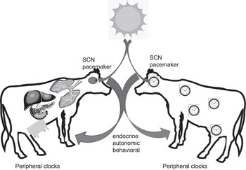
Outputs from the master clock regulate and synchronize peripheral clocks that are located in every tissue of the body (Damiola, Reference Damiola2000; Yamazaki et al., Reference Yamazaki, Numano, Abe, Hida, Takahashi, Ueda, Block, Sakaki, Menaker and Tei2000; Schibler et al., Reference Schibler, Ripperger and Brown2003; Sheward et al., Reference Sheward, Maywood, French, Horn, Hastings, Seckl, Holmes and Harmar2007). These tissue clocks in turn drive the circadian expression of local transcriptomes, thereby coordinating metabolism and physiology of the entire animal (Figure 1). Thus, external and internal cues are integrated by pacemaker neurons within the SCN and set the master clock which in turn provides a ‘reference time’ for all peripheral tissue clocks. The role of SCN-synchronized peripheral clocks in coordinating xenobiotic detoxification, cell division and nutrient metabolism is important to health, and disturbances to circadian timing are recognized as factors in major systemic illness (Bass and Takahashi, Reference Bass and Takahashi2010).
Schematic represents how circadian system coordinates internal physiology with the environment. The primary pacemaker, suprachiasmatic nuclei, is entrained to solar time through retinal afferents and synchronizes and maintains tissue clocks through endocrine, autonomic and behavioral (feeding-related) cues. Adapted from Hastings et al. (Reference Hastings, O'Neill and Maywood2007).

Core molecular clock genes that drive circadian rhythms contain positive and negative elements that form auto-regulatory feedback loops
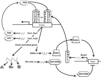
Control of the circadian clock consists of oscillatory feedback activities of core molecular clock genes. In mammals, the core clock genes are conserved, and the positive loop consists of BMAL1 (aka ARNTL), CLOCK and NPAS2 gene products, whereas the negative loop consists of the Per and Cry gene products (Darlington, Reference Darlington1998; Gekakis, Reference Gekakis1998; Kume, Reference Kume1999; Yagita, Reference Yagita2000). CLOCK and BMAL1 (or NPAS2 and BMAL1) proteins activate transcription of numerous target genes, including their own repressors, Pers and Crys thus forming a transcriptional feedback loop. BMAL1 expression is also regulated by two of its transcriptional targets, nuclear receptors Rev-erbα (NR1D1) and Rorα (RORA/NR1F1), which repress or activate, respectively, BMAL1 transcription (Preitner et al., Reference Preitner, Damiola, Luis Lopez, Zakany, Duboule, Albrecht and Schibler2002; Guillaumond et al., Reference Guillaumond, Dardente, Giguère and Cermakian2005). The CLOCK/BMAL1 (NPAS2/CLOCK) complex also drives rhythmic expression of numerous other genes (Figure 2; Brown and Schibler, Reference Brown and Schibler1999; Noshiro et al., Reference Noshiro, Usui, Kawamoto, Kubo, Fujimoto, Furukawa, Honma, Makishima, Honma and Kato2007). Both external and internal cues can directly or indirectly affect the expression of core clocks genes in the SCN and peripheral clocks, and thus the molecular components of circadian control systems provide novel avenues for therapeutic intervention (Hastings et al., Reference Hastings, O'Neill and Maywood2007).
Transcriptional and translational autoregulatory feedback loops that regulate circadian rhythms in cells. The positive loop consists of BMAL1 and CLOCK gene products or BMAL1 and NPAS2 gene products and the negative loop consists of the Per and Cry gene products. CLOCK and BMAL1 proteins heterodimerize to activate transcription of numerous target genes, including their own repressors, PERs and CRYs thus forming a transcriptional feedback loop. BMAL1 expression is also regulated by two of its transcriptional targets, nuclear receptors Rev-erbα and Rorα, which repress or activate, respectively, BMAL1 transcription by competing for the same promoter element and forming a secondary interlocked feedback loop.

Global temporal expression profiles of SCN, liver, adipose and heart tissues in mammals revealed that ∼3% to 10% of expressed transcripts in the genome are under circadian control (Akhtar et al., Reference Akhtar, Reddy, Maywood, Clayton, King, Smith, Gant, Hastings and Kyriacou2002; Panda, Reference Panda2002; Storch et al., Reference Storch, Lipan, Leykin, Viswanathan, Davis, Wong and Weitz2002; Ando et al., Reference Ando, Yanagihara, Hayashi, Obi, Tsuruoka, Takamura, Kaneko and Fujimura2005). Many of the tissue-specific clock-regulated genes were involved in rate-limiting steps critical for organ function. In the rodent liver, 35% of clock-regulated genes direct biosynthesis and metabolism and 10% regulate gene transcription (Akhtar et al., Reference Akhtar, Reddy, Maywood, Clayton, King, Smith, Gant, Hastings and Kyriacou2002). These include transcription factors and nuclear hormone receptors that regulate carbohydrate and lipid homeostasis (Brewer et al., Reference Brewer, Lange, Baler and Anzulovich2005; Yang et al., Reference Yang, Kurotani, Yamada, Kimura and Gonzalez2006). Thus, many rate-limiting hepatic enzymes controlling glycolysis, gluconeogenesis, fatty acid metabolism and amino acid metabolism are regulated by the circadian system (Oishi et al., Reference Oishi, Fukui and Ishida2000; Froy, Reference Froy2007).
Recent studies in our laboratory and others have shown the existence of a circadian clock in the mammary gland (Metz et al., Reference Metz, Qu, Laffin, Earnest and Porter2006; Casey et al., Reference Casey, Patel, Dykema, Dover, Furge and Plaut2009; Maningat et al., Reference Maningat, Sen, Rijnkels, Sunehag, Hadsell, Bray and Haymond2009).
Using transcriptome analysis we investigated coordinated gene expression changes in mammary, liver and adipose during the transition from pregnancy to lactation (Casey et al., Reference Casey, Patel, Dykema, Dover, Furge and Plaut2009). For this study total RNA was isolated from mammary, liver and adipose tissues that were collected from rat dams on day 20 of pregnancy (P20; n = 5) and day 1 of lactation (L1; n = 5). We found that multiple pathways and gene sets related to energy homeostasis were changed in peripheral tissues at the onset of lactation. Molecular signatures of mammary, liver and adipose were enriched with gene sets associated with central nervous system reception, integration and response to environmental and internal stimuli. To gain insight into what was stimulating these changes, we examined genes commonly upregulated at the onset of lactation clustered in the gene ontology transcription regulator activity. There were 112 genes commonly upregulated among mammary, liver and adipose during the transition from pregnancy to lactation in this ontology, including the core clock genes ARNTL (aka BMAL1), NPAS2 and CLOCK, the clock regulator RORA, as well as SREBF2. SREBF2 is a sterol receptor-binding protein transcription factor that activates enzymes important to de novo lipid synthesis. When we examined genes commonly downregulated in mammary, liver and adipose tissues during the pregnancy to lactation transition, we found that rhythmic process was significantly enriched with downregulated genes including NR1D1, DBP and BHLHB2 (all three are well-characterized transcriptional targets of CLOCK/BMAL1). In summary, during the transition from pregnancy to lactation there was a coordinated induction in expression of the positive limb core clock genes and their regulators (BMAL1(ARNTL), NPAS2, CLOCK and RORA) and a suppression in expression of regulators of the negative limb of core clock genes (BHLHB2, NR1D1, CSNK1E) across all three tissues (Casey et al., Reference Casey, Patel, Dykema, Dover, Furge and Plaut2009). These data suggest that coordinated changes in the circadian system occur during the transition from pregnancy to lactation. Circadian sampling (collecting tissues every 4 h over a 24-h period) to describe temporal changes in gene expression showed that expression rhythms for BMAL1 (Arntl) and PER1 increased in amplitude during the transition from pregnancy to lactation in mice (Metz et al., Reference Metz, Qu, Laffin, Earnest and Porter2006). Changes in amplitude reflect the relative strength of the underlying pacemaker (Bass and Takahashi, Reference Bass and Takahashi2010), and thus suggest that changes in the mammary core clock reflect the increase in metabolic capacity of this tissue. Global expression analysis of RNA isolated from milk fat globules of human breast milk revealed that ∼7% of the genes expressed in lactating breast show circadian patterns including core clock and metabolic genes (Maningat et al., Reference Maningat, Sen, Rijnkels, Sunehag, Hadsell, Bray and Haymond2009), suggesting that the mammary core clock, directly or indirectly, regulates a set of genes important to its metabolic output, milk.
Preliminary studies in our laboratory suggest that cows also have functional mammary clocks. Using an approach pioneered by Balsalobre (Balsalobre et al., Reference Balsalobre, Damiola and Schibler1998), we found that treating bovine mammary cells (MAC-T) with media supplemented with 50% serum for 2 h resulted in a synchronized circadian pattern of expression of the core clock gene BMAL1 (Casey, T. and Plaut K, unpublished results). Second, we found that expression of BMAL1 was significantly induced when prolactin was added to bovine mammary explant culture (unpublished results), suggesting that the mammary clock is responsive to lactogens. Third, expression of core clock genes BMAL1 and PER2 showed circadian patterns of expression in RNA isolated from milk fat globule crescents of mid-lactation cows (unpublished results), which correlated with circadian changes in expression of acetyl-CoA carboxylase (ACACA) as well as percent milk fat (unpublished results). These findings suggest that the mammary clock in cows may be responsible for the diurnal variation in milk composition (Kuhn et al., Reference Kuhn, Carrick and Wilde1980; Nielsen et al., Reference Nielsen, Ingvartsen and Larsen2003; Barkova et al., Reference Barkova, Nazarenko and Zhdanova2005; Lubetzky et al., Reference Lubetzky, Littner, Mimouni, Dollberg and Mandel2006; Cubero et al., Reference Cubero, Narciso, Terron, Rial, Esteban, Rivero, Parvez, Rodriguez and Barriga2007; Lubetzky et al., Reference Lubetzky, Mimouni, Dollberg, Salomon and Mandel2007).
The mammary circadian clock may not only respond to systemic cues, but may also be regulated by local signals. Serotonin (5-HT), which acts as both a neurotransmitter and hormone that entrains circadian clocks, has been proposed to be a feedback inhibitor of lactation (Stull et al., Reference Stull, Pai, Vomachka, Marshall, Jacob and Horseman2007). The gene coding for tryptophan hydroxylase 1, the rate-limiting enzyme for serotonin synthesis, is expressed in bovine mammary epithelial cells and is upregulated by prolactin in vitro (Matsuda et al., Reference Matsuda, Imaoka, Vomachka, Gudelsky, Hou, Mistry, Bailey, Nieport, Walther, Bader and Horseman2004). Addition of serotonin to mammosphere cultures reduced the expression of α-lactalbumin and casein genes. In contrast, inhibiting serotonin synthesis or blocking its receptor increased milk protein gene expression (Hernandez et al., Reference Hernandez, Stiening, Wheelock, Baumgard, Parkhurst and Collier2008). Further, intramammary infusion of serotonin reduced milk synthesis, whereas blocking the receptor increased milk synthesis in multiparous Holstein cows (Hernandez et al., Reference Hernandez, Stiening, Wheelock, Baumgard, Parkhurst and Collier2008). Somatotropin treatment of mice decreased mammary expression of tryptophan hydroxylase 1 (Hadsell et al., Reference Hadsell, Parlow, Torres, George and Olea2008). Further studies will be needed to definitively link the mammary clock with serotonin's effect on milk protein synthesis and mammary involution.
Metabolic function and circadian clocks are tightly interwoven such that clocks drive metabolic processes and various metabolic parameters affect clocks (Green et al., Reference Green, Takahashi and Bass2008). In rodents, peripheral liver clocks are synchronized by hormones reflecting the integrated nature of the circadian system (i.e. melatonin and glucocorticoids) and nutrient status (i.e. glucagon and insulin) (Ruiter et al., Reference Ruiter, La Fleur, van Heijningen, van der Vliet, Kalsbeek and Buijs2003; Kennaway et al., Reference Kennaway, Owens, Voultsios and Varcoe2006; Kohsaka and Bass, Reference Kohsaka and Bass2007; Kohsaka et al., Reference Kohsaka, Laposky, Ramsey, Estrada, Joshu, Kobayashi, Turek and Bass2007; Stokkan et al., 2001). This hormonal pattern coordinates liver metabolism to the appropriate time of day and nutritional status of the animal. Rodents with clock gene mutations have dysfunctional glucose homeostasis, insulin secretion and sensitivity and fat and cholesterol metabolism (Rudic et al., Reference Rudic, McNamara, Curtis, Boston, Panda, Hogenesch and Fitzgerald2004; Turek et al., Reference Turek, Joshu, Kohsaka, Lin, Ivanova, McDearmon, Laposky, Losee-Olson, Easton, Jensen, Eckel, Takahashi and Bass2005). Similarly, single nucleotide polymorphisms in Clock and Bmal1 genes in humans are associated with abnormal hepatic fat and glucose metabolism (Sookoian et al., Reference Sookoian, Castano, Gemma, Gianotti and Pirola2007; Woon et al., Reference Woon, Kaisaki, Braganca, Bihoreau, Levy, Farrall and Gauguier2007).
Similar to other organisms the circadian system appears to play a role in ruminant metabolism. Cows display distinct circadian patterns of intake (DeVries et al., Reference DeVries, von Keyserlingk and Beauchemin2003) and plasma concentrations of metabolic hormones including insulin, somatotropin, cortisol, melatonin, and triiodothyronine show diurnal patterns of secretion (Hedlund et al., Reference Hedlund, Lischko, Rollag and Niswender1977a and Reference Hedlund, Doelger, Tollerton, Lischko and Johnson1977b; Bitman et al., Reference Bitman, Kahl, Wood and Lefcourt1994; Lefcourt et al., Reference Lefcourt, Bitman, Kahl and Wood1993, Reference Lefcourt, Akers, Wood and Bitman1994, Reference Lefcourt, Bitman, Wood and Akers1995 and Reference Lefcourt, Huntington, Akers, Wood and Bitman1999). Circadian oscillation in plasma glucose, nonesterified fatty acids, β-hydroxybutyrate and urea nitrogen have also been observed in cows (Bitman et al., Reference Bitman, Wood and Lefcourt1990; Lefcourt et al., Reference Lefcourt, Huntington, Akers, Wood and Bitman1999). Studies carried out at the molecular level revealed that DBP mRNA expression, a clock-regulated transcription factor that directs hepatocyte metabolism (Oishi et al., Reference Oishi, Fukui and Ishida2000; Noshiro et al., Reference Noshiro, Usui, Kawamoto, Kubo, Fujimoto, Furukawa, Honma, Makishima, Honma and Kato2007), showed a circadian pattern that was altered when cattle were injected with somatotropin (Eleswarapu and Jiang, Reference Eleswarapu and Jiang2005). These daily cycles in hormones and metabolites likely help coordinate timing of intake with metabolism.
Feed availability is a peripheral clock Zeitgeber.
Although the LD cycle is the most reliable and strongest external signal that synchronizes (entrains) biological rhythms with the environment, food availability has also been shown to entrain biological rhythms of the peripheral clock in the liver (Stokkan et al., Reference Stokkan, Yamazaki, Tei, Sakaki and Menaker2001). When food is available only for a limited time each day, rats increase their locomotor activity 2 to 4 h before the onset of food availability (Stokkan et al., Reference Stokkan, Yamazaki, Tei, Sakaki and Menaker2001). This food anticipatory behavior occurs in other mammals, including goats, and is often accompanied by increases in body temperature, secretion of corticosterone, gastrointestinal motility and activity of digestive enzymes (Stokkan et al., Reference Stokkan, Yamazaki, Tei, Sakaki and Menaker2001; Piccione et al., Reference Piccione, Caolaa and Refinetti2003; Mendoza, Reference Mendoza2007; Bass and Takahashi, Reference Bass and Takahashi2010).
Microarray analysis of the impact of two acute restricted feeding regimens (4 v. 10 days) with identical body weight (BW) loss (19%) on hepatic gene expression in rats showed that the two regimens led to distinct patterns of differentially expressed genes in liver. Transcription profiles of 4-day restricted rats suggested that they were in an early phase of metabolic adaptation to feed restriction. Ten days of feed-restricting rats induced changes in gene expression associated with long-term metabolic adaptation to nutrient restriction as well as changes in the core clock genes PER1, PER2 and ARNTL (BMAL1; Pohjanvirta et al., Reference Pohjanvirta, Boutros, Moffat, Linden, Wendelin and Okey2008). These findings suggest that molecular clocks in the liver are reset to the animal's nutrient status as well nutrient availability in their environment.
To systematically dissect the role that food, feeding pattern and the circadian oscillator play in determining rhythmic gene expression in liver, global transcriptional changes in response to fasting and re-feeding were measured in wild-type (WT) C57Bl6 mice and oscillator-deficient mice (cry1 −/−;cry2 −/−; Cry mutant). Circadian gene expression profiles of these mice were examined under three different conditions: ad libitum, daytime-restricted feeding and prolonged fasting. In oscillator-deficient Cry mutant mice, restricted feeding restored 24-h rhythms in gene expression in 617 transcripts, which were identified as the ‘food-only’ oscillating transcripts. A total of 368 circadian clock-driven transcripts were identified under fasting conditions in WT mice. When WT mice were restricted to daytime feeding, 4960 transcripts displayed rhythmic expression (Vollmers et al., Reference Vollmers, Gill, DiTacchio, Pulivarthy, Le and Panda2009), showing that there is a clear synergy between the circadian and metabolic systems in the regulation of rhythmic transcription in the liver. These studies also suggest that there is an independent but interactive organization that links metabolic controls with circadian clocks. This organization likely allows organisms to coordinate tissue responses to predictable changes in their energy state (i.e. diurnal variation in fasting-feeding) through peripheral clocks. In addition, thousands of transcripts can be altered in response to unpredictable changes in the energy state through the uncoupling of metabolic and circadian timers.
Photoperiod manipulation has clear physiological and production effects in cattle.
A long-day photoperiod (LDPP), characterized as 16 h light (L) and 8 h dark (D), has been shown to hasten puberty and increase BW gain, feed efficiency, mammary parenchyma and prolactin concentration relative to animals exposed to short-day photoperiod (SDPP; 8L : 16D; Peters et al., Reference Peters, Chapin, Emery and Tucker1981; Petitclerc et al., Reference Petitclerc, Chapin and Tucker1984; Zinn et al., Reference Zinn, Chapin and Tucker1986). LDPP also increases milk yield without increased consumption of feed (Peters et al., Reference Peters, Chapin, Leining and Tucker1978), and exposure to SDPP during the dry period enhances subsequent lactation performance (Dahl, Reference Dahl2008). Photoperiod effects on lactation are due in part to changes in mammary cell proliferation as well as immune and metabolic capacity of the animal (Dahl et al., Reference Dahl, Auchtung and Kendall2002; Auchtung et al., Reference Auchtung, Kendall, Salak-Johnson, McFadden and Dahl2003 and Reference Auchtung, Rius, Kendall, McFadden and Dahl2005).
Soay sheep, which maintain many WT characteristics, show circadian patterns of hepatic BMAL1 and PER2 mRNA expression, and photoperiod manipulation resulted in changes in amplitude of BMAL1 expression and phase of PER2 expression (Andersson et al., Reference Andersson, Johnston, Messager, Hazlerigg and Lincoln2005). The shifting pattern of PER2 mRNA correlated with daily rhythms in plasma cortisol secretion, suggesting that cortisol regulates timing of PER2 expression in the ovine liver (Andersson et al., Reference Andersson, Johnston, Messager, Hazlerigg and Lincoln2005). Photoperiodic treatment of cattle altered liver mRNA levels of multiple metabolic enzymes known to show circadian patterns, including ACACA, phosphoenolpyruvate carboxykinase and fatty acid synthase (Dahl, Reference Dahl2008).
Studies focused on understanding the endocrine effects of altering photoperiod have shown that circulating levels of prolactin increased when heifers and dairy cows were exposed to LDPP, melatonin decreased, but levels of growth hormone and glucocorticoids did not change (Peters and Tucker, Reference Peters and Tucker1978; Peters et al., Reference Peters, Chapin, Emery and Tucker1981). The increased prolactin concentrations are believed to account in part for the increased mammary parenchymal weight in prepubertal and post-pubertal heifers exposed to LDPP compared to heifers exposed to SDPP, and these effects carried through to production, as heifers exposed to LDPP produced more milk during their first lactation than SDPP (Petitclerc et al., Reference Petitclerc, Kineman, Zinn and Tucker1985; Rius and Dahl, Reference Rius and Dahl2006). However, and importantly, when prolactin was administered exogenously to dairy cows there was no effect on their milk production (Plaut et al., Reference Plaut, Bauman, Agergaard and Akers1987) suggesting that other mechanisms are stimulating the increase in milk yield in lactating cows exposed to LDPP. Slow-release melatonin implants administered during LDPP for 12 weeks decreased plasma prolactin levels and reduced milk yield by 23%. Melatonin also reduced concentrations of lactose in milk, but increased concentrations of fat, protein and casein (Auldist et al., Reference Auldist, Turner, McMahon and Prosser2007). These results suggest that seasonal variation in milk quality and quantity may be due to changes in photoperiod mediated by increased concentrations of plasma melatonin in association with decreased concentrations of plasma prolactin.
Increasing exposure to light reduces the duration of melatonin secretion, and thus the duration of melatonin secretion is shorter during the LDPP. In several species, including sheep and deer, pinealectomy abolishes the photoperiod effect on prolactin concentrations, suggesting that there is a regulatory relationship of melatonin on prolactin levels. However, this regulatory relationship is not as clear cut in cattle. Pinealectomy or melatonin fed/infusion did not affect prolactin plasma concentrations in bull calves (Stanisiewski et al., Reference Stanisiewski, Ames, Chapin, Blaze and Tucker1988a and Reference Stanisiewski, Chapin, Ames, Zinn and Tucker1988b). In contrast, melatonin fed to prepubertal heifers in the middle of LDPP lowered mean serum prolactin by 27%, and decreased DNA content and concentration in mammary parenchyma (Sanchez-Barcelo et al., Reference Sanchez-Barcelo, Mediavilla, Zinn, Buchanan, Chapin and Tucker1991). When melatonin was fed to heifers to mimic an SDPP, Insulin-like growth factor-I (IGF-I) induction by long days was suppressed, but there was no effect on milk yield in cows (Dahl et al., Reference Dahl, Buchanan and Tucker2000). Slow-release melatonin decreased mean concentrations of plasma prolactin by 23%, but did not affect IGF-1 concentrations (Auldist et al., Reference Auldist, Turner, McMahon and Prosser2007). Immediate effects of exogenous melatonin (1 to 4 h) on dairy cows caused a significant increase in the levels of total cholesterol and triglycerides, slight increases in glucose and insulin levels, and a significant decrease in the concentration of free fatty acids, but had no effect on the activity of liver enzymes (Darul and Kruczynska, Reference Darul and Kruczynska2004).
IGF-I may also mediate the galactopoietic response to LDPP. Relative to SDPP, there are higher plasma IGF-I concentrations occur during LDPP (Dahl et al., Reference Dahl, Elsasser, Capuco, Erdman and Peters1997). Higher basal and rbST-stimulated plasma IGF-I concentrations occurred in LDPP (i.e. summer months). These increases in plasma IGF-1 occurred despite large decreases in feed intake and energy balance due to thermal stress (Collier et al., Reference Collier, Miller, McLaughlin, Johnson and Baile2008). Collier et al. (Reference Collier, Miller, McLaughlin, Johnson and Baile2008) suggested that the observed seasonal patterns in plasma IGF-I may be indicative of seasonal differences in the coupling of the somatotropin-IGF axis, as there was no evidence for an uncoupling of the somatotropin-IGF-I axis in summer despite an induced negative energy balance during thermal stress.
The increase in mammary parenchyma due to LDPP may also be mediated in part by changes in the mammary clock. In rodents, expression of core clock genes and c-Myc and Cyclin D1, which regulate cell division and show circadian patterns of expression, change with mammary development and differentiation. Expression of the clock genes PER1 and BMAL1 were elevated in differentiated HC-11 cells, whereas PER2 mRNA levels were higher in undifferentiated cells (Metz et al., Reference Metz, Qu, Laffin, Earnest and Porter2006). Similarly, in vivo PER1 and BMAL1 mRNA levels were elevated in late pregnant and lactating mammary tissues from mice, whereas PER2 expression was higher in proliferating virgin and early pregnant glands. In both HC-11 cells and mammary glands, elevated Per2 expression was positively correlated with c-Myc and Cyclin D1 mRNA levels, whereas Per1 and Bmal1 expression changed in conjunction with β-casein mRNA levels (Metz et al., Reference Metz, Qu, Laffin, Earnest and Porter2006). These data suggest that circadian clock genes may play a role in mammary gland development and differentiation.
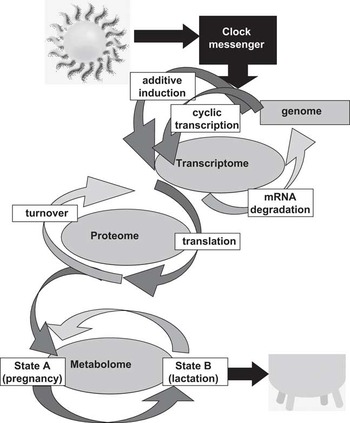
Endocrine and autonomic changes due to altering photoperiod clearly play a part in the production changes evident in cattle. These changes in the endocrine system are likely regulated hierarchically by changes in the master clock of the circadian system that elicits both endocrine changes and autonomic outputs that set and synchronize peripheral clocks to the environment (Figure 3). In turn, peripheral clocks likely play a key role in setting the metabolic output of the organ, including mammary, liver and adipose tissue. Inputs and outputs of the circadian system need to be identified so that a clear understanding of environment–gene interaction can be understood to maximize dairy production in varying environments.
Illustration of how molecular clocks affect metabolic output, as modified from (Casey et al., Reference Casey, Patel, Dykema, Dover, Furge and Plaut2009). We envision that the master clock receives physiological cues during the transition from pregnancy to lactation. These cues are integrated and the master clock responds by sending out signals to peripheral tissues which result in coordinated changes in hormonal milieu and metabolic rhythms to accommodate for the increased metabolic demand of lactation. Photoperiod length and nutrient status of dam serve as inputs to the circadian system as well, and thus influence the outputs, that is, metabolic capacity of the dam. As long-day photoperiod (LDPP) is usually coincident with greater feed availability in nature, ruminants likely evolved with a circadian system pre-programmed to take advantage of these seasonal changes. Thus, a pre-set mechanism (that includes seasonal changes in melatonin levels) adjusts circadian rhythms in a manner that increases mammary metabolic output during LDPP.

Conclusion
The circadian system functions to coordinate metabolism and physiological processes and receive and respond to environmental cues. In order to initiate lactation coordinated changes in metabolism and hormonal milieu must occur. We envision that the master clock receives and responds to physiological cues during the transition from pregnancy to lactation to coordinate changes in metabolism and hormonal milieu necessary to initiate milk synthesis. Altering photoperiod causes changes in feed efficiency, cell proliferation and endocrine milieu that are indicative of homeostatic adjustments in response to the animal's environment. The SCN functions as a key site for environment–gene interactions in the cow. Changes in photoperiod induces changes in clock genes, which in turn alters autonomic signals and the endocrine milieu to prepare the animal for seasonal changes in feed availability or night/day patterns of nutrient use. By identifying environmental, physiological and nervous inputs that affect changes in expression of core clock genes centrally and/or peripherally we will be able to effectively alter the clock genes to maximize productive efficiency in the dairy cow (Figure 3).
Acknowledgments
We would like to acknowledge the financial support or NASA Competitive Grant: NCC2-1373.