The development of an aorta–right atrial fistula is a very rare complication of aortic isthmic atresia. We described a case of aorta–right atrial communication associated with isthmic atresia.
Case report
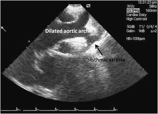
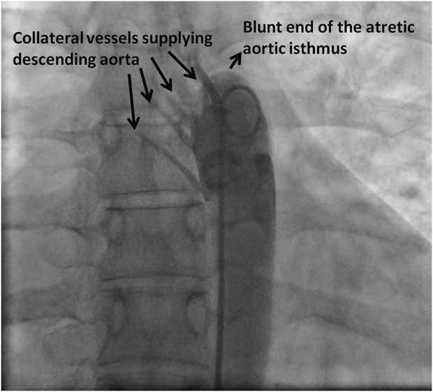
A 19-year-old woman was admitted to our hospital with the complaints of dyspnoea and headache. Heart and respiratory rates were 68 bpm and 18/min, respectively. Her blood pressure was 190/80 mmHg in both arms, but it was 95/65 in right leg. Her femoral pulses were weak. We heard grade 3/6 continuous murmur mainly in the third intercostal space at left sternal border radiating throughout the precordium. Transthoracic echocardiography demonstrated aorta–right atrial shunt in the parasternal short-axis imaging window (Video 1). The right heart chambers were found to be dilated. The descending thoracic aorta was found to be atretic at the isthmic region, with dilatation of the aortic arch and its major branches (Fig 1). There was no gradient in the descending aorta with continuous wave (CW) Doppler. Cardiac catheterisation from the right femoral vein demonstrated complete obstruction of the descending thoracic aorta with collateral formation (Fig 2). The systolic and diastolic blood pressures in the descending thoracic aorta were 95/65 mmHg, respectively. Cardiac catheterisation was performed via right upper extremity. Aortography confirmed the atresia of aortic isthmus with dilatation of the aortic arch and its branches (Video 2). A pigtail catheter was directly introduced into the right atrium through the ruptured sinus of Valsalva. Aortography and left ventriculography confirmed the rupture of the sinus of Valsalva and showed the shunt from the aorta to the right atrium (Video 3). The systolic and diastolic blood pressures in the aortic arch were 95/65 mmHg, respectively. We opted for surgical treatment, but she refused to undergo surgery.
Suprasternal imaging demonstrates aortic isthmic atresia and dilatation of aortic arch and its branches.

Descending aortography performed via the femoral route shows isthmic atresia and collateral flow into the descending aorta.

Discussion
Aorta–right atrial shunt is a rare manifestation of acquired and congenital heart diseases. Rupture of sinus of Valsalva aneurysm and prosthetic aortic valvular endocarditis may cause aorta–right atrial shunt.Reference Alozie, Kische, kaminski and Ince 1 , Reference Ozer, Deniz, Atalar, Aksoyek, Ovunc and Kes 2 Chest trauma, aortic dissection causing aorta–right atrial shunt, and congenital fistula between the aorta and the right atrium were also reported.Reference Chang, Chu and Lee 3 – Reference Hsu, Ho and Wang 5 High pressure in the ascending aorta and possibly weakness in the ascending aortic wall were thought to be the probable mechanisms of aorta–right atrial communication in our case.
Another important issue regarding the isthmic obstruction is hypertension. Coarctation of the aorta is one of the secondary causes of hypertension. The theories of hypertension in coarctation of the aorta are mechanical obstruction leading to high pressure in upper extremities, neural theory involving the distensibility characteristics of the aorta and sensitivity of carotid baroreceptors, and renal theory.Reference Perloff 6 We think that similar mechanisms are responsible for the development of hypertension in aortic isthmic atresia. This case demonstrates the importance of physical examination, which may lead to diagnosis of complex medical conditions. Aortic isthmic obstruction should be suspected in case of hypertension in young adults.
The role of echocardiography and Doppler colour flow imaging is paramount in the correct diagnosis. Non-invasive imaging modalities such as computed tomography and cardiac magnetic resonance imaging are useful for the diagnosis and demonstration of complete anatomical abnormalities. We preferred cardiac catheterisation for the confirmation of the diagnosis and evaluation of haemodynamic derangement.
To our knowledge, aorta–right atrial communication is very rarely associated with aortic isthmic atresia. The treatment of choice should be surgical correction. The treatment of isthmic atresia by a covered stent and closure of aorta–right atrial communication by occluder devices may be alternative means of treatment.