Hypertrophic cardiomyopathy is a heterogeneous monogenic heart disease with a prevalence of at least one in 500 in the general population.Reference Maron, Gardin, Flack, Gidding, Kurosaki and Bild 1 The disease is recognised as a major cause of arrhythmic sudden cardiac death, heart failure, and atrial fibrillation, in which sudden cardiac death is the most common and devastating consequence.Reference Maron and Maron 2 , Reference Zhao, Wang, Gao, Li and Brewer 3 In most cases, the first clinical manifestation of hypertrophic cardiomyopathy is sudden cardiac death, without any warning signs and symptoms.Reference Maron 4 Therefore, it is very important to make a timely and accurate diagnosis of hypertrophic cardiomyopathy. Genetic testing, which is now commercially available, is a powerful tool to definitively diagnose an affected genetic status. The American College of Cardiology Foundation/American Heart Association guideline for the diagnosis and treatment of hypertrophic cardiomyopathy recommends that the relatives of family members known to have hypertrophic cardiomyopathy should undergo genetic testing to identify those with unrecognised disease, and all allele carriers should be followed-up closely with electrocardiography and echocardiography examinations.Reference Gersh, Maron and Bonow 5
At least 11 genes have been identified to date that primarily encode >1500 cardiac sarcomere and sarcomere-related single-nucleotide polymorphisms that are critical for the basic contractile function of the heart;Reference Semsarian, Ingles, Maron and Maron 6 , Reference Maron, Maron and Semsarian 7 however, the pathogenic genes involved in hypertrophic cardiomyopathy are still in need of further exploration. In addition, because of the existence of a “genotype-positive phenotype-negative” subgroup, such that asymptomatic gene carriers show no evidence of the disease phenotype – that is, left ventricle hypertrophy – the value of the currently available clinical genetic tests for hypertrophic cardiomyopathy diagnosis and prognosis evaluation is limited.Reference Gray, Ingles and Semsarian 8 To improve clinical diagnosis and genetic counselling, it is critical to further elucidate the clinicopathogenic characteristics of hypertrophic cardiomyopathy and to identify novel pathogenic abnormalities. In the present study, we performed a gene sequencing analysis of a Chinese hypertrophic cardiomyopathy family to screen for potential genetic abnormality, and we also investigated the prognostic features of the hypertrophic cardiomyopathy patients affected by this abnormality.
Materials and methods
Clinical evaluation
The diagnosis of hypertrophic cardiomyopathy was made by two-dimensional echocardiography according to the American College of Cardiology Foundation/American Heart Association practice guideline.Reference Gersh, Maron and Bonow 5 In the absence of other causes of left ventricular hypertrophy, a maximal left ventricular wall thickness ⩾13 mm was diagnosed as hypertrophic cardiomyopathy. In this study, the proband was a 32-year-old female. A total of 14 living hypertrophic cardiomyopathy family members who gave their informed consent were enrolled for this study, which included eight males and six females. Their ages ranged from 4 to 68 years, with a mean age of 39.1 years (median age, 42 years). All family members were screened using medical history, physical examination, electrocardiography, and two-dimensional echocardiography. In addition, blood samples were collected from each family member for DNA extraction.
Target sequence capture sequencing
To identify the pathogenic gene within the genome of this hypertrophic cardiomyopathy family, we analysed 26 candidate pathogenic genes – MYH7, MYBPC3, TNNT2, TNNI3, MYL2, MYL3, TPM1, ACTC1, MYH6, TNNC1, TTN, ACTN2, TCAP, VCL, ANKRD1, CAV3, CSPR3, LDB3, MYOZ2, NEXN, JPH2, PLN, CASQ2, CALR3, PRKAG2, and LAMP2 – in the proband using target sequence capture sequencing technique. In brief, genomic DNA was extracted using the Tiangen RelaxGene Blood DNA System (Tiangen, Cat #DP319, Beijing, China). Next, the DNA (0.1–0.5 μg) was fragmented to an average length of 250 bp using ultrasound and enriched using AMPureXP beads (Agencourt, Beckman Coulter, California, United States of America). The ends of the fragmented DNA were repaired and adenylated, and universal compatible adaptors were ligated. The target fragments were enriched using the Agilent SureSelect® Capture Library (Agilent, Santa Clara, California, United States of America) after eight cycles of polymerase chain reaction. Thereafter, the library was re-enriched during 12 cycles of polymerase chain reaction amplification. A multiplexed paired-end sequencing run of 147 cycles was carried out using the TruSeq SBS Kit v5 (Illumina, San Diego, California, United States of America) on the Genome Analyzer IIx (GAIIx) (Illumina). CLC Genomics Workbench software (CLC Bio, Aarhus, Denmark) was used for duplicate read removal, read mapping against the human genome reference sequence (NCBI database, GRCh37/hg19), coverage analysis, and variant calling and annotation. Data analysis was restricted to variants in the coding regions. Variation was defined using a loci sequencing depth ⩾25×and a base variation proportion ⩾20%.
Confirmation with Sanger sequencing
A putative pathogenic sequence variant was further verified in 14 hypertrophic cardiomyopathy family members using Sanger sequencing. The primers for sequencing the MYH7 gene were as follows: the forward primer 5'-CAAAGCCAGGATCAGAACCCAG-3' and the reverse primer 5'-GGAGTCAATGGAAAAGAGATGT-3'.
Literature reviewing work
We used the identified sequence variant in a comprehensive bibliographical search – in PubMed and Google Scholar – to collect all available clinical information of families and individuals who carried such an allele. The references cited in these reports were also evaluated to include any potential missing cases. All allele carriers were included for further evaluation.
Statistical analysis
Prism 5.0 software (GraphPad, La Jolla, California, United States of America) was used for statistical analysis. Survival analysis was performed using the Kaplan–Meier method. A log-rank test was performed for intergroup comparisons. A p value of <0.05 was considered to be statistically significant.
Results
Clinical and genetic features of the hypertrophic cardiomyopathy family
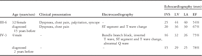
Of the 14 hypertrophic cardiomyopathy family members who underwent echocardiography, three had positive results: the proband, the proband’s son, and the proband’s father (Fig 1a). The clinical features of the proband and the proband’s son are described in Table 1. The proband’s father was asymptomatic, showing no electrocardiographic abnormalities. In addition, II-3 and II-5 complained of intermittent chest pain, but both the electrocardiography and the echocardiography findings were normal.
Echocardiograms and genetic tests of the hypertrophic cardiomyopathy family members. ( a ) Interventricular septal thickening was observed in three members via echocardiogram (septum thickness: the proband, 25 mm; the proband’s son, 16 mm; the proband’s father, 15 mm). ( b ) Genetic sequencing identified the c.2146 G>A (Gly716Arg) sequence variant of the MYH7 gene in the proband and the proband’s son. ( c ) The alignment of the MYH7 gene in different species showed a high conservation of pro716 amino acids.

Clinical features of the two hypertrophic cardiomyopathy family members affected by the Gly716Arg Allele.

EF=ejection fraction; IVS=interventricular septal thickness in diastole; LA=left atrium; LV=left ventricle
The target sequence capture sequencing identified a c.2146 G>A mutation in the MYH7 gene of the proband. No other sequence variants were identified. The alteration was within exon 19 of the MYH7 gene, which resulted in substitution of Glycine with Arginine at the 716th amino acid of MYH7 encoding the protein β-myosin heavy chain. This result was confirmed with Sanger sequencing (Fig 1b). In addition, of the other 13 family members who underwent Sanger sequencing, only one – the proband’s son – had the same genotype. Homology comparison showed that the pro716 amino acid is highly conserved between different species (Fig 1c). Although the proband’s father was previously an athlete, based on his genotype, we diagnosed him with physiological cardiac hypertrophy.
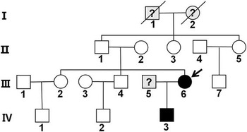
A pedigree of the family was drawn using clinical and genetic manifestations (Fig 2). Both the father and the mother of the proband were not carriers of the allele; thus, the mutation was classified as “de novo”.
Pedigrees of the hypertrophic cardiomyopathy family. Symbols denote sex and disease status. boxes=male; circles=female; closed=affected; open=unaffected; slashed=deceased;?=unknown; arrow=proband.

Survival analysis
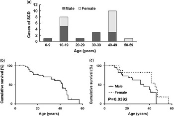
A total of eight reports on the Gly716Arg sequence variant were identified.Reference Anan, Greve and Thierfelder 9 – Reference Garcia-Giustiniani, Arad and Ortiz-Genga 16 After careful review, we obtained detailed descriptions for 43 allele carriers, including the two patients in this study. The total cohort comprised 25 males and 18 females, with a mean age of 28.9 years (median age, 32 years; with a range from 4 to 57 years). We found that all 43 allele carriers harboured typical hypertrophic cardiomyopathy manifestations. Further survival analysis revealed that the median survival age of this cohort was 42 years, with 25 cases who experienced sudden cardiac death. Interestingly, sudden cardiac death mainly occurred in the second and the fifth decade of life, and the male carriers tended to undergo sudden cardiac death at younger ages compared with females. The log-rank test showed that the female carriers had a longer life expectancy than males (median survival, 45 years versus 35 years) (Fig 3). With regard to treatment, two patients underwent heart transplantation in this cohort.Reference Garcia-Giustiniani, Arad and Ortiz-Genga 16 Drug treatment and implantable defibrillator intervention were not reported.
Epidemiological features of hypertrophic cardiomyopathy patients with the Gly716Arg allele. ( a ) Age and sex distribution of 25 patients who died from sudden cardiac death (SCD); two peak ages were observed: one in the second and one in the fifth decade of life. ( b ) Descriptive overall survival curve of 43 reported cases of hypertrophic cardiomyopathy. ( c ) Kaplan–Meier curves indicate that females have a longer life expectancy than males.

Discussion
In the present study, we identified a Gly716Arg sequence variant using target sequence capture sequencing, and then verified the presence of this allele using Sanger sequencing in a Chinese hypertrophic cardiomyopathy family. Our main finding was the second hypertrophic cardiomyopathy case identified with a “de novo” Gly716Arg mutation.Reference Rai, Ahmad and Bahl 13 Moreover, we have re-reviewed the cases of 43 different patients reported in eight different studies and our present study. Our results suggest that the Gly716Arg allele carriers have an extremely poor prognosis, and the females seem to have a longer life expectancy than males. To the best of our knowledge, this study is the largest cohort study of the Gly716Arg allele. These findings are similar to a previous study that showed that the Gly716Arg allele caused a “malignant” phenotype.Reference Ackerman, VanDriest and Ommen 11
MYH7 is the first and most commonly identified pathogenic gene responsible for hypertrophic cardiomyopathy.Reference Geisterfer-Lowrance, Kass and Tanigawa 17 , Reference McNally, Golbus and Puckelwartz 18 The gene encodes β-myosin heavy chain, which is 1935 amino acids in length and constitutes the head and tail domains of Myosin II. According to previous reports, MYH7 mutations can be detected in ~40% of all hypertrophic cardiomyopathy cases;Reference Van Driest, Jaeger and Ommen 19 , Reference Richard, Charron and Carrier 20 however, a “de novo” missense mutation of the MYH7 gene is rather uncommon. Our finding suggests that the risk of inherited “sporadic” mutations should be taken into account.
Most MYH7 mutations result in amino acid substitutions in the globular head of the protein and affect the binding sites for adenosine triphosphate, actin, and essential or regulatory light chains.Reference Richard, Charron and Carrier 20 Previously, at least 40 variant alleles have been identified.Reference Van Driest, Jaeger and Ommen 19 The c.2146 G>A allele lies in the converter domain of the 19th exon of the MYH7 gene, which results in the Gly716Arg alteration and anomalous adenosine triphosphatase function. Other alleles including Arg712Leu, Asp717Gly, Arg719Trp/Gln, Arg721Lys, Arg723Cys/Gly/His, Ala728Val, Pro731Leu, Arg733Glu/Arg, Gln734Glu, Ile736Met/Thr, Gly741Trp/Arg, Ala742Glu, Val763Gly/Met, Phe764Leu, and Gly768Arg are also frequently detected in this area.Reference Garcia-Giustiniani, Arad and Ortiz-Genga 16 , Reference Richard, Charron and Carrier 20 On the basis of the results of previously published studies and our results, most of these alleles appear to be associated with severe hypertrophic cardiomyopathy and a high risk of sudden cardiac death.Reference Anan, Greve and Thierfelder 9 , Reference Abchee and Marian 21 – Reference Miller, Colegrave and Peckham 24 Moreover, our results show that the patients with the Gly716Arg allele have a median survival age of only 42 years, which is shorter than the median survival ages associated with the Arg719Trp, Gly768Arg, Ile736Thr, and Arg723Gly alleles but longer than those ages associated with the Arg719Gln and Asp717Gly alleles.Reference Garcia-Giustiniani, Arad and Ortiz-Genga 16 Another interesting finding is that the female carriers appear to have a longer life expectancy than the male carriers, which was not reported for other alleles. The detailed mechanisms underlying this observation are still unclear. As this disorder is inherited as an autosomal-dominant trait, we speculate that some sex hormones and some molecules produced by the sex chromosome may interact with the cardiac β-myosin heavy chain gene. A larger sample size is needed to further confirm this finding.
Hypertrophic cardiomyopathy is regarded as a disease caused by autosomal-dominant mutations in genes encoding protein components of the sarcomere and its constituent myofilament elements; however, pathogenic mutations can only be identified in fewer than 50% of clinically affected probands, and genetic defects in a substantial proportion of patients are unrecognised at present. In addition, the clinical utility of some variants is still uncertain and they have virtually no clinical use for family screening. Therefore, the clinical utility of genetic testing varies according to the pathogenicity of the mutations. The genotype–phenotype data available at this time suggest that all Gly716Arg allele carriers manifest severe cardiovascular disorders – that is, there are no “genotype-positive phenotype-negative” cases associated with the Gly716Arg allele.
The treatment strategy for hypertrophic cardiomyopathy depends on the clinical presentations.Reference Gersh, Maron and Bonow 5 For patients without symptoms – genotype-positive phenotype-negative – no treatment is needed. For patients diagnosed with hypertrophic cardiomyopathy over 10 years, implantable defibrillator is now established as the most effective treatment. It is the only strategy capable of prolonging life and altering the natural history of hypertrophic cardiomyopathy. Anti-arrhythmic or rate-control drugs, warfarin, cardioversion, and radiofrequency ablation are recommended for patients with atrial fibrillation and stroke. Myectomy is recommended for patients with drug-refractory heart failure symptoms and outflow obstruction. Heart transplantation is usually performed in patients with ejection fraction <50%. Nevertheless, the long-term effects of these treatment strategies are still unclear for Gly716Arg allele carriers. It seems that without timely intervention measures, progressive heart failure and sudden cardiac death are inevitable. Therefore, close follow-up and timely intervention are very important for such patients.
Acknowledgements
We are grateful to the laboratory team of National center for Cardiovascular Diseases for their kind help.
Financial Support
The study was supported by the Applied Fundamental Research grant of Qingdao government (No. 13-1-4-141-jch) and the grant from the National Natural Science Foundation of China (No. 81502272).
Conflicts of Interest
None.
Ethical Standards
The authors assert that all procedures contributing to this work comply with the ethical standards of the relevant national guidelines of China on human experimentation and with the Helsinki Declaration of 1975, as revised in 2008, and has been approved by the institutional committees of Qingdao University.