Introduction
Leptin and ghrelin are the tissue hormones considered to take part in the regulation of energy metabolism. Leptin is synthesized particularly by fat cells in the adipose tissue. It is also sythesized in the stomach, skeletal muscle and in the liver. Leptin regulates appetite suppression and food intake. It also stimulates energy expenditure. This hormone plays the role of a signal that informs the central nervous system of the body’s fat status (Meier and Gressner, Reference Meier and Gressner2004). The body fat mass is the particular factor that determines plasma leptin concentration in humans (Speakman et al., Reference Speakman, Stubbs and Mercer2002). In horses, peripheral concentration of plasma leptin is also related to fat mass and body condition score (Buff et al., Reference Buff, Dodds, Morrison, Whitley, McFadin, Daniel, Djiane and Keisler2002; Kearns et al., Reference Kearns, McKeever, Roegner, Brady and Malinowski2006; Gordon et al., Reference Gordon, McKeever, Betros and Manso Filho2007a). Plasma leptin concentration not only reflects body fat mass but can also play a role as a sensor of energy imbalance (Gordon and McKeever, Reference Gordon and McKeever2005). The plasma concentration of this hormone shows the diurnal variation attributed to feeding time and insulin response (Steelman et al., Reference Steelman, Michael-Eller, Gibbs and Potter2006). Short-term feed deprivation leads to a decrease in the mean plasma concentration of leptin in mares (Buff et al., Reference Buff, Morrison, Ganjam and Keisler2005 and Reference Buff, Johnson, Wiedmeyer, Ganjam, Messer and Keisler2006). The changes in circulating leptin concentration according to the nutritional status indicate that the production of this protein by fat tissue is modulated by several factors (McManus and Fitzgerald, Reference McManus and Fitzgerald2000). It has been demonstrated that the synthesis of leptin in humans is stimulated by food intake, insulin and glucocorticoids. It is also suppressed by fasting, as well as by cAMP and β3-adrenoreceptor agonists like norepinephrine (Meier and Gressner, Reference Meier and Gressner2004). Dotsch et al. (Reference Dotsch, Meissner and Rascher2003) showed that the main function of leptin is not only to cause an anorectic effect but also to increase the oxidation of hepatic fatty acid.
The changes in the plasma leptin concentration according to several physiological variables are still not clear. In humans, leptin level is higher in females than in males. It is also higher in adults than in children. This information is not fully supported in the study of horses (Buff et al., Reference Buff, Dodds, Morrison, Whitley, McFadin, Daniel, Djiane and Keisler2002). The studies of mares suggest that leptin may be an important link between nutrition and reproductive status (Gentry et al., Reference Gentry, Thompson, Gentry, Davis, Godke and Cartmill2002). Leptin can also play an important role in the regulation of lipid metabolism during the periparturient period (Kędzierski et al., 2008).
Ghrelin is the peptide hormone produced primarily in the epithelial cells that line the fundus of the stomach. Generally, the physiological and metabolic role of ghrelin is contrary to the relation of that to leptin. In humans, plasma ghrelin concentration increases significantly before a meal and decreases within 1 h after food intake (Meier and Gressner, Reference Meier and Gressner2004). In cases of low-caloric diets and chronic exercise, ghrelin levels were increased; and in states of obesity or after glucose challenge, the concentrations of this peptide were reduced (Meier and Gressner, Reference Meier and Gressner2004; Gordon and McKeever, Reference Gordon and McKeever2006). Ghrelin stimulates the release of the growth hormone from the pituitary gland and increases appetite, food intake, body weight (BW), energy balance and especially fat tissue by decreasing fat oxidation in the liver (Matyjek et al., Reference Matyjek, Kapica, Puzio, Bąblewska and Zabielski2004; Barazzoni et al., Reference Barazzoni, Bosutti, Stabel, Cattin, Roder, Visintin, Cattin, Biolo, Zanetti and Guarnieri2005). The value of this peptide decreases over the period of childhood and puberty (Broglio et al., Reference Broglio, Benso, Castiglioni, Gottero, Prodam, Destefanis, Gauna, Van Der Lely, Deghenghi, Bo, Arvat and Ghigo2003).
The correlation of this parameter with BW, age and body condition in horses is still unclear. It is known that the plasma ghrelin concentration is greater in fit, young Standardbred racehorses with a low percent of fat mass and a low body condition score than in 11-year-old unfit mares (Gordon et al., Reference Gordon, McKeever, Betros and Manso Filho2007a). The exercise test leads to changes in plasma ghrelin concentration determined during the effort and a few hours after the end of the effort (Gordon et al., Reference Gordon, McKeever, Bokman, Betros, Manso Filho, Liburt and Streltsova2006 and Reference Gordon, McKeever, Betros and Manso Filho2007a).
Standardbred foals begin training when they are 1-year olds. At this time, they are still in the growing period. Training and workload particularly influences their metabolism (Kędzierski et al., 2007). The aim of this study was to evaluate the effects of exercise on plasma concentrations of leptin and ghrelin according to the age of Standardbred foals. The effect of sex and season of the year was also analyzed.
Material and methods
Horses
The effect of acute exercise was studied in a group of 42 clinically healthy young Standardbred horses in the trotters training center during two experimental periods. The two periods were: (1) in December and (2) in July (Table 1). Ten horses were present in both experimental periods: first in winter as 1.5-year-old foals and then in summer as 2-year-old foals. Prior to the start of the observation period, the horses had been conditioned with a standard training program. The type of training and its intensity were consistent with the principles of the trainer preparing the horses for races.
Demographic summary of the Standardbred trotters investigated in December and in July

n = number of horses in the group.
*Prior to the day of study the horses investigated completed a training program.
Exercise test protocol
In days of the study, all horses took part in a training session composed of a 5 min warm up, a 30 min trot to a distance of 7000 to 11 000 m with a mean speed of 4.0 to 6.2 m/s on the sand track and a 10 min cool down in a slow trot. Ambient condition in December: temperature +4°C, relative humidity 45% to 55%, velocity of wind 2 to 3 m/s, and in July: +20°C, 40% to 85% and 4 to 5 m/s. Three blood samples from a jugular vein were collected via venipuncture from each horse. These samples were taken at rest, after the end of the training session and 30 min after the end of the effort. The samples were collected in EDTA-vacutainer tubes and immediately centrifuged for the plasma separation. The obtained plasma was stored at −20°C until further analysis.
Blood analysis
Plasma leptin concentration was determined using a multi-species leptin radioimmunoassay (RIA) kit (Linco Research, St Louis, MO, USA), previously validated for use with horses (McManus and Fitzgerald, Reference McManus and Fitzgerald2000), demonstrating an assay coefficient of variation (CV) of 8.5%. The lowest level of leptin that can be detected by this assay is 1 ng/ml. The total ghrelin plasma concentration was determined using a commercial RIA kit (Linco Research) demonstrating the sensitivity at the level of 93 pg/ml. The results were expressed as humans equivalents of immunoreactive ghrelin (Gordon and McKeever, Reference Gordon and McKeever2005). Plasma triacylglycerides (TG) concentration was measured by the colorimetric method using an enzymatic diagnostic kit (Cormay, Lublin, Poland) and expressed as mmol/l.
Statistical analysis
The results were presented as mean ± standard error (s.e.). Comparisons between the consecutive groups of horses were analyzed using the Multiple HSD Tukey test (ANOVA). The correlation coefficient was also used to compare plasma leptin, ghrelin and TG concentrations. The statistical significance was accepted at the level P ⩽ 0.05.
Results
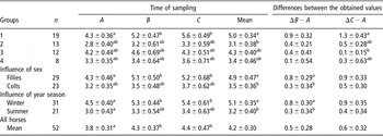
Plasma leptin concentrations in all the analyzed groups of horses are summarized in Table 2. The mean concentration of leptin in blood plasma measured just after exercise and after 30 min rest increased significantly. This rise was significant in Group 1, whereas in Groups 2, 3 and 4 the increase did not reach the level of statistical significance. The differences between values determined immediately after the end of the exercise and at rest (ΔB − A) and between data obtained 30 min after the end of the exercise and at rest (ΔC − A) were also investigated. The value of ΔC − A decreased with age and/or training, and was significantly higher in Group 1 in compared with Group 3. The mean values of leptin determined in December were significantly higher than those obtained in July. The plasma concentration of this peptide varied not only with the season of the year but also with the sex of trotters. These values were higher in fillies than in colts. The ΔB − A value was also significantly higher in fillies than in colts. The results obtained 30 min after the end of the effort did not reach a level of statistical significance.
Plasma leptin concentration (ng/ml) in Standardbred trotters during exercise test

n = number of horses in the group.
A = samples taken at rest; B = immediately after the end of exercise; C = after 30-min rest.
Values are mean ± s.e.
a,bMean values, in compared groups, with different superscripts differ at P ⩽ 0.05 according to Multiple Tukey HSD test (ANOVA).
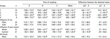
Plasma ghrelin concentrations in the horses studied are shown in Table 3. The exercise significantly increased the mean plasma concentration of ghrelin measured just after the end of the effort. The effect of exercise measured as ΔB − A was higher in Group 1 than in Group 3. The value of ΔC − A was also significantly higher in Group 1 than in Group 3. It is also important to note that ΔB − A and ΔC − A in Groups 3 and 4 had minus values. Generally, plasma ghrelin concentration in groups investigated in December and in June showed the same seasonal distributions as the leptin level. Sex had no significant effect on plasma ghrelin concentration.
Plasma ghrelin concentration (pg/ml) in Standardbred trotters during exercise test

n = number of horses in the group.
A = samples taken at rest; B = immediately after the end of exercise; C = after 30-min rest.
Values are mean ± s.e.
a,b,cMean values, in compared groups, with different superscripts differ at P ⩽ 0.05 according to Multiple Tukey HSD test (ANOVA).
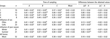
The concentration of plasma TG rose during the investigated exercise and decreased after 30 min of rest. The value of plasma TG increase was generally higher in fillies than in colts, and higher in winter than in summer (Table 4).
Plasma triacylglycerols (TG) concentration (mmol/l) in Standardbred trotters during exercise test

n = number of horses in the group.
A = samples taken at rest; B = immediately after the end of exercise; C = after 30-min rest.
Values are mean ± s.e.
a,bMean values, in compared groups, with different superscripts differ at P ⩽ 0.05 according to Multiple Tukey HSD test (ANOVA).
A positive correlation between the plasma concentrations of leptin and TG measured just after the effort (r = 0.65) was found, but it was less pronounced in the differences ΔB − A calculated for these parameters (r = 0.47).
Discussion
Individual variability of plasma leptin concentration was observed in our study. High standard deviations are typical for horses in good body condition and can be explained by genetic heterogeneity (Gentry et al., Reference Gentry, Thompson, Gentry, Davis, Godke and Cartmill2002). The physical effort the investigated trotters were submitted to was typical for daily training session. This physical effort led to the increase in the plasma leptin concentration determined immediately after exercise. This increase remained elevated throughout the following 30 min rest after training sessions. The direct effect of exercise on the plasma leptin concentration in horses has not been reported. An increase of leptin value was indicated 180 min after 5 min of lunging only in mares selected for high leptin concentration (Cartmill et al., Reference Cartmill, Thompson, Storer, Gentry and Huff2003). In the study of Gordon et al. (Reference Gordon, McKeever, Betros and Manso Filho2007b), short-term high-intensity exercise did not lead to changes in the leptin level during the first 60 min after the test, but the decrease was shown in this hormone plasma concentration 24 h later. Piccione et al. (Reference Piccione, Bertolucci, Foa and Caola2004) did not observe any changes in the plasma leptin concentration in exercised athletic horses. In humans, a decrease or a rise in the plasma leptin concentration measured after the exercise tests on the level of 75% VO2max were reported (Kraemer et al., Reference Kraemer, Durand, Acevedo, Johnson, Kraemer, Gimpel and Castracane2003; Desgorces et al., Reference Desgorces, Chennaoui, Gomez-Merino, Drogou, Bonneau and Guezennec2004). In the present study, the exercise was not considered intensive and it lasted about 45 min. It is possible that the intensity and duration of the performed effort were enough to increase the plasma leptin concentration in foals. During the bout of exertion, a significant increase in plasma TG level was also observed. These changes reflect the increased rate of lipolysis (Poso et al., Reference Poso, Viljanen-Tarifa, Soveri and Oksanen1989). During exercise in horses, the part of free fatty acids (FFA) liberated in the process of lipolysis is oxidized and the remainder is used for resynthesis of TG. The high value of circulating TG represents the predominance of TG resynthesis over the FFA utilization during the exercise. The high increase in plasma TG level in exercise conditions can suggest that during the increased rate of lipolysis, the FFA oxidation is hindered. In this light, the increased level of leptin as a hormone increasing the hepatic FFA oxidation can play a positive role in the process of energy metabolism regulation. It is known that during exercise the level of plasma insulin decreases and norepinephrine, glucocorticoids and cAMP increase. They are the factors that can regulate the leptin synthesis (Meier and Gressner, Reference Meier and Gressner2004). The precise mechanism involving the rise in plasma leptin concentration during exercise is unknown.
The present study demonstrates a significant effect of age on the changes in the plasma leptin concentration with exercise. The values of ΔC − A for leptin were the greatest in the group of the youngest trotters. The tendency of these values to decrease with age was seen in consecutive groups. The tendency to the stabilization of leptin levels during the training period was reported in humans (Desgorces et al., Reference Desgorces, Chennaoui, Gomez-Merino, Drogou, Bonneau and Guezennec2004). It was also demonstrated that psychological stress rises the serum leptin concentration in humans (Otsuka et al., Reference Otsuka, Yatsuya, Tamakoshi, Matsushita, Wada and Toyoshima2006). In this light, it is probable that high increase in the plasma leptin level shown in 1.5-year-old foals is partially associated with stress, which appeared during the initial months of training.
A high correlation coefficient between leptin and TG values after exercise (r = 0.65) was found. Similar results were reported in the study of non-diabetic humans, in which the plasma leptin concentration was directly and highly associated with plasma TG level (Chan et al., Reference Chan, Watts, Ng, Uchida, Sakai, Yamashita and Barrett2005).
The results mentioned above indicate that during exercise the lipolysis and the leptin release from adipose tissue occur simultaneously.
The results of the present study show that plasma leptin concentration is higher in fillies than in colts and that the post-exercise increase of this parameter is higher in young mares. Buff et al. (Reference Buff, Dodds, Morrison, Whitley, McFadin, Daniel, Djiane and Keisler2002) reported a lower level of leptin in mares than in stallions. In their work, however, the adult Quarter horses studied were 8- to 24-years old. On the contrary, horses investigated in the present study were of a different breed, younger and submitted to race training. The feeding and breeding conditions were also not comparable. The marked sexual dimorphism in plasma leptin responses to physical exercise was shown in the study of humans (Sandoval et al., Reference Sandoval, Galassetti, Tate, Neill and Davis2003). The mechanism of leptin secretion is amenable to gonadal steroids (Kraemer et al., Reference Kraemer, Durand, Acevedo, Johnson, Kraemer, Gimpel and Castracane2003; Puder et al., Reference Puder, Monaco, Sen Gupta, Wang, Ferin and Warren2006).
The seasonal variables found in the present work were also different from that reported previously. The study of stud mares indicated the tendency to decrease in plasma leptin concentration in the period from summer to winter and the same trends were seen in values of BW and percent of body fat mass (Gentry et al., Reference Gentry, Thompson, Gentry, Davis, Godke and Cartmill2002; Buff et al., Reference Buff, Messer, Cogswell, Johnson, Keisler and Ganjam2007). However, these results were obtained in animals that were not exercised. In trained male rowers, a significant decrease in plasma leptin concentration was shown according to training intensity (Jurimae et al., Reference Jurimae, Maestu and Jurimae2003). In our study, plasma TG values calculated as differences between levels obtained just after the end of the effort and at rest were lower in summer than in winter. With leptin this was similar. The decrease in plasma TG concentration determined at rest and after exercise in consecutive phases of training was reported previously (Kędzierski and Podolak, Reference Kędzierski and Podolak2002). Probably, the fluctuations observed here, in plasma leptin concentration according to the season of the year, reflected the changes in lipid metabolism regulation with the year-long training process.
In the exercise-studied trotters, exercise also influenced the plasma ghrelin concentration determined after an effort. The trend of reported changes was associated with the age of investigated horses. In the paper of Gordon et al. (Reference Gordon, McKeever, Betros and Manso Filho2007b), exercise-induced alternations in plasma concentration of ghrelin were seen only many hours after the short-term, high-intensity exercise in old mares. But during interval exercise tests, the increase in plasma ghrelin concentration was shown just after the end of the effort (Gordon et al., Reference Gordon, McKeever, Bokman, Betros, Manso Filho, Liburt and Streltsova2006). It can be ascertained, that 45-min training exercise induced changes in plasma ghrelin concentration in young trotters investigated here. Ghrelin is one of the glucoregulatory hormones, which induces an increase in the plasma glucose level that is followed by a reduction in insulin secretion (Broglio et al., Reference Broglio, Arvat, Benso, Gottero, Muccioli, Papotti, Van Der Lely, Deghenghi and Ghigo2001 and Reference Broglio, Benso, Castiglioni, Gottero, Prodam, Destefanis, Gauna, Van Der Lely, Deghenghi, Bo, Arvat and Ghigo2003). The results of the Broglio et al. (Reference Broglio, Arvat, Benso, Gottero, Muccioli, Papotti, Van Der Lely, Deghenghi and Ghigo2001) study suggest that ghrelin could directly stimulate glycogenolysis, which takes part also in exercise conditions. Surprisingly, the exercise had decreased effect or no effect on plasma ghrelin in adult humans (Kraemer et al., Reference Kraemer, Durand, Acevedo, Johnson, Kraemer, Hebert and Castracane2004; Ghanbari-Niaki, Reference Ghanbari-Niaki2006; Vestergaard et al., Reference Vestergaard, Dall, Lange, Kjaer, Christiansen and Jorgensen2007). In the present study, a similar effect can be observed in 2-year old and older trotters. In 1.5-year-old foals, the plasma ghrelin level increased after exercise similar to leptin. In rats, ghrelin expression can be stimulated by leptin administration (Toshinai et al., Reference Toshinai, Mondal, Nakazato, Date, Murakami, Kojima, Kangawa and Matsukura2001). It is also probable that in the group of the youngest foals investigated here, the increase in plasma leptin concentration induced the rise in plasma ghrelin concentration. Ghrelin might also integrate the energy balance by the decrease of lipid oxidation in the liver, and favors fat utilization in muscles (Barazzoni et al., Reference Barazzoni, Bosutti, Stabel, Cattin, Roder, Visintin, Cattin, Biolo, Zanetti and Guarnieri2005). The appearance of a low level of this peptide after exercise promotes the utilization of fat in the liver. Moreover, the FFA exposure suppresses circulating ghrelin concentration in humans (Gormsen et al., Reference Gormsen, Gjedsted, Gjedde, Vestergaard, Christiansen, Jorgensen, Nielsen and Moller2006). Plasma FFA concentration increased by about 40-fold in studied foals during exercise (Kędzierski et al., 2007). The tendency to decrease in plasma ghrelin concentration shown in 2.5 to 3-year-old trotters can help in oxidation of FFA released in the process of lipolysis. This phenomenon leads to a more economical use of glucose, which is necessary in these conditions to replenish the glycogen store (Hyyppa et al., Reference Hyyppa, Rasanen and Poso1997). The increase of the ghrelin level observed here in the youngest trotters after exercise is rather the result of a high leptin level. Our previous studies indicated that the regulation of lipid metabolism during exercise is a process formed in the consecutive months of the training process. In this light, a downward trend in plasma ghrelin concentration determined in studied trotters after exercise was also connected to a sustained process of training.
In summary, the training induced changes in plasma leptin and ghrelin concentrations in Standardbred foals. The post-exercise increase in plasma concentrations of these hormones was the greatest in the group of the youngest trotters. The changes in plasma leptin and ghrelin concentrations during bouts of exertion tend to decrease with age and/or with training.