Introduction
Ototoxic drugs and excessive noise exposure can cause cochlear and vestibular hair cell damage, resulting in hearing loss and balance disorders. In mammalian inner ear hair cells, spontaneous recovery from damage is limited.Reference Warchol, Lambert, Goldstein, Forge and Corwin1 Several reports have suggested that stem or progenitor cells have the potential to undergo transdifferentiation, i.e. to change lineage, resulting in an altered cell phenotype.Reference Bjornson, Rietze, Reynolds, Magli and Vescovi2–Reference Mezey, Chandross, Harta, Maki and McKercher4 These findings suggest that cell therapy, unlike transplantation, is not irreversible.
In vertebrates, the inner ear is the sensory organ responsible for hearing and the perception of balance and acceleration. It is composed of a closed epithelial layer which is differentiated into specific regions containing the sensory transducing elements, hair cells and supporting cells.
Several reports have suggested that the proteins myosin VIIa and Brn-3c may be used as specific markers of the hair cell immunophenotype, and that pan-cytokeratin may be similarly used for the supporting cell immunophenotype.Reference Weir, Rivolta and Holley5–Reference Kojima, Tamura, Nishida and Ito9
Transplantation of multipotential stem cells into the inner ear may enable the recovery of auditory function. The neural stem cell is a multipotential cell which can differentiate into neurons or glial cells when isolated from the cerebral cortex or hippocampus.Reference Reynolds, Tetzlaff and Weiss10–Reference Palmer, Takahashi and Gage12
In this study, we used immunofluorescence and immunohistochemical techniques to investigate whether neural progenitor cells derived from the fetal mouse central nervous system (CNS) could be induced to generate inner ear sensory epithelial phenotypes.
Materials and methods
Experimental animal and medium
Adult SPF (SPF grade animal) Kumming mice (provided by Anhui Medical University) were mated at 7 p.m. (female:male ratio = 2:1). At 8 a.m. the next day, the female mice were examined. If a yellow-white vaginal secretion was detected, the mouse was labelled as pregnant. This time point was specified as day 0.5. If no secretion was detected, the mouse was examined again at 8 p.m.
Neural stem cell (NSC) growth-promoting medium was produced by mixing Dulbecco's Modified Eagle Media: Nutrient Mixture F-12. (DMEM/F12) serum-free medium with B27 (Gibco, American), 20 ng/ml (fiber growth factor) (Peretech, American) and 20 ng/ml (Epidermal Growth Factor) (Peretech).
By adding 10 per cent fetal bovine serum (Hyclone, American) and 10 mg/ml (Transforming Growth Factor) (Peretech), the growth-promoting medium was converted into induction medium.
Neural stem cell primary culture
At day 14.5, the pregnant mice were sacrificed and whole mice embryo brains were removed and transferred into calcium- and magnesium-free Hanks medium. This CNS tissue was mechanically dissociated in DMEM/F12 by trituration. The cell suspension was then filtered using a nylon grid with 200 holes per cm2, and centrifuged at 1000 rpm for 10 minutes. Cells were then resuspended in growth-promoting medium at a concentration of 6 × 10⁵ cells/ml. This cell suspension was transferred into untreated culture flasks and incubated at 37° in a humid atmosphere containing 5 per cent CO2.
After 3 to 4 days, induction medium was added to the culture flasks.
After 7 days, neurospheres (round, floating neural stem cell colonies) were identified in the culture medium.
Neural stem cell processing
When each culture flask contained 50–200 cells, its contents were centrifuged at 1000 rpm for 10 minutes. The cell precipitate was then mechanically removed (using a glass pipette) and resuspended, forming a single cell suspension. Cells were counted and then transferred into two culture flasks labelled P1.
Differentiation and identification of neural stem cells
The neurospheres (P2 or P4) (P means passage) were transferred to poly-l-lysine (Sigma, Shanghai, China) coated cover-glasses in six-well culture plates, and were cultured in induction medium for three weeks.
The resulting cells were fixed in 4 per cent paraformaldehyde in phosphate buffer saline. Immunofluorescence and immunohistochemical analyses were then conducted. The following primary antibodies were used: anti-nestin (1:200; Sigma), glial fibrillary acid protein (1:200; Sigma), myosin VIIa (1:200; SantaCruz, Shanghai, China), Brn-3c (1:200; SantaCruz) and pan-cytokeratin (1:200; Santa Cruz). The following secondary antibodies were used: biotin-labelled antibody (1:200; Vector, Shanghai, China), fluorescein isothiocyanate (FITC)-labelled antibody (1:200; Sigma) and cy3-labelled antibody (1:200; Sigma). Hoechst 33342 (Sigma) was used for nuclear staining.
Results
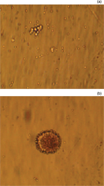
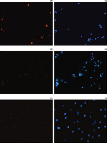
Although the primary cell cultures contained many dead cells and much cellular debris, they also contained living cells, which were round and transparent and tended to accumulate at the bottom of the culture flask (although a few were attached to the culture flask sides). After 7 days of culture, the neural stem cells formed round, floating colonies, termed neurospheres (Figure 1). Most of the neural stem cells expressed nestin epitopes, but not Brn-3c or myosin VIIa (Figures 1 and 2).
Photomicrographs showing (a) round cells seen after 24-hour culture (contrast; ×10), and (b) neurospheres seen after 7-day culture (contrast; ×20).

(a) Immunofluorescence images for nestin. (b) & (c) Immunofluoresence images for Brn-3c and myosin VIIa, respectively, showing no cellular immunoreactivity. (d)–(f) Nuclear staining images in the same field (Hoechst 33342; ×20).

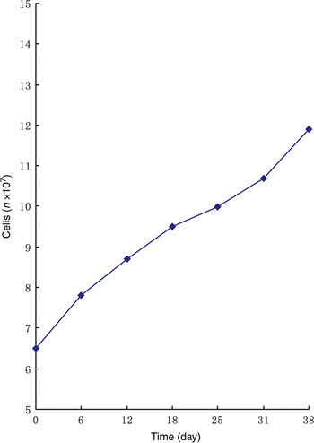
The neural stem cells were processed by centrifugation, resuspension and counting (Figure 3); this procedure was repeated six times. At the end of this series of procedures, the neural stem cells had the same shape as the primary cells first examined, and expressed nestin epitopes.
The cell growth curve.

After incubation in the induction medium, the cells did not form neurospheres. The cell is in the medim added serum. So the cells attached to the poly-l-lysine-coated cover glass.
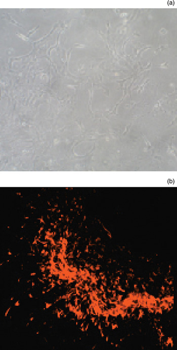
Approximately 7 days later, the cells showed irregular shapes, a large cell body, abundant cytoplasm, many elongated apophyses, and round or ovoid karyons which leaned to one side i.e. cells gater together; the cells also expressed glial fibrillary acidic protein (GFAP) epitopes (Figure 4).
Photomicrographs of cells growing on poly-l-lysine pre-coated cover-glass (a) after 7-day culture (contrast; ×20), and (b) after 7-day culture, using cy3 immunofluorescence staining to show GFAP immunoreaction (×10).

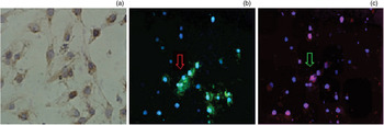
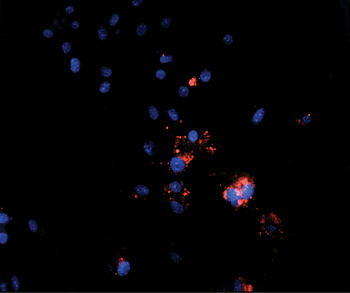
After three weeks' incubation in induction medium, the cells were seen to express myosin VIIa and Brn-3c (Figure 5), but not the supporting cell marker pan-cytokeratin. However, after four weeks' incubation in induction medium, expression of pan-cytokeratin was observed (Figure 6).
Photomicrographs of cells growing on poly-l-lysin pre-coated cover-glass, after three-week culture, showing: (a) myosin VIIa immunoreaction (contrast; ×40); (b) myosin VIIa immunoreaction (green) and Hoechst 33342 nuclear staining (blue) (contrast; ×20); and (c) Brn-3c immunoreaction (red) and Hoechst 33342 nuclear staining (blue), in the same field as Figure 5(b) (arrow indicates cell staining for both myosin VIIa and Brn-3c) (contrast; ×20).

Photomicrograph of cells growing on poly-l-lysine pre-coated cover-glass, after four-week culture, showing pan-cytokeratin immunoreaction (red) and Hoechst 33342 nuclear staining (blue) (contrast; ×20).

Discussion
Stem cells have many distinctive characteristics, including the ability to reproduce as new, undifferentiated stem cells, the potential for transdifferentiation along a new cell lineage, and fissionability (The stem cell has abillty of differentiation).Reference Weiss, Reynolds, Vescovi, Morshead, Craig and van der Kooy13, Reference Reynolds and Weiss14
Neural stem cells are multipotential cells; when isolated from the cerebral cortex or hippocampus, they can differentiate into neurons or glial cells.Reference Reynolds, Tetzlaff and Weiss10–Reference Palmer, Takahashi and Gage12
In the current study, we did not use enzymatic digestion, but rather used a glass pipette to mechanically dissociate the tissue. Because the fetal mouse brain is very soft, it is difficult to control the timing of enzymatic digestion. (We had tried to use enzymatic digestion in an earlier experiment, but found our cell yields were much lower.)
In fact, there were many impurity and other cells in the primary cells, because of the serum-free medium. The other cells, like blood cells and fiber cells, will gradually die. But the stem cells will survive because of the b-FGF and EGF.Reference Kuhn, Winkler, Kempermann, Thal and Gage15, Reference Kitchens, Snyder and Gottlieb16
When cell cultures were centrifuged, the old culture medium fluid (the supernate) was not discarded; rather, this fluid was added back into the new cell suspension. This was done to retain any growth factors secreted by the neural stem cells to aid further cell reproduction, and also to ensure that no neural stem cells were lost.
Inner hair cells transduce mechanical stimulation, caused by sound pressure or gravity, into electrical signals. Myosin VIIa (one of the myosin superfamily proteins) and Brn-3c (one of the POU (nuclear factor) family transcription factors) have been used as marker proteins to identify hair cell immunophenotypes, and pan-cytokeratin has been used as a marker protein to identify supporting cell immunophenotypes.Reference Weir, Rivolta and Holley5, Reference Lawlor, Marcotti, Rivolta, Kros and Holley6, Reference Malgrange, Belachew, Thiry, Nguyen, Rogister and Alvarez17 Likewise, nestin has been used as a marker protein to identify neural stem cells.Reference Johansson, Lothian, Molin, Okano and Lendahl18–Reference Yamaguchi, Saito, Suzuki and Mori20 In the current study, we used nestin to identify the P1 passage 1 neural stem cells. After three-week incubation in induction medium, we observed expression of myosin VIIa; however, we did not observe expression of the supporting cell marker pan-cytokeratin. This result was the same as that reported by Kojima et al. Reference Kojima, Tamura, Nishida and Ito9 These results may be related to the following factors: the embryonic development of the mouse; the presence of growth factor(s) in the induction medium; and/or the incubation period in the induction medium. For this reason, we prolonged the induction medium incubation period to four weeks. After this, we did finally observe expression of pan-cytokeratin, but only in a few cells.
Clarke et al. have demonstrated that adult mice neural stem cells can give rise to cells of all germ layers, both in vitro and in vivo.Reference Clarke, Johansson, Wilbertz, Veress, Nilsson and Karlström21 Furthermore, Echeverri and Tanaka have shown that adult axolotl neural stem cells can switch their ectodermal lineage to a mesodermal lineage, and in this way contribute to axolotl tail generation.Reference Echeverri and Tanaka22 In our study, we found the neural stem cells had the potential to generate hair cell and supporting cell immunophenotypes.
• This study investigated whether hair cell immunophenotypes can be derived from the central nervous system
• Neural progenitors obtained from fetal rat central nervous system cells generated cell immunophenotypes which expressed the hair cell marker proteins myosin VIIa and Brn-3c and the supporting cell marker pan-cytokeratin, in vitro
• Neural progenitors may be useful material for cell transplantation therapy aiming to replace damaged inner ear hair cells
This finding indicates that neural stem cells may be a useful material for transplantation therapy aiming to replace damaged inner ear hair cells.
Acknowledgements
We thank Dr Zhimin Zhai for instruction in the technique of cell culture. We also thank the Stem Cell Transplantation Laboratory of Anhui Province Hospital.