The first description of a direct communication between the left ventricle and the right atrium was reported by Buhl in 1857.Reference Buhl1 The first successful closure of such a defect, using hypothermia and inflow occlusion, was reported in 1957 from the Hospital of the University of Pennsylvania.Reference Kirby, Johnson and Zinsser2 It was Gerbode and his colleagues,Reference Gerbode, Hultgren, Melrose and Osborn3 however, who collected together the first series of patients undergoing surgical correction of left ventricular-to-right atrial shunts. Frank Gerbode, born in 1907, and living until 1984, was a noted surgeon at Stanford University. Along with John Osborn, he performed the first surgical correction of an atrial septal defect on the West Coast of the United States of America under bypass, using a heart-lung machine of their own design.Reference Hill4 During the 40 years of his career, Gerbode contributed over 200 peer-reviewed articles to the cardiothoracic surgical literature, trained over 200 surgeons, developed computerized monitoring for critically ill patients in collaboration with International Business Machines, and founded The Institute of Medical Sciences, which is now The Medical Research Institute of San Francisco, California.
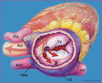
In their initial study, Gerbode and his colleaguesReference Gerbode, Hultgren, Melrose and Osborn3 described two routes for blood to travel from the left ventricle to the right atrium (Fig. 1). The more common, indirect, type of communication occurs via a perimembranous ventricular septal defect when there is an additional defect in the leaflets of the tricuspid valve. In this variant, the shunting is initially below the attachment of the leaflets of the tricuspid valve.Reference Riemenschneider and Moss5 Such indirect left ventricular-to-right atrial shunts accounted for 4 of the 5 patients included in the initial series.Reference Gerbode, Hultgren, Melrose and Osborn3 For the purpose of this review, we excluded patients with such indirect left ventricular-to-right atrial shunts from our analysis.
The illustration shows the direct and indirect variants of ventriculo-atrial communications. Legend: Ao = aorta, CS = coronary sinus, IVC = inferior caval vein, PA = pulmonary artery, RA = right atrium, SVC = superior caval vein, TV = tricuspid valve, VSD = ventricular septal defect.

In the second of the options described by Gerbode and his associates, the blood in the left ventricle flows directly to the right atrium through a congenital defect in the atrioventricular component of the membranous septum, this communication being above the hinge of the leaflets of the tricuspid valve. It is this type of defect, accounting for but 1 patient in the initial series,Reference Gerbode, Hultgren, Melrose and Osborn3 which is now defined as the Gerbode defect in The International Congenital Heart Surgery Nomenclature and Database Project of The Society of Thoracic Surgeons.Reference Jacobs, Burke, Quintessenza and Mavroudis6 Our current review is concerned with this rare malformation.
Methods
Ours was a retrospective case series. We used the computerized database for cardiac surgical patients at Children’s Memorial Hospital in Chicago to identify patients who had undergone surgical closure of a Gerbode defect between 1990 and the present. The Institutional Review Board at Children’s Memorial Hospital, Chicago, Illinois approved this retrospective study and granted a waiver of informed consent on June 27, 2008.
Patient Demographics and Diagnosis
We identified 6 patients, 2 male and 4 female, who had undergone closure of a direct ventriculo-atrial defect between the years 1990 and 2008 at Children’s Memorial Hospital. The median age at time of repair was 1.6 years, with a range from 0.4 to 19 years. All patients were symptomatic at presentation. In 3 patients there was evidence of congestive cardiac failure, 2 had failed to thrive, and 2 had exercise intolerance. In 4, there were associated cardiac diagnoses. In 1 patient with a patent arterial duct, the aortic arch was right-sided. A persistent left superior caval vein was present in 3 patients, 2 also having an anomalous left hepatic vein. In 1 patient, this hepatic vein drained directly to the right atrium, and in the other patient it drained directly to the coronary sinus.
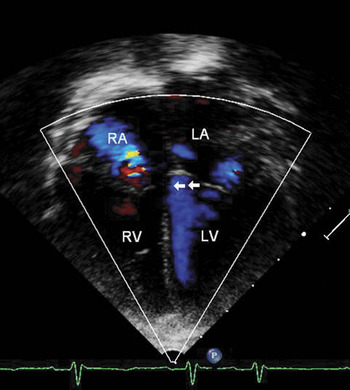
All patients were diagnosed by echocardiography (Fig. 2). The gradient as measured echocardiographically ranged from 65 to 150 millimetres of mercury, with a mean gradient of 109 millimetres of mercury. Echocardiography also showed dilation of the right atrium in all patients.
The echocardiogram in one of our patients as seen in the four-chamber view, with added colour Doppler interrogation. Abbreviations: LA = left atrium, LV = left ventricle, RA = right atrium, RV = right ventricle. The arrows demonstrate flow across the Gerbode defect.

Surgical Technique
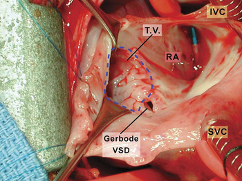
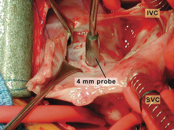
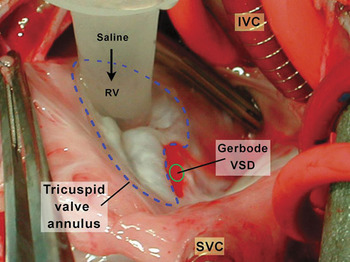
All patients were repaired via a median sternotomy using continuous cardiopulmonary bypass, bicaval venous cannulation, aortic cross clamping, cold blood cardioplegia, and insertion of a patch to close the defect. In Figure 3, we show the typical anatomy of the defect as seen through the opened right atrium from the head of the operating table. In Figure 4, we show a probe of 4 millimetres passed across the defect, and in Figure 5, it can be seen that insufflation of the right ventricle with cold saline shows the tricuspid valve to be competent. The fact that no saline passes through the defect indicates that the congenital defect is directly between the left ventricle and the right atrium. In all cases, the defect was closed by placing pledget-supported Ticron sutures around its circumference, using the sutures to secure a patch of 0.4-mm thick Gore-Tex® (W.L. Gore & Assoc., Flagstaff, AZ).
The surgical view of the opened right atrium. Abbreviations: IVC = inferior caval vein, Gerbode VSD = true Gerbode ventriculo-atrial defect, RA = right atrium, SVC = superior caval vein, TV = tricuspid valve.

A probe of 4 millimetres has been passed into the Gerbode defect. Abbreviations: IVC = inferior caval vein, mm = millimetre, SVC = superior caval vein.

Insufflation of saline into the right ventricle shows the integrity of the tricuspid valve. Abbreviations: IVC = inferior caval vein, Gerbode VSD = true Gerbode ventriculo-atrial defect, RV = right ventricle, SVC = superior caval vein.

Results
There was no operative or late mortality. The size of the ventriculo-atrial defect ranged from 4 to 8 millimetres, with a mean size of 6.2 ± 2.0 millimetres. In all patients, the opening was on the atrial aspect of the hinge of the tricuspid valve. The mean period of cardiopulmonary bypass was 90.5 ± 11.3 minutes, the mean time required for aortic cross-clamping was 54.8 ± 6.9 minutes, and the mean postoperative stay was 4 days. All patients were in normal sinus rhythm. There was no residual ventriculo-atrial shunting, and no patients required a reoperation. Postoperative echocardiography demonstrated at most trivial tricuspid valvar regurgitation.
Discussion
The direct variant of the Gerbode ventriculo-atrial defect is a very rare congenital anomaly.Reference McKay, Battistessa, Wilkinson and Wright7 We have encountered only 6 of these defects at our institution in the past 19 years. Over the course of this same time period, we have repaired over 400 isolated perimembranous, muscular, and doubly committed juxtaarterial ventricular septal defects. Given the rarity of the true Gerbode defect, it is interesting that most of these patients had associated cardiovascular anomalies.
All patients in our series were diagnosed by echocardiography. A Gerbode defect should be suspected when echocardiographic interrogation reveals an unusually dilated right atrium. The other diagnostic feature is the high Doppler gradient between the left ventricle and the right atrium, the mean gradient in our patients being 109 millimetres of mercury. The gradient reflects the notable difference between the left ventricular systolic pressure and the low right atrial pressure.
Left ventricular-to-right atrial shunting causes most of these patients to have significant symptoms, and all of our patients had either congestive cardiac failure, failure to thrive, or exercise intolerance. Spontaneous closure of these defects must be very rare. Our experience shows that surgical closure is safe, so we recommend closure of all direct Gerbode defects. Although the defect may in some cases be anatomically small, in all cases we used a patch to prevent recurrence.
Although the atrioventricular conduction axis penetrates through the atrioventricular membranous septum, all patients in our series were in normal sinus rhythm subsequent to placement of the patch. We encountered no residual ventriculo-atrial defects, no reoperations, and no significant tricuspid valvar insufficiency. The Gerbode ventriculo-atrial defect, therefore, is very rare, but if encountered can be closed surgically with excellent results.