Introduction
The family Allocreadiidae Looss, 1902 is a widely distributed group of digenean parasites primarily inhabiting the intestines of freshwater fishes, with a particular affinity for cypriniforms, salmonids, percids, and acipenserids (Caira and Bogéa Reference Caira, Bogea, Jones, Bray and Gibson2005). The systematics of the family has a long and complex history, traditionally based on morphological characters such as the distribution of vitelline follicles, position and ratios of internal organs, and the morphology of the genital apparatus (Caira Reference Caira1989; Koval Reference Koval and Skryabin1966). However, over the past two decades, the advent of molecular phylogenetics has revolutionised our understanding of allocreadiid diversity and evolution. Numerous studies have provided phylogenetic reconstructions based on the nuclear 28S ribosomal RNA (rRNA) gene and internal transcribed spacer (ITS) region and the mitochondrial cytochrome c oxidase subunit 1 (cox1) gene, leading to the description of new species (Atopkin et al. Reference Atopkin, Sokolov, Shedko, Vainutis and Orlovskaya2018; Aydogdu et al. Reference Aydogdu, Vainutis, Voronova and Aydogdu2023; Vainutis Reference Vainutis2020, Reference Vainutis2024; Vainutis et al. Reference Vainutis, Voronova and Urabe2021, Reference Vainutis, Voronova, Urabe and Kazarin2023), the resurrection of previously synonymised genera (Atopkin et al. Reference Atopkin, Sokolov, Vainutis, Voropaeva, Shedko and Choudhury2020; Vainutis et al. Reference Vainutis, Voronova and Urabe2021), and the recognition of cryptic diversity within widely distributed taxa (Petkevičiūtė et al. Reference Petkevičiūtė, Stunžėnas and Stanevičiūtė2023; Sokolov et al. Reference Sokolov, Khasanov and Lebedeva2023). A landmark synthesis by Bogatov and Vainutis (Reference Bogatov and Vainutis2022), based on a comprehensive phylogenetic tree of 45 valid species, formally proposed an East Asian origin for the family, with subsequent dispersal events to North America via Beringia and to Europe via Transbaikalia and Yakutia.
Despite these significant advances, our understanding of the timing of these evolutionary events remains fragmentary. While molecular clock methods have been successfully applied to estimate divergence times for other trematode groups, such as the superfamily Troglotrematoidea (Vainutis et al. Reference Vainutis, Voronova, Duscher, Shchelkanov and MYu2022), specific divergence time estimates for Allocreadiidae are largely lacking. The only available estimates within the family are for the Neotropical genus Margotrema Lamothe-Argumedo, 1972, which were calculated to have diverged from its common ancestor approximately 6.53 million years ago (Ma) (Martínez-Aquino et al. Reference Martínez-Aquino, Ceccarelli, Eguiarte, Vázquez-Domínguez and Pérez-Ponce de León2014). However, these estimates are restricted to a single, relatively young genus and do not provide information on the deeper nodes of the family tree, such as the origin of the family itself or the divergence of its major Palearctic genera like Acrolichanus Ward, 1917, Stephanophiala Nicoll, 1909, Bunodera Railliet, 1896, Allocreadium Looss, 1900, and Crepidostomum Braun, 1900. Testing the biogeographical hypotheses proposed by Bogatov and Vainutis (Reference Bogatov and Vainutis2022) requires placing them in a temporal framework. For instance, their suggestion that the divergence of Bunodera spp. was linked to the radiation of Perciformes approximately 75 Ma, and that of Allocreadium spp. to the earlier diversification of Cypriniformes around 95 Ma, remains a hypothesis that needs to be tested with molecular dating.
To address this gap, we provide new molecular and morphological data for two poorly known East Asian species of the genus Allocreadium: A. hasu (Roitman Reference Roitman1963) and A. pseudaspii (Akhmerov Reference Akhmerov1960). Both species are redescribed in detail based on newly collected material from the Russian Far East and Japan (Honshu), including their morphology and genetic characterisation using fragments of the 28S rRNA and cox1 mtDNA genes. The inclusion of these novel sequences significantly expands the existing molecular dataset for Palearctic allocreadiids.
The primary aims of this study are therefore: (1) to provide a formal redescription of A. hasu and A. pseudaspii; (2) to reconstruct the phylogenetic relationships within Allocreadiidae using an expanded dataset that includes newly generated 28S and cox1 sequences for these and other East Asian species; and (3) to estimate the divergence times for key nodes within the family, using a calibrated molecular clock approach. Specifically, we will test the temporal framework of the biogeographic scenarios proposed by Bogatov and Vainutis (Reference Bogatov and Vainutis2022) and provide the first comprehensive timeline for the origin and diversification of this important family of fish parasites.
Material and methods
Material collection
Eleven adult individuals of Allocreadium pseudaspii were collected from the intestines of six infected Lagowski’s minnows, Rhynchocypris lagowskii, from the Bolshaya Ussurka River basin, Dalnerechensky District, Primorye, Russia (45°58′52.0″ N, 134°00′51.1″ E). A total of 42 adult individuals of Allocreadium hasu were collected from the intestines of Opsariichthys uncirostris from Lake Biwa at the mouth of the Seri River, near Hikone City, Shiga Prefecture, Japan (35°16′29.6″ N, 136°14′04.7″ E). Collected samples, previously identified under a microscope, were rinsed in saline and fixed in 70% and 96% ethanol for morphological and molecular studies, respectively. All specimens were stored at 4°C. Trematode specimens used for the morphological study were overstained in alum carmine, dehydrated in a graded ethanol series, cleared in clove oil, and mounted in Canada balsam. All measurements are given in millimetres (mm), except those of eggs and the prepharynx, which are given in micrometres (μm).
DNA extraction, amplification, and sequencing
Genomic DNA was extracted from 11 adult individuals of Allocreadium spp. using the HotSHOT alkaline lysis method (Truett Reference Truett and Kieleczawa2006). A 28S rDNA fragment was amplified using the forward primer U178 (5′-GCA CCC GCT GAA YTT AAG-3′) and reverse primer L1642 (5′-CCA GCG CCA TCC ATT TTC A-3′) (Lockyer et al. Reference Lockyer, Olson and Littlewood2003). A fragment of cox1 mtDNA from both A. pseudaspii and A. hasu was amplified using the forward primer JB3 (5′-TTT TTT GGG CAT CCT GAG GTT TAT-3′) and reverse primer JB4.5 (5′-TAA AGA AAG AAC ATA ATG AAA ATG-3′) (Bowles et al. Reference Bowles, Blair and McManus1992). The amplification protocol for 28S rDNA was performed under the following conditions: 2 min denaturation hold at 94 °C, 40 cycles of 30 s at 94 °C, 30 s at 52 °C, 2 min at 72 °C, and a 7 min extension hold at 72 °C. The amplification protocol for Cox1 mtDNA: 1 min denaturation hold at 94 °C, 30 cycles of 15 s at 94 °C, 30 s at 50 °C, 2 min at 72 °C, and a 7 min extension hold at 72 °C. Each polymerase chain reaction (PCR) included a negative control and positive controls, using both primers to detect possible contamination. PCR products were sequenced directly using the BigDye Terminator Cycle Sequencing Kit (Applied Biosystems, USA), according to the manufacturer’s instructions, and the internal sequencing primers 3S (5′-CGG TGG ATC ACT CGG CTC GTG-3′) (Bowles et al. Reference Bowles, Blair and Mcmanus1995), 1200F (5′-CCC GAA AGA TGG TGA ACT ATG C-3′), 1200R (5′-GGG CAT CAC AGA CCT G-3′), and 900F (5′-CCG TCT TGA AAC ACG GAC CAA G-3′) (Lockyer et al. Reference Lockyer, Olson and Littlewood2003). The PCR products were read with an ABI 3500 genetic analyser at the A. V. Zhirmunsky National Scientific Center of Marine Biology FEB RAS. In total, 30 novel sequences were obtained for two Allocreadium species and Acrolichanus sp. (Table 1).
Geographic localities where the studied material was collected.

Figure 1. Long description
The left panel is a large-scale map centered on the Sea of Japan. To the West and Northwest are South Korea, North Korea, and China. To the North is the Russia Primorsky region, marked with a red dot. To the East and Southeast are the Japanese islands of Hokkaido and Honshu. A blue dot is located on the western coast of Honshu.
Two inset panels on the right provide localized detail.
1. The top inset zooms into the red dot in Russia. It shows the Bolshaya Ussurka River. A red dot marks the Bolshaya Ussurka River channel at coordinates 45 degrees 58 minutes 52.1 seconds North, 134 degrees 00 minutes 50.7 seconds East. A scale bar indicates 200 meters.
2. The bottom inset zooms into the blue dot in Japan. It shows Biwa Lake and the island of Honshu. A blue dot marks the Seri River mouth at coordinates 35 degrees 16 minutes 29.5 seconds North, 136 degrees 14 minutes 07.0 seconds East. A scale bar indicates 2 kilometers.
A general scale bar at the bottom right of the main map indicates 200 kilometers.
Information on the species, host, site of infection, geographic origin, and genetic data (partial 28S rRNA and cox1 mtDNA sequences) used in the phylogenetic reconstructions

Table 1. Long description
The table consists of five columns.
Column 1, Species:
- Allocreadium hasu
- Allocreadium pseudaspii
- Acrolichanus species
Column 2, Host or Site of infection:
- Opsariichthys uncirostris forward slash intestine
- Rhynchocypris lagowskii forward slash intestine
- Huso dauricus forward slash intestine
Column 3, Locality:
- Japan, Honshu Island, Shiga Prefecture, Biwa Lake basin, Seri River mouth
- Russia, Primorsky region, Dalnerechensky District, Bolshaya Ussurka River
- Russia, Khabarovsk region, Amur River
Column 4, GenBank submission 28S r R N A gene:
- P P 4 6 8 3 2 6 through P P 4 6 8 3 3 4
- P P 4 6 8 3 2 4, P P 4 6 8 3 2 5
- P Z 5 3 8 9 2 7 through P Z 5 3 8 9 3 4
Column 5, GenBank submission cox 1 m t D N A gene:
- P P 4 6 8 5 1 3 through P P 4 6 8 5 2 1
- P P 4 6 8 5 1 1, P P 4 6 8 5 1 2
- No data provided for Acrolichanus species.
Alignment and phylogenetic analyses
DNA sequences were assembled with SeqScape v. 2.6 software and aligned using the MEGA X Alignment Explorer with default options (Kumar et al. Reference Kumar, Stecher, Li, Knyaz and Tamura2018). Genetic divergence was estimated using genetic p-distance values calculated by including all substitution types in MEGA X software. Phylogenetic analyses were conducted using the Bayesian inference (BI) algorithm in MrBayes 3.1.2 software (Huelsenbeck et al. Reference Huelsenbeck, Ronquist, Nielsen and Bollback2001). The BI algorithm was performed using the GTR+I+G model (general time-reversible model with gamma distribution), selected using jModelTest v. 2.1.5 software (Darriba et al. Reference Darriba, Taboada, Doallo and Posada2012). The resulting tree was rooted with the outgroup taxa. Bayesian inference was applied using the following nucleotide substitution parameters: nst = 6 and rates = gamma. The Markov chain Monte Carlo (MCMC) algorithm was performed using 500,000 generations in two independent runs. The first 25% of generations were discarded as burn-in.
The maximum composite likelihood (MCL) estimate of the pattern of nucleotide substitution was calculated in MEGA X using the Tamura–Nei model and pairwise deletion for gaps and missing data (Tamura and Nei Reference Tamura and Nei1993). This analysis involved 25 nucleotide sequences of Allocreadium species.
The alignment of partial 28S rDNA sequences, trimmed to the length of the shortest sequence, was 1,145 bp long. For phylogenetic reconstruction of Allocreadium spp., 42 sequences of the 28S rRNA gene fragment were used. Of these, 83 partial sequences of the 28S rRNA gene were downloaded from GenBank (National Center for Biotechnology Information [NCBI]). The outgroup taxa are presented with the species of the families Callodistomidae and Gorgoderidae.
Estimation of molecular clock rates
The program BEAST (1.8.2) was used to estimate the evolutionary rates and approximate age of the Trematoda lineage (Drummond et al. Reference Drummond, Suchard, Xie and Rambaut2012) based on the 28S rDNA sequences (see Table 1). We estimated mean node ages and their 95% highest posterior densities (HPDs) under a relaxed-molecular-clock (uncorrelated lognormal) assumption using Bayesian inference (BI); a uniform Yule tree prior, appropriate for hierarchical rather than reticulate relationships, was chosen. We applied the optimal HKY+G model through BEAUti v. 1.8.2 in the BEAST package (Drummond et al. Reference Drummond, Suchard, Xie and Rambaut2012). In the absence of specific information on substitution rates for the gene fragments in our species, we applied lognormal distributions with a mean of 1 and standard deviation of 0.33, allowing for auto-optimisation as the runs progressed. The first detection of trematode eggs in coprolites of Early Cretaceous archosaurs was used as a deep calibration point (~125.93 million years ago [Ma]) (Poinar and Boucot Reference Poinar and Boucot2006). This corresponded to the consensus estimate of the date of the most recent common ancestor (MRCA) of trematodes. BEAST uses MCMC to average over tree space. Following a burn-in of the initial 10% of cycles, divergence times were sampled once every 1,000 generations from 10 million MCMC iterations. Tracer v. 1.6, a graphical tool for visualising and diagnosing MCMC output, was used to check chain convergence to the stationary distribution and to summarise parameter estimates, errors, and confidence intervals (Rambaut et al. Reference Rambaut, Drummond, Xie, Baele and Suchard2018). The simulation was repeated three times and generated analogous results.
Results
Redescription of Allocreadium hasu Ozaki, 1926 (Figure 2a–c)
Description: Based on 10 adult specimens. Body elongate-ovate, rapidly attenuated anteriorly in forebody, fairly small; 3.79–4.72 (3.87) mm long. Widest part of body in ovarian region or between anterior testis and seminal receptacle – 1.05–1.26 (1.17) mm. Forebody 0.57–0.87 (0.69) mm long. Hindbody 2.74–3.34 (2.96) mm long. Tegument smooth. Oral sucker subterminal, round; 0.24–0.34 (0.28) mm long by 0.27–0.34 (0.31) mm wide. Eyespot pigment and short prepharynx present, but not consistently observed in all individuals. Prepharynx short, nearly 50 μm (n=1). Pharynx pear-shaped, 0.15–0.17 (0.159) mm long by 0.15–0.17 (0.159) mm wide. Oesophagus bifurcating dorsal to ventral sucker, 0.25–0.31 mm long. Caeca ending some distance anterior to posterior extremity of body. Ventral sucker large, round, located in anterior third of body, larger than oral sucker, 0.46–0.51 (0.488) mm long by 0.44–0.53 (0.472) mm wide. Testes large, deeply indented irregularly, tandem, in middle third of hindbody; anterior testis 0.53–0.81 (0.64) mm long by 0.47–0.89 (0.58) mm wide, posterior testis 0.51–0.86 (0.64) mm long by 0.49–0.69 (0.56) mm wide. Cirrus pouch clavate to elliptical, overlapping ventral sucker, 0.42–0.49 (0.446) mm long by 0.14–0.25 (0.19) mm wide. Seminal vesicle tubular, convoluted, in posterior half to two-thirds of cirrus pouch. Ejaculatory duct fairly long, sinuous. Genital atrium small. Genital pore at midlevel of esophagus. Ovary usually 3-lobed (rarely subglobular), medium-sized, 0.18–0.34 (0.245) mm long by (0.238) mm wide. Seminal receptacle flask-shaped, usually submedian, 0.25 – 0.37 (0.334) mm long by 0.21–0.32 (0.265) mm wide. Laurer’s canal short. Ootype postovarian. Uterus between anterior testis and intestinal bifurcation, overlapping intestines. Eggs oval, operculated, 64–86 (73) μm long by 43 – 60 (51) μm wide. Vitelline follicles mostly dorsal to intestines, distributed from bifurcal level to posterior extremity of body. Excretory vesicle extending to midlevel of post-testicular region beyond intestinal ends, 0.58–0.64 mm long (n=2). Excretory pore posterodorsal.
Line drawings of adult worms of two Allocreadium species: (a) Allocreadium hasu entire body, ventral view; (b) cirrus pouch; (c) posttesticular space of body; (d) Allocreadium pseudaspii entire body, ventral view; (e) cirrus pouch; (f) posttesticular space of body; (g) ovarian complex. Scale bar: a – 1000 μm; b – 100 μm; c – 300 μm; d – 500 μm; e – 200 μm; f – 500 μm; g – 200 μm. Abbreviations: t – testis; in – intestine; ev – excretory vesicle.

Figure 2. Long description
A series of seven line drawings labeled a through g.
Panel a shows the entire body of Allocreadium hasu in ventral view. It features an oral sucker at the anterior end, followed by a pharynx, a ventral sucker, and internal organs including a lobed ovary and two large tandem testes. A vertical scale bar at the bottom right indicates 1000 micrometers.
Panel b is a detailed view of the cirrus pouch of A. hasu, showing internal seminal vesicles. A vertical scale bar indicates 100 micrometers.
Panel c shows the posttesticular space of A. hasu. The posterior end of a testis labeled t is at the top. Two intestinal branches labeled in run down the sides, and a central excretory vesicle labeled e v leads to the posterior pore. A vertical scale bar indicates 300 micrometers.
Panel d shows the entire body of Allocreadium pseudaspii in ventral view. It is more elongated than A. hasu, with a prominent oral sucker, ventral sucker, and a dense field of vitelline follicles along the lateral margins. A vertical scale bar indicates 500 micrometers.
Panel e shows the cirrus pouch of A. pseudaspii, which is elongated and curved. A vertical scale bar indicates 200 micrometers.
Panel f shows the posttesticular space of A. pseudaspii, with labels t for testis, in for intestine, and e v for excretory vesicle. A vertical scale bar indicates 500 micrometers.
Panel g illustrates the ovarian complex of A. pseudaspii, showing the branching duct system connecting the ovary to surrounding vitelline follicles. A vertical scale bar indicates 200 micrometers.
Taxonomic summary
Host : Opsariichthys uncirostris (type), Xenocyprididae, Cypriniformes.
Locality : Japan, Honshu Island, Shiga Prefecture, Biwa Lake basin, Seri River mouth.
Site of infection: intestine.
Other hosts in Japan: Gnathopogon elongatus elongatus, Nipponocypris temminckii, Zacco platypus.
Type locality: Japan, Shiga Prefecture, Biwa Lake basin, Biwa Lake.
Other localities in Japan: Hyogo Prefecture – Asago River (Shimazu Reference Shimazu1988). Hiroshima Prefecture – Eno River at Yoshida, Yoshida-cho, Akitakata City; Saijo River at Ooya, Saijo-cho, Shobara City (Shimazu Reference Shimazu1988, 2016a). Oita Prefecture – Chikugo River at Kobuchi Bridge, Miyoshikobuchi-machi, Hita City; Ooyama River at Seiwa Bridge, Ooyama-machi, Hita City (Shimazu Reference Shimazu2016a).
Voucher specimens: five topotypes (accession no. T4) were deposited in the helminthological collection of the Far Eastern State Technical Fisheries University, Vladivostok, Russia. Deposition date: 11 March 2024.
ZooBank registration Life Science Identifier (LSID): urn:lsid:zoobank.org:act:BAE9C6AA-896A-4E0D-8AEE-9040E68972E9.
Sample size: 42 adult specimens were found in O. uncirostris.
Remarks on morphology
The trematodes identified as Allocreadium from Lake Biwa in this study are morphologically and morphometrically fully consistent with Allocreadium hasu Ozaki, 1926 (Shimazu Reference Shimazu2016a). Given that A. hasu is the closest genetic relative of A. anastasii (0.97%) and A. khankaiense (0.09%) (Supplementary Table 1), forming a monophyletic species group with them, it differs from them at the interspecific level. Allocreadium hasu is distinguished from A. khankaiense and A. anastasii by a combination of characters: larger and deeply indented irregular testes vs. entire rounded or oval testes; three-lobed ovary vs. rounded or suboval; anterior border of vitelline fields at the posterior margin of the ventral sucker; and absence of vitelline follicles in the testicular region (original data; Shimazu Reference Shimazu2016a; Vainutis Reference Vainutis2020, Reference Vainutis2024; Vainutis et al. Reference Vainutis, Voronova, Urabe and Kazarin2023). Morphological features uniting all three species as a single group are: dorsal bifurcation of the intestine relative to the ventral sucker, and position of the cirrus pouch at the level of the ventral sucker.
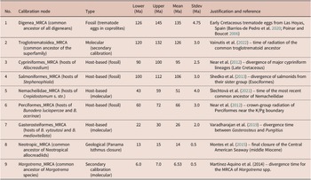
Calibrations used in this study

Table 2. Long description
The table consists of eight columns: No., Calibration node, Type, Lower (M a), Upper (M a), Mean (M a), Stdev (M a), and Justification and reference.
* Row 1: Digenea underscore M R C A; Fossil (trematode eggs); Lower 126, Upper 145, Mean 135, Stdev 4.75; Early Cretaceous trematode eggs from Spain.
* Row 2: Troglotrematoidea underscore M R C A; Molecular (secondary); Lower 120, Upper 132, Mean 126, Stdev 3.0; Vainutis et al. (2022).
* Row 3: Cypriniformes underscore M R C A; Host-based (fossil); Lower 90, Upper 100, Mean 95, Stdev 2.5; Near et al. (2012).
* Row 4: Salmoniformes underscore M R C A; Host-based (fossil); Lower 100, Upper 112, Mean 106, Stdev 3.0; Shedko et al. (2013).
* Row 5: Nemacheilidae underscore M R C A; Host-based (molecular); Lower 43, Upper 59, Mean 51, Stdev 4.0; Šlechtová et al. (2021).
* Row 6: Perciformes underscore M R C A; Host-based (fossil); Lower 60, Upper 72, Mean 66, Stdev 3.0; Near et al. (2012).
* Row 7: Gasterosteiformes underscore M R C A; Host-based (molecular); Lower 22, Upper 30, Mean 26, Stdev 2.0; Varadharajan et al. (2019).
* Row 8: Neotropic underscore M R C A; Geological (Panama Isthmus closure); Lower 13, Upper 15, Mean 14, Stdev 0.5; Montes et al. (2015).
* Row 9: Margotrema underscore M R C A; Secondary calibration (molecular); Lower 6.0, Upper 7.0, Mean 6.53, Stdev 0.5; Martínez-Aquino et al. (2014).
Redescription of Allocreadium pseudaspii (Akhmerov, Reference Akhmerov1960) Bychovskaya-Pavlovskaya, 1962 (Figure 2d–g)
Description: Based on five adult specimens. Relatively small trematodes. Body elongate fusiform, 3.07 – 3.57 (3.32) mm long. Widest part of body in ovarian region – 0.6–0.85 (0.71) mm wide. Forebody 0.65–0.75 (0.695) mm long. Hindbody 1.85–2.45 (2.175) mm long. Tegument smooth. Anterior end with distinct collar-like elevation adjacent to oral sucker, in some specimens divided into small lobes. Oral sucker subterminal, round, 0.31–0.36 (0.328) mm long by 0.28–0.34 (0.31) mm wide. Prepharynx short, 30–54 (35) μm long. Pharynx pear-shaped or sub-spherical, 0.15–0.18 (0.165) mm long by 0.17–0.20 (0.187) mm wide. Oesophagus slender, 0.41–0.46 (0.46) mm long, reaching posterior margin of ventral sucker, bifurcating into thick caeca terminating blindly near posterior end of body at varying distance – 0.32–0.35 (0.338) mm long. Ventral sucker round, larger than oral sucker, situated in anterior third of body, 0.35–0.37 (0.36) mm long by 0.35–0.43 (0.39) mm wide. Forebody with a small cluster of eyespot remnants in the pharyngeal or post-pharyngeal region. Reproductive system well developed. Testes oval, tandem, in posterior half of body; posterior testis longer than anterior. Anterior testis 0.31–0.41 (0.345) mm long by 0.26–0.41 (0.33) mm wide. Posterior testis 0.34–0.47 (0.38) mm long by 0.29–0.42 (0.35) mm wide. Seminal ducts reaching proximal end of cirrus pouch at level of posterior margin of ventral sucker. Cirrus pouch thin-walled, 0.45–0.56 (0.508) mm long by 0.20–0.22 (0.206) mm wide, enclosing seminal vesicle looping several times, continuing into prostatic complex with distinct, large prostatic glands, and weakly armed cirrus. Genital pore submedian, slightly dextral. Ovary oval, about 1.5 times smaller than testis, dextral, 0.23–0.29 (0.266) mm long by 0.19–0.26 (0.228) mm wide. Seminal receptacle small, flask-shaped, postovarian, pretesticular, sometimes at level of anterior testis, producing distinct notch on its anterior margin, 0.18–0.28 (0.223) mm long by 0.13–0.15 (0.144) mm wide. Uterus with numerous eggs (nearly 100–150) between ventral sucker and anterior testis, with one uterine loop extending ventrally beyond anterior testis and reaching anterior margin of posterior testis. Eggs small, oval, operculated, 55–80 (64) μm long by 40–60 (50) μm wide. Vitelline follicles large, irregular in shape, closely adjacent. Posterior border of vitellarium densely filling posttesticular space; lateral fields extending anteriorly to level slightly behind or at level of posterior margin of pharynx, covering caeca ventrally without confluencing on ventral and dorsal sides; occasionally entering inter-testicular space. Thick vitelline reservoir passing between ovary and seminal receptacle dorsally. Excretory vesicle tubular, bifurcating immediately behind posterior testis. Excretory pore terminal.
Taxonomic summary
Synonyms: Allocreadium elongatum (Akhmerov, Reference Akhmerov1960) Bychovskaya-Pavlovskaya, 1962; Neoallocreadium elongatum Akhmerov, Reference Akhmerov1960; Neoallocreadium pseudaspii Akhmerov, Reference Akhmerov1960.
New host examined: Rhynchocypris lagowskii Dybowski, 1869, Leuciscidae, Cypriniformes.
Site of infection: intestine.
New locality: Russia, Primorsky region, Dalnerechensky District, Bolshaya Ussurka River.
Type host: Pseudaspius leptocephalus (Pallas, 1776), Leuciscidae, Cypriniformes.
Other hosts: Chanodichthys mongolicus (Basilewsky, 1855), Xenocyprididae, Cypriniformes.
Type locality: Russia, Khabarovsky region, Amur River basin, Udyl Lake (near Elabuga village).
Other localities: Russia, Khabarovsky region, Amur River (as A. elongatum) (Akhmerov Reference Akhmerov1960).
New material: Five specimens (accession no. T3) deposited in the helminthological collection of the Far Eastern State Technical Fisheries University, Vladivostok, Russia (deposition date: 11 March 2024).
Zoobank registration LSID: urn:lsid:zoobank.org:act:E724A7F2-47B4-4A99-B73B-0E2C94909C8A.
Sample size: Two juvenile and eight adult specimens were isolated from five individuals of Rhynchocypris lagowskii.
Remarks on nomenclature
The name elongatum has been applied to two different species within the genus Allocreadium. Park (Reference Park1939) originally described Macrolecithus elongatus from Korea, which was later transferred to Allocreadium as A. elongatum (Park, Reference Park1939) Shimazu, Reference Shimazu1988. Akhmerov (Reference Akhmerov1960) described Neoallocreadium elongatum from the Amur Basin, which was also later transferred to Allocreadium. This created a secondary homonymy within the genus Allocreadium. To avoid this homonymy and following the Principle of Priority (ICZN Article 23), the later name A. elongatum (Akhmerov, Reference Akhmerov1960) must be rejected as a junior homonym of A. elongatum (Park Reference Park1939). Therefore, we recognise Allocreadium pseudaspii (Akhmerov Reference Akhmerov1960) as the valid name for the Amur Basin species, treating A. elongatum (Akhmerov, Reference Akhmerov1960) as its junior synonym. This treatment is consistent with the most recent taxonomic revisions (Vainutis Reference Vainutis2024; Vainutis et al. Reference Vainutis, Voronova, Urabe and Kazarin2023).
Remarks on morphology
A mirror situation is observed when analysing another Far Eastern species, Allocreadium pseudaspii. Morphometrically and morphologically, our worms from the Bolshaya Ussurka River are identical to Allocreadium pseudaspii originally described from the Amur River (Akhmerov Reference Akhmerov1960). The presence of eyespot remnants is also fully consistent with the original description of the species by Akhmerov (Reference Akhmerov1960). However, the phylogenetic position of this species deserves special attention – its nesting within the group of European species, which includes A. isoporum, A. crassum, and A. dogieli. According to the dichotomous key to Palearctic Allocreadium spp. (Vainutis Reference Vainutis2024), A. pseudaspii occupies a distinct position based on the extension of vitelline follicles: the anterior border of its vitelline fields reaches the middle of the forebody, whereas in A. dogieli this border is within the acetabular region, and in the closely related A. isoporum and A. crassum, it is in the posterior part of the body, usually immediately behind the ventral sucker.
The two species described in this work, A. hasu and A. pseudaspii, demonstrate interspecific differences relative to each other, which include the following characters: esophageal bifurcation dorsal to ventral sucker vs. posterior to ventral sucker; anterior border of vitelline follicles at the level of the posterior margin of the ventral sucker vs. at the level of the posterior margin of the pharynx; proximal end of cirrus pouch at midlevel of ventral sucker vs. at posterior margin of ventral sucker; testes lobed vs. entire oval; ovary 3‑lobed vs. entire rounded; excretory vesicle reaching midlevel of post‑testicular space vs. posterior margin of posterior testis. These character variations fall within the range of interspecific differences among species of the genus Allocreadium. The remaining characters reflect the similarity between A. hasu and A. pseudaspii and are consistent with the general morphology of the genus Allocreadium (Aydogdu et al. Reference Aydogdu, Vainutis, Voronova and Aydogdu2023; Dos Santos et al. Reference Dos Santos, Gilbert, Avenant-Oldewage and Dumbo2021; Koval Reference Koval and Skryabin1966; Petkevičiūtė et al. Reference Petkevičiūtė, Stunžėnas and Stanevičiūtė2023; Sokolov et al. Reference Sokolov, Khasanov and Lebedeva2023; Vainutis Reference Vainutis2020, Reference Vainutis2024; Vainutis et al. Reference Vainutis, Voronova, Urabe and Kazarin2023).
Genetic data
Phylogenetic analysis based on the 28S rRNA gene
New partial 28S sequences were obtained for Allocreadium hasu (nine sequences, 1734 bp) and A. pseudaspii (two sequences, 1734 bp). Based on the sequencing data length (1269 bp), genetic p-distances within the genus Allocreadium for the 28S gene ranged from 0.09% to 6.83% (Supplementary Table 1). Allocreadium hasu and A. pseudaspii differed from each other by 3.54%. Allocreadium hasu was closer to A. khankaiense (0.09%), and A. pseudaspii was closer to European A. crassum (0.62%) and A. dogieli (0.35%). Both species differed from other Allocreadium spp. as follows: 0.97–6.21% (A. hasu) and 1.77–6.12% (A. pseudaspii).
Three phylogenetic reconstructions were performed based on the available sequence lengths of different species using the Bayesian Inference method. Thus, fragment lengths of 2757 bp (14 species), 1145 bp (19 species), and 509 bp (21 species) were used (Figures 3 and 4). In each case, the standard distribution of Allocreadium species was maintained: a species group (A. bursense, A. macrolecithum, A. pseudoisoporum), an independent position of A. hemibarbi, a sister species group (A. khankaiense, A. hasu, and A. anastasii), and separate species A. pseudaspii.
Phylogenetic tree reconstructed with 2757 bp of the 28S rRNA gene using Bayesian inference, showing relationships among 14 allocreadiid species (including nine Allocreadium spp.). One species of the Gorgoderidae was used as rooting outgroup. Numbers on branches indicate posterior probabilities.

Figure 3. Long description
The tree originates from a root on the left. At the bottom, a single branch leads to the outgroup P V 8 3 3 2 0 6 Phyllodistomum umblae, labeled Gorgoderid outgroup. Above this, the main Allocreadiidae clade splits into several groups.
Moving from top to bottom within the main clade:
1. A large group labeled Allocreadium contains several sub-clades. The top-most sub-clade includes species hasu (P P 4 6 8 3 2 6 through P P 4 6 8 3 3 4) with a posterior probability of 0.99. Below it are khankaiense (M K 2 1 1 2 1 4, M K 2 1 1 2 1 5), anastasii (M K 2 1 1 2 0 9, M K 2 1 1 2 1 0), and pseudaspii (P P 4 6 8 3 2 4, P Z 5 3 7 3 5 3 through P Z 5 3 7 3 5 5).
2. Further down the Allocreadium branch are hemibarbi (M K 2 1 1 2 2 0, M K 2 1 1 2 2 1), an unnamed Allocreadium sp. (P Z 2 1 2 4 3 4, P Z 2 1 2 4 3 5), bursense (O K 0 4 5 5 2 1, O K 0 4 5 5 2 2), macrolecithum (O R 0 6 6 2 2 8, O R 0 6 6 2 2 9), and pseudoisoporum (M K 2 5 8 6 8 5, M K 2 5 8 6 8 6).
3. Below the Allocreadium clade is a single branch for Pseudoparacreptotrema yaguezani (P P 5 4 8 2 2 4).
4. Next is the Crepidostomum clade, containing oschmarini (P Z 2 1 0 6 1 6, P Z 2 1 0 6 1 7), achmerovi (M K 8 1 8 6 1 7, M K 8 1 8 6 1 8), and metoecus (M K 8 1 8 6 1 0, M T 1 9 6 3 5 5).
5. The final group at the bottom of the Allocreadiidae clade is Acrolichanus, containing two Acrolichanus sp. samples (P Z 5 3 8 9 2 7, P Z 5 3 8 9 3 0).
Most internal nodes are marked with a posterior probability of 1. A scale bar at the bottom left indicates a genetic distance of 0.02.
Phylogenetic tree reconstructed with different lengths of the 28S rRNA gene (a – 1145 bp, b – 509 bp) using Bayesian inference, illustrating relationships among 71 (a) and 73 (b) allocreadiid species, including 19 (a) and 21 (b) Allocreadium spp. Outgroup taxa comprise species of the Gorgoderidae and Callodistomidae. Numbers on branches indicate posterior probabilities.

Figure 4. Long description
A two-panel figure showing phylogenetic trees labeled A and B.
Panel A (Left):
- Title: 1145 b p.
- Structure: A large blue-shaded box contains the Allocreadiidae ingroup. The tree branches from a single root on the left toward numerous species names on the right.
- Top Clades: Includes genera such as Creptotrema, Wallinia, and Pseudoparacreptotrema.
- Middle Clades: Includes Crepidostomum and Stephanophiala.
- Bottom Clades: A dense cluster of Allocreadium species, including A. pseudaspii and A. hasu.
- Outgroups: Below the blue box, the tree includes a Callodistomid outgroup (Prosthenhystera spp.) and Gorgoderid rooting taxa (Nagmia, Plesiochorus, Degeneria, Phyllodistomum, and Gorgodera).
- Scale bar: 0.02 at the bottom left.
Panel B (Right):
- Title: 509 b p.
- Structure: Similar topology to Panel A but with slightly different branch lengths and posterior probability values.
- Ingroup: The blue-shaded Allocreadiidae ingroup contains 73 species. Notable differences include the placement of Bunodera species within the Crepidostomum clade.
- Outgroups: Identical Callodistomid and Gorgoderid taxa as in Panel A.
- Scale bar: 0.04 at the bottom left.
In both panels, decimal numbers (posterior probabilities) are positioned at the nodes, ranging from 0.52 to 1. Species names are listed as a combination of GenBank accession numbers and binomial nomenclature.
The phylogenetic tree was reconstructed from 2757 bp of the 28S rRNA gene using Bayesian Inference (Figure 3). The gorgoderid species Phyllodistomum umblae served as the rooting outgroup. The basal position within the ingroup (Allocreadiidae) was occupied by Acrolichanus sp., which branched off directly from the ancestral node. Three species of Crepidostomum and Pseudoparacreptotrema yaguezani formed a well-supported monophyletic group (posterior probability 1.00). This group was recovered as sister to the genus Allocreadium, which was represented by nine species. Within Allocreadium, the basal positions were occupied by a group (pseudoisoporum, bursense, macrolecithum). The species pseudaspii formed a separate branch, which was sister to a subclade comprising khankaiense, anastasii, and hasu. All branches within the tree received maximal statistical support, with posterior probability values ranging from 0.99 to 1.00.
Phylogenetic trees were reconstructed from different lengths of the 28S rRNA gene using Bayesian inference: Tree A based on 1145 bp (71 allocreadiid species, 19 Allocreadium spp.) and Tree B based on 509 bp (73 allocreadiid species, 21 Allocreadium spp.) (Figure 4a,b). The rooting group consisted of five gorgoderid species and four species of Prosthenhystera (Callodistomidae) serving as the outgroup. In tree A (1145 bp), the genus Acrolichanus occupied the basal position within Allocreadiidae. Crepidostomum chaenogobii was maximally distant from both Crepidostomum sensu stricto and Crepidostomum sensu lato, and was placed as a close relative of Acrolichanus with maximal statistical support (posterior probability, PP = 1.00). The genus Allocreadium was recovered as sister to Bunodera, but with moderate support (PP = 0.86). In tree B (509 bp), an alternative topology was observed: the genus Stephanophiala occupied the basal position. Acrolichanus appeared in the subsequent branch but with low statistical support (PP = 0.72). In this reconstruction, Allocreadium formed a separate, well-supported clade (PP = 1.00), while Bunodera occupied an intermediate position between Crepidostomum chaenogobii and Crepidostomum sensu stricto, although this placement received very low support (PP = 0.52, 0.56, 0.55). The resolution of Allocreadium species was largely congruent between the two trees. Notably, tree B included two Indian species, A. tori and A. schizothoracis, which formed a sister relationship with the African species A. apokryfi at the terminal position within the genus.
The molecular clock phylogeny of trematodes of the suborder Xiphidiata was estimated based on a 1139 bp fragment of the 28S rRNA gene for 106 species from six superfamilies (Figure 5). The most recent common ancestor (MRCA) of the superfamily Gorgoderoidea was dated to 133 Ma (Lower Cretaceous), while its type family, the Gorgoderidae, diverged at 119 Ma within the same period. The ancestral node of the families Allocreadiidae and Callodistomidae was dated to 110 Ma (Lower Cretaceous). The core of the Allocreadiidae ingroup comprises five basal genera, the majority of whose species are distributed across the Holarctic: Allocreadium, Acrolichanus, Bunodera, Crepidostomum, and Stephanophiala. The genus Prosthenhystera (Callodistomidae) was erroneously recovered within Allocreadiidae as sister to Acrolichanus, although with weak statistical support (posterior probability, PP = 0.87). The divergence of the common ancestor of four Prosthenhystera species began approximately 23 Ma, at the Oligocene–Miocene boundary. Although the definitive hosts of Acrolichanus are sturgeons (Acipenseridae) – among the most ancient of ray-finned fishes – Acrolichanus occupies an intermediate position in the chronological record of the five basal allocreadiid genera. The divergence of its MRCA, which also gave rise to the genus Prosthenhystera, began at 58 Ma. The most ancient of all allocreadiid genera is Stephanophiala, whose divergence began approximately 96 Ma (Upper Cretaceous), following the lower divergence point of its salmonid hosts at 100 Ma. The evolutionary trajectory of the type genus Allocreadium (93 Ma) nearly coincides with the midpoint of divergence of the MRCA of its typical cypriniform hosts at 95 Ma. Subsequently, towards the end of the Cretaceous (76 Ma), a separate lineage began to form, currently comprising the single species Crepidostomum chaenogobii, which is associated with gobiid and gasterosteid fishes. This species exhibits a sister relationship to the lineage that gave rise to the genera Acrolichanus and Prosthenhystera. The divergence of the genus Bunodera coincides with that of C. chaenogobii. Within this genus, the lineage of species parasitizing percid fishes also appeared in the Paleocene, similar to Acrolichanus, and represents the most ancient lineage (62 Ma for B. luciopercae, B. acerinae), whereas the lineage associated with gasterosteids is considerably younger, dating to approximately 26 Ma (Oligocene) for B. vytautasi and B. mediovitellata. The genus Bunodera formed a separate lineage within a clade comprising Allocreadium, Acrolichanus, and Crepidostomum chaenogobii. However, the structure of the entire clade lacks statistical support (PP = 0.33), due to the erroneous placement of Prosthenhystera within it. The divergence of the youngest genus, Crepidostomum sensu stricto, comprising five species, began approximately 49 Ma (early Eocene). This genus forms a sister relationship with the lineage of American allocreadiid species (PP = 0.96), including the North American Crepidostomum sensu lato species, whose divergence occurred in two independent phases, with separation into groups sensu lato A (C. affine, C. illinoiense) and sensu lato B (C. cornutum, C. auritum) at approximately 19 Ma and 9 Ma, respectively.
Phylogenetic tree with estimated divergence times. Species divergence was estimated by a Bayesian model using BEAST with a relaxed molecular clock and is given in million years. Abbreviations on the timescale: epochs of the Cenozoic era: Pal – Paleocene; Olig – Oligocene; Pl – Pliocene; P – Pleistocene; H – Holocene. The animals in the figure are typical representatives of the corresponding epochs.

Figure 5. Long description
A phylogenetic tree with horizontal branches extending from a root on the left toward species names on the right.
* The tree is divided into color-coded clades: a large blue-shaded region at the top contains Allocreadiidae and Gorgoderoidea. Below this are smaller clades in green (Opecoeloidea), yellow (Brachylaimoidea and Troglotrematoidea), light green (Plagiorchioidea), and pink (Microphalloidea). At the bottom, a single orange branch represents the rooting taxon Haplorchis pumilio.
* Horizontal blue bars on the branches indicate confidence intervals for divergence times, with decimal values representing posterior probabilities.
* Below the tree is a horizontal X-axis timescale ranging from negative 240 to 0, representing million years ago.
* Under the timescale, colored bars represent geological periods: Triassic (purple), Jurassic (brown), Cretaceous (light green), Paleogene (orange), and Neogene (yellow). These are grouped into the Mesozoic and Cenozoic eras.
* The bottom-most layer features black silhouettes of representative fauna for each epoch: dinosaurs like a long-necked sauropod and a theropod appear under the Mesozoic; mammals such as a primitive horse, a primate, a mammoth, and a human silhouette appear under the Cenozoic.
* Specific epoch abbreviations at the far right of the Cenozoic include Pal for Paleocene, Olig for Oligocene, Pl for Pliocene, P for Pleistocene, and H for Holocene.
Phylogenetic analysis based on the cox1 mtDNA gene
New partial cox1 mtDNA gene sequences were obtained for Allocreadium hasu (nine sequences, 447 bp) and A. pseudaspii (two sequences, 447 bp). Interspecific genetic distances (uncorrected p-distances) between A. hasu and other congeners ranged from 18.36% to 30.22%. Across the nine Allocreadium species analysed, interspecific distances varied from 18.36% to 39.78%.
Phylogenetic relationships were reconstructed using Bayesian inference (BI) based on 383 bp of the cox1 mtDNA gene (Figure 6). The resulting tree exhibited topologies that diverged from the well-resolved 28S rRNA phylogeny, including Allocreadium and Crepidostomum s. str. The tree showed two monophyletic groups comprising the genera Allocreadium (11 species) and Crepidostomum sensu stricto (three species), though internal relationships of some Allocreadium species were not fully resolved.
Phylogenetic tree reconstructed using Bayesian inference based on 383 bp of the cox1 mtDNA gene, illustrating relationships among 11 species of Allocreadium and three species of Crepidostomum. Numbers on branches indicate posterior probabilities. The rooting outgroup is represented by one species of the Callodistomidae.

Figure 6. Long description
The tree begins at a root on the left with a scale bar of 0.05 below it.
At the bottom, separated by a vertical line, is the rooting outgroup: K C 8 9 9 8 4 8 Prosthenhystera sp. 1 from the Callodistomidae family.
Above the outgroup is the Allocreadiidae ingroup, enclosed in a light blue shaded box. This ingroup splits into two main branches.
The lower branch of the ingroup contains four Crepidostomum species: 15-1 C. nemachilus, 1970-3 C. sp., 1-1 C. metoecus, and C N 1-1 C. achmerovi, with posterior probabilities of 0.77 and 1 at their nodes.
The upper branch of the ingroup contains several clades of Allocreadium. Moving from bottom to top within this section:
- A clade with O R 9 4 5 2 1 9 and O R 9 4 5 2 2 0 A. hemibarbi (probability 1).
- A clade with O M 9 1 4 8 4 9 and O M 9 1 4 8 5 0 A. pseudoisoporum (probability 1).
- A clade with A S P 1 4-1 and A S P 1 4-2 A. pseudaspii (probability 1).
- A larger complex clade (probability 0.94) containing A. gotoi, A. bursense, A. macrolecithum, A. anastasii, A. hasu, A. khankaiense, and A. lobatum.
Specific terminal nodes at the top include O R 9 8 7 8 4 7 and N C _ 0 8 6 7 1 7 A. lobatum. Posterior probability values are labeled at every major node, ranging from 0.56 to 1.
Discussion
Current research continues a series of studies on the type genus of the family Allocreadiidae in the Russian Far East, including descriptions of five new species (Aydogdu et al. Reference Aydogdu, Vainutis, Voronova and Aydogdu2023; Vainutis Reference Vainutis2020, Reference Vainutis2024; Vainutis et al. Reference Vainutis, Voronova, Urabe and Kazarin2023), genotyping of A. khankaiense and Allocreadium sp. 1 (=A. anastasii) (Vainutis Reference Vainutis2024; Vainutis et al. Reference Vainutis, Voronova and Urabe2021, Reference Vainutis, Voronova, Urabe and Kazarin2023), hypotheses about coevolution with cypriniform hosts (Bogatov and Vainutis Reference Bogatov and Vainutis2022), and biogeographic distribution of A. khankaiense in faunistic complexes of the Palaearctic realm (Vainutis et al. Reference Vainutis, Belyaev, Afanasyev and Fomina2024). The morphological and genetic diagnosis of A. hasu and A. pseudaspii presented herein clarifies the phylogenetic relationships within the genus Allocreadium.
Molecular clock estimates are subject to considerable uncertainty, particularly in the absence of reliable fossil calibrations (Loker Reference Loker2025; Poinar and Boucot Reference Poinar and Boucot2006). The broad confidence intervals observed for many nodes (Figure 5) reflect this uncertainty and should be taken into account when interpreting the divergence times presented here. Our estimates are intended as working hypotheses rather than precise chronological statements.
We acknowledge that the use of host-based calibrations implicitly assumes some degree of phylogenetic congruence between parasites and their definitive hosts. However, host-switching events are well documented in trematodes and cannot be excluded a priori (Blasco-Costa et al. Reference Blasco-Costa, Hayward, Poulin and Balbuena2021). Our estimates should therefore be interpreted with caution, as they represent minimum divergence ages under the assumption that the observed parasite-host associations are at least partly conserved. Future studies incorporating cophylogenetic analyses will be necessary to test the degree of congruence between allocreadiid and fish host phylogenies.
Morphological analysis
The morphological and morphometric analyses confirmed that the trematodes from Lake Biwa are fully consistent with Allocreadium hasu Shimazu, 2016, and those from the Bolshaya Ussurka River correspond to Allocreadium pseudaspii (Akhmerov, Reference Akhmerov1960) Bychovskaya-Pavlovskaya, 1962 (for details, see Results). The presence of eyespot remnants in A. pseudaspii is consistent with the original description (Akhmerov Reference Akhmerov1960). The key morphological differences between A. hasu and A. pseudaspii – including the position of the esophageal bifurcation, the anterior extent of vitelline follicles, and the shape of the testes – are stable and fall within the range of interspecific variation observed in the genus Allocreadium (Koval Reference Koval and Skryabin1966; Vainutis Reference Vainutis2020, Reference Vainutis2024; Vainutis et al. Reference Vainutis, Voronova, Urabe and Kazarin2023).
Divergence of Allocreadium
The 28S gene fragment showed high similarity of Allocreadium hasu to A. khankaiense found in three rivers of the southern Primorsky region: the Komissarovka River (Vainutis Reference Vainutis2020), the Artyomovka River (Vainutis et al. Reference Vainutis, Voronova and Urabe2021), and the Pavlovka River (Vainutis et al. Reference Vainutis, Voronova, Urabe and Kazarin2023). A relationship between these species is also evident through one of their definitive hosts, Opsariichthys uncirostris, which was found infected with A. khankaiense in the Komissarovka River (unpublished data 2020). The host O. uncirostris is distributed in the Russian Far East, Korea, China, Taiwan, and Southeast Asia, but Allocreadium hasu has not been recorded in these regions. In Lake Biwa, A. gotoi and A. hasu are common parasites. In Japan, O. uncirostris is naturally distributed in Lake Biwa and the Mikata Five Lakes, and A. hasu has also been recorded from Nipponocypris temminki, Zacco platypus, and Gnathopogon elongatus on Kyushu Island (Shimazu Reference Shimazu2016a). The main question is whether Allocreadium hasu, a typical parasite of Japan, is distributed in the rivers and lakes of the southern Russian Far East. According to Sokolovsky and Epur (Reference Sokolovsky and Epur2008) and Barabanshchikov et al. (Reference Barabanshchikov, Nazarov and Svirsky2006), Opsariichthys uncirostris inhabits the Tumen River and Lake Khanka. According to our unpublished data, A. khankaiense also infects O. u. amurensis in the Komissarovka River (collections from June 2020), whereas no cases of A. hasu introduction have been recorded to date. Examination of O. u. amurensis in the Bolshaya Ussurka River (July 2019, July 2020) revealed neither A. khankaiense nor A. hasu nor any other representatives of the genus Allocreadium.
Allocreadium pseudaspii, first recorded by us in the Bolshaya Ussurka River, has a unique phylogenetic position despite its East Asian locality. It is possible that, as in the case of Crepidostomum metoecus and Stephanophiala farionis, a distant relative of Allocreadium crassum had a close relationship with A. pseudaspii and was transported by salmonid fishes from the Asian region to Northwestern Europe. Cases of infection of salmonid fishes with Allocreadium species have been noted earlier, but so far have not been confirmed by a combination of genetic and morphological data. The most likely scenario is dispersal through natural cypriniform hosts via Siberian water bodies. It is now inadmissible to argue unidirectionally about the divergence and dispersal of Allocreadium spp. from Asia to Europe, since new genetic data for A. transversale (Petkevičiūtė et al. Reference Petkevičiūtė, Stunžėnas and Stanevičiūtė2023) and A. papilligerum (Sokolov et al. Reference Sokolov, Khasanov and Lebedeva2023) have revealed genetic relatedness and their placement in a single species group with the East Asian A. pseudoisoporum and A. macrolecithum (Vainutis Reference Vainutis2024). Thus, the Trans-Eurasian group containing three European species (A. isoporum, A. crassum, A. dogieli) does not necessarily have a common ancestor in East Asia; the ancestor could also have been located in what is now Eastern Europe. Until recently, the definitive host of Allocreadium crassum was unknown, but Petkevičiūtė et al. (Reference Petkevičiūtė, Stunžėnas and Stanevičiūtė2023) showed that the common roach Rutilus rutilus and the rudd Scardinius erythrophthalmus serve in this role. Both genera belong to the subfamily Leuciscinae (family Leuciscidae), as does the genus Rhynchocypris, some species of which may facilitate a wider distribution of trematodes. After a series of studies, it should be emphasised that the lake minnow Rhynchocypris percnura and Lagowski’s minnow Rhynchocypris lagowskii are often definitive hosts of Allocreadium spp. (at least four species) in various rivers of the southern Russian Far East (Vainutis Reference Vainutis2020, Reference Vainutis2024). In addition, A. pseudaspii infects leuciscids R. lagowskii (according to this study) and Pseudaspius leptocephalus (Akhmerov Reference Akhmerov1960). It is evident that the spread of Allocreadium spp. occurs more efficiently through infection of Rhynchocypris spp. due to their greater mobility and adaptability.
The radiation time of the common gorgoderoid ancestor and the family Allocreadiidae (Figure 5) corresponded to the Hauterivian age of the Lower Cretaceous and the Albian age of the Lower Cretaceous, respectively. The origin of these trematodes indicates their later origin relative to the superfamily Troglotrematoidea, representatives of which were previously mistakenly assigned to Gorgoderoidea. Phylogenetic analysis shows that Gorgoderoidea reliably includes seven families (Figure 4). In this study, the divergence of the MRCA of Troglotrematoidea, as well as of the families Troglotrematidae and Paragonimidae separately, is dated earlier (nearly 150 Ma) than calculated by Vainutis et al. (Reference Vainutis, Voronova, Duscher, Shchelkanov and MYu2022) and Voronova et al. (Reference Voronova, Vainutis, Tabakaeva, Sapotsky, Kakareka, Volkov, Galkina and Shchelkanov2022). This may indicate the initial stages of divergence of the Troglotrematoidea MRCA at the Jurassic–Cretaceous boundary.
Given the established parasite–host relationships of allocreadiids at the adult stage with fish, it should be clarified that Allocreadium spp. is associated not simply with cyprinid fishes but with the entire order of cypriniform fishes (Cypriniformes). According to the latest classification, this order includes 23 families, more than half of which can serve as definitive hosts of adult Allocreadium spp. in various regions of the Palaearctic, forming a complex network of distribution of these trematode species and an equally complex evolutionary history. The divergence time of the common ancestor of Allocreadium species (nearly 93 Ma) coincides with that of the common ancestor of Cypriniformes (nearly 95 Ma), meaning that the MRCA of Allocreadium spp. was associated with some ancient cypriniform species no earlier than 93 Ma. This timeline corresponds to the Turonian age of the Late Cretaceous – a period of divergence and widespread distribution of ammonites and the separation of the western part of Eurasia by the Turgai Strait. Such separation could have provided isolation of the early MRCA of Allocreadium spp. and its descendants in the eastern part of ancient Eurasia. This could explain the formation of the common trans-Eurasian group of species from European (A. transversale, A. papilligerum) and Asian (A. pseudoisoporum, A. bursense, A. macrolecithum) taxa. A possible scenario is that a separate population of the MRCA of this group was later naturally introduced after the reunification of the European and Asian subcontinents. Later introduction due to human activity cannot be ruled out either. The youngest species of this group, A. pseudoisoporum, was likely secondarily introduced into the Asian region from Europe, as it is genetically close to A. papilligerum (0.18%), and the divergence of their common ancestor is dated to about 3 Ma in the Piacenzian age of the Pliocene. Previously, we assumed that the initial divergence of the common ancestor of A. khankaiense and A. anastasii could have been associated with the separation of the Palaeo-Amur into the independent Amur and Razdolnaya river basins at the Pliocene–Pleistocene boundary (Vainutis Reference Vainutis2020; Vainutis et al. Reference Vainutis, Voronova, Urabe and Kazarin2023). However, according to our calculations, the earliest divergence of these species could have occurred earlier, about 12 Ma, in the Serravallian age of the Miocene. In this case, the accumulation of unique morphological characters in different populations of A. khankaiense and A. anastasii (Vainutis Reference Vainutis2020, Reference Vainutis2024; Vainutis et al. Reference Vainutis, Voronova, Urabe and Kazarin2023) could be partially explained by their earlier dispersal in the Amur and Razdolnaya basins.
Further interpretation of these hypotheses may be complicated by the divergence time of the first intermediate hosts of allocreadiids – bivalve molluscs of the family Sphaeriidae, most commonly Sphaerium spp. and Pisidium spp. According to Bespalaya et al. (Reference Bespalaya, Vinarski, Aksenova, Babushkin, Gofarov, Kondakov, Konopleva, Kropotin, Mabrouki, Ovchankova, Palatov, Sokolova, Shevchenko, Travina, Taybi, Soboleva, Zubrii and Bolotov2024), the MRCA of sphaeriids diverged around 54 Ma (Ypresian age of the Eocene), which not only challenges the hypothetical molecular dating of the MRCA of Allocreadium spp. (nearly 93 Ma) but also casts doubt on the molecular dating of the MRCA of the whole Allocreadiidae (nearly 110 Ma). Probably, in the future, the onset of divergence of the MRCA of allocreadiids should be reassessed if molluscs from other families with a more ancient origin than Sphaeriidae are not identified as their first intermediate hosts. The role of sphaeriid molluscs as first intermediate hosts in the early evolution of Allocreadiidae remains uncertain. Given that Sphaeriidae diverged approximately 54 Ma (Bespalaya et al. Reference Bespalaya, Vinarski, Aksenova, Babushkin, Gofarov, Kondakov, Konopleva, Kropotin, Mabrouki, Ovchankova, Palatov, Sokolova, Shevchenko, Travina, Taybi, Soboleva, Zubrii and Bolotov2024), whereas the MRCA of Allocreadiidae is estimated at approximately 110 Ma, it is possible that the earliest allocreadiids used other molluscan groups as first intermediate hosts. Alternatively, the association with sphaeriids may have been acquired secondarily. This question requires further investigation, including broader screening of potential molluscan hosts.
Divergence of Stephanophiala and Crepidostomum sensu stricto
In this context, the choice of the calibration point for the MRCA of Crepidostomum s. str. based on the divergence time of nemacheilid fishes (nearly 51 Ma) is well justified. First, it does not contradict the origin time of sphaeriid mollusks, which at the time of the MRCA emergence (nearly 49 Ma) had been diverging for about 5 million years. Second, the choice of the divergence of the Nemacheilidae MRCA is justified by the association of four species of Crepidostomum s. str. with fishes of the genus Barbatula: B. barbatula – C. oschmarini (Petkevičiūtė et al. Reference Petkevičiūtė, Stunžėnas, Zhokhov, Poddubnaya and Stanevičiūtė2018; Zhokhov and Pugacheva Reference Zhokhov and Pugacheva1998); B. toni – C. metoecus, C. nemachilus, C. achmerovi (Vainutis et al. Reference Vainutis, Voronova and Urabe2021). At the same time, C. brinkmanni is a confirmed parasite of salmonid fishes in Western Europe (Faltýnková et al. Reference Faltýnková, Pantoja, Skírnisson and Kudlai2020; Soldanova et al. Reference Soldánová, Georgieva, Roháčová, Knudsen, Kuhn, Henriksen, Siwertsson, Shaw, Kuris, Amundsen, Scholz, Lafferty and Kostadinova2017). The divergence of C. brinkmanni (nearly 5 Ma) occurs much later than that of the Crepidostomum s. str. MRCA. Despite the more ancient evolutionary history of salmonid fishes, their infection with C. brinkmanni could have begun secondarily as a result of the transition of the MRCA of the trans-Eurasian group of the genus (also including the cosmopolitan C. metoecus) to salmonid fishes. At the same time, the younger type species C. metoecus (nearly 2 Ma) retains association with various fish species from the families Nemacheilidae, Salmonidae, Gasterosteidae, Cottidae, and Gobiidae, which is collectively confirmed by morphological and genetic data (Atopkin et al. Reference Atopkin and Shedko2014; Shimazu Reference Shimazu2016b; Vainutis et al. Reference Vainutis, Voronova and Urabe2021). Consequently, C. brinkmanni may be associated with or adapted to the habitat conditions of nemacheilid fishes in the European region.
As the typical definitive hosts of Stephanophiala are salmonid fishes, we associate the divergence of this genus with the parallel evolution of its salmonid hosts. According to some estimates, the onset of the evolutionary history of salmonids is dated to between 70 and 96 Ma, following their divergence from Esociformes (Zhivotovsky Reference Zhivotovsky2015). Molecular dating has expanded the estimated divergence period of Salmoniformes and Esociformes (Near et al. Reference Near, Eytan, Dornburg, Kuhn, Moore, Davis, Wainwright, Friedman and Smith2012; Shedko et al. Reference Shedko, Miroshnichenko and Nemkova2013); therefore, a midpoint of 106 Ma was adopted as the calibration point. Since the centre of origin of salmonids is usually associated with the North Pacific, we propose that the genus Stephanophiala originated in the Asian subcontinent, subsequently dispersing via marine salmonids to the European subcontinent and into North America (Figure 7c,d). Dispersal through freshwater connections from northern Asia to North America via the Bering land bridge is also plausible (Figure 7e).
Evolutionary scheme of divergence events and distribution of Allocreadiidae across the globe during different geological epochs; modified after Bogatov and Vainutis (Reference Bogatov and Vainutis2022). Abbreviations: Gg – Gorgoderoidea; Gr – Gorgoderidae; Op – Opecoelidae; Al – Allocreadium; Ac – Acrolichanus; Bu – Bunodera spp.; Bp – percid associated Bunodera; Bg – gasterosteid-associated Bunodera; Cc – Crepidostomum chaenogobii; Cr – Crepidostomum s. str.; Csl – Crepidostomum s. lat.; St – Stephanophiala; ASA – South American allocreadiids; MRCA – most recent common ancestor. Arrows mark possible pathways of Allocreadiidae dispersal.

Figure 7. Long description
The figure consists of six panels labeled a through f, showing continental drift and parasite distribution.
* Panel a, 150 M a: The supercontinent is largely intact. G g plus O p M R C A is located in the Northeast landmass.
* Panel b, 100 M a: Continents begin to separate. A red arrow points to the Turgai strait in the North. In the Northeast, G r plus A l are shown with radiating red dispersal arrows. A fish silhouette labeled S m is off the East coast.
* Panel c, 75 M a: Further separation of landmasses. Turgai strait is still marked. A l and S t are distributed across the Northern landmasses. C c is marked with a green dot in the Northeast. A l with a question mark appears in the Southern landmass.
* Panel d, 50 M a: Continents approach modern shapes. In the Northeast, labels include A l, S t, A c, B u, and C c. Red arrows indicate dispersal between these groups.
* Panel e, 25 M a: In the Northwest (North America), labels include A l, A c, B g, C r, and A S A M R C A. In the Northeast (Eurasia), labels include A l, B p, A c, C r, B g, S t, and C c. A l is also shown in Africa and India with southward dispersal arrows.
* Panel f, Present time: Modern continental arrangement. North America contains C r, A c, S t, A l, and C s l. South America contains A l and A S A. Eurasia and Africa contain C r, S t, B p, A c, B g, C c, and A l. Red arrows indicate ongoing connectivity and dispersal pathways across the Northern Hemisphere.
Divergence of Bunodera and Crepidostomum chaenogobii
As with previous allocreadiids, the origin of the genus Bunodera may also be linked to the divergence of its typical hosts – percid fishes – in North America and Europe. The two lineages of Bunodera originated in the Paleogene, but their divergence points are separated by approximately 36 million years. The hypothetical divergence point of the MRCA of the percid-associated species (B. luciopercae, B. acerinae) is dated slightly later (62 Ma) than the midpoint of percid divergence – in the Paleocene. The gasterosteid-associated group diverged later (approximately 26 Ma) – in the Oligocene. The status of other Bunodera species remains uncertain: some parasitise gasterosteids (B. eucaliae, B. inconstans), while B. sacculata infects both percids and gasterosteids, with the latter being the closest relative of B. vytautasi (Atopkin et al. Reference Atopkin, Sokolov, Shedko, Vainutis and Orlovskaya2018). Longer sequences for these species are needed to include them in a comprehensive divergence analysis; however, we do know that B. eucaliae and B. inconstans are exclusively parasites of gasterosteids – endemics of North America (Choudhury and León-Règagnon Reference Choudhury and León-Règagnon2005). The divergence and dispersal of early Bunodera spp. occurred in Eurasia from approximately 50 Ma to the present, with gradual dispersal of individual groups across the Bering land bridge (Figure 7e) into North America, where the formation of unique endemic taxa (B. eucaliae, B. inconstans, B. sacculata) became possible through adaptation to new hosts under novel environmental conditions. Most likely, the amphipacifictropical B. mediovitellata also originated in the waters of Northeast Asia. Furthermore, B. luciopercae is known to infect grayling (Thymallus spp.) in the European region (Atopkin et al. Reference Atopkin, Sokolov, Shedko, Vainutis and Orlovskaya2018), indicating adaptation to the internal environment of salmonid fishes. This fact creates an additional dispersal scenario for Bunodera via the amphi-Atlantic region, although it does not exclude the Beringian route.
Crepidostomum chaenogobii, which typically clusters with the genus Acrolichanus, is characterised by a restricted distribution in the rivers of Hokkaido Island (Japan) (Ayer and Katahira Reference Ayer and Katahira2015; Shimazu Reference Shimazu2016b) and a single stream on Sakhalin Island (Russia) (Vainutis et al. Reference Vainutis, Voronova and Urabe2021), and is associated with gobiid (Hokkaido) and gasterosteid fishes (Hokkaido, Sakhalin). Its divergence from Acrolichanus (76 Ma) suggests that the common ancestor originated and subsequently developed in Northeast Asia, becoming isolated on islands between 75 Ma and the present (Figure 7c–f). Since the origin of the first Gobioidei is dated to 100 Ma (McCraney et al. Reference McCraney, Thacker, Faircloth, Harrington, Near and Alfaro2025), a host switch from gobiids to gasterosteids must have occurred later, within the 75–0 Ma interval, as the first divergence point of gasterosteids is estimated at approximately 30 Ma (Varadharajan et al. Reference Varadharajan, Rastas, Löytynoja, Matschiner, Calboli, Guo, Nederbragt, Jakobsen and Merilä2019).
Biogeographic implications and evolutionary scenario
The evolutionary history of the family Allocreadiidae represents a complex pathway of species divergence, involving ecological isolation at various stages, the spread of early species across isolated landmasses, subsequent ecological overlap within shared hosts, and parallelism mediated by fish hosts at the adult stage. Building upon the previous hypothesis on the origin of the family (Bogatov and Vainutis Reference Bogatov and Vainutis2022) and new divergence time estimates (Figure 5), we propose a modified hypothesis for the divergence, evolution, and dispersal of its species (Figure 7). According to our data, the split between the common ancestors of the superfamilies Opecoeloidea and Gorgoderoidea began approximately 150 Ma in the Upper Jurassic (Figure 7a). Given that many species of these superfamilies are currently distributed across Eurasia, we consider the most plausible scenario to be their origin in ancient Eurasia (Figure 7b), which was subsequently divided into European and Asian subcontinents following the formation of the Turgai Strait around 100 Ma, until their reunification sometime between 50 and 25 Ma (Figure 7c–e).
Almost contemporaneously with Stephanophiala, the divergence of a parallel allocreadiid lineage began, which evolved in association with cypriniform fishes – the MRCA of the genus Allocreadium (Al, Figure 7c, d). The distribution of A. macrolecithum and A. bursense in Northeast and West Asia, respectively, suggests that the genus Allocreadium originated in the Asian subcontinent. Moreover, numerous phylogenetic reconstructions, including our own (Figures 3–5), indicate that species of this group (the trans‑Eurasian group) are closer to the common ancestor. Although an earlier hypothesis proposed a South Asian origin for this genus, given the high species diversity in India (Bogatov and Vainutis Reference Bogatov and Vainutis2022), our molecular clock estimates indicate a more ancient origin, dating to a period when the Indian subcontinent was not yet connected to Eurasia and lay in the Southern Hemisphere. In the present study, the African species A. apokryfi is positioned close to the trans-Eurasian lineage, and the divergence of its MRCA began after its separation from the remaining species of the genus (69 Ma), which form distinct species groups, in some cases associated with specific regions (e.g., Northeast Asia – A. hasu, A. khankaiense, A. anastasii). At the time of divergence of the trans-Eurasian group MRCA (approximately 100–75 Ma), Africa and the Asian and European subcontinents were separated by marine barriers. Thus, the divergence of A. apokryfi itself began later, after the merging of the continents, roughly between 50 and 25 Ma (Figure 7d,e). Similarly, the divergence of the Indian species (A. tori, A. schizothoracis), which are sister to A. apokryfi (Figure 4b), must have occurred after the collision of the Indian subcontinent with Eurasia, also within the 50–25 Ma interval (Figure 7d).
Subsequently, the common ancestor of Crepidostomum-like allocreadiids (sensu stricto and sensu lato) spread into the territory of modern North America, a process most likely facilitated by the existence of the Bering land bridge (Figure 7e). Vainutis et al. (Reference Vainutis, Voronova and Urabe2021) showed the division of four North American Crepidostomum species into two independent genetic lineages, one of which (sensu lato B) is more closely related to the genus Margotrema. In our reconstructions, this relationship is statistically highly supported and demonstrates the independent position of C. cornutum and C. auritum as a separate genus (Figure 5). The divergence between the two Crepidostomum s. l. lineages is estimated at 10 million years, with C. cornutum and C. auritum being older than the sister genus Margotrema. Earlier, Martínez‑Aquino et al. (2014) showed that the divergence of Margotrema spp. occurred approximately 6.5 Ma, a finding consistent with our results (Figure 5).
Furthermore, our estimations indicate that the most recent common ancestor of all South American allocreadiid species (ASA), comprising eight genera, dates to approximately 15 Ma – corresponding to the upper node of the Panama Isthmus formation (Montes et al. Reference Montes, Cardona, Jaramillo, Pardo, Silva, Valencia, Ayala, Pérez-Angel, Rodriguez-Parra, Ramirez and Niño2015). Thus, at the time of the emergence of the first South American allocreadiid lineage, the Panama Isthmus was either absent or in its very early stages of formation. Consequently, the restriction of ASA to the waters and fish of South America may be explained by dispersal via anadromous fish, or alternatively, this process may have begun much later, after the isthmus had fully formed. The genus Creptotrema, which occupies a terminal position on the tree, is not the youngest among allocreadiids overall; its MRCA diverged approximately 11 Ma, whereas the youngest representative of the family is Creptotrematina, at about 5 Ma. This observation is consistent with the region representing the terminal point of allocreadiid expansion.
Thus, the evolutionary history of allocreadiids is best explained by linear divergence of ancestral forms from Eurasia into North America via the Bering land bridge, also through amphitropical or amphiatlantic fishes, later through Central America across the Panama Isthmus into South America. We can distinguish two main evolutionary lineages of allocreadiids. One lineage includes the genera Allocreadium, Acrolichanus, Bunodera, and Crepidostomum chaenogobii; the second lineage is represented by Stephanophiala, Crepidostomum s.s., Crepidostomum s.l., Margotrema, Megalogonia, and eight Neotropical genera: Creptotrema, Creptotrematina, Paracreptotrema, Paracreptotrematoides, Pseudoparacreptotrema, Wallinia, Martorellia, and Mesoamericatrema (Figure 5). At the same time, separate secondary lineages within already formed genera (A. pseudaspii, A. pseudoisoporum) or those forming new generic lineages spread deep into regions previously inhabited by more ancient species (Figure 7).
Our divergence time estimates incorporated calibration points based on geological data in some cases and on molecular data in others, and the deviation from existing hypotheses is only weakly expressed. Overall, when using a large number of calibrations for such a taxonomically diverse and complex group as the Allocreadiidae, some of the earlier hypotheses concerning the origin of allocreadiids in general (Bogatov and Vainutis Reference Bogatov and Vainutis2022) and of their individual representatives (Martínez‑Aquino et al. 2014) are supported.
Conclusion
Morphological and genetic data confirm Allocreadium hasu and A. pseudaspii as valid species. Allocreadium hasu from Lake Biwa is genetically close to Far Eastern A. khankaiense and A. anastasii but remains restricted to Japan despite the wide distribution of its host Opsariichthys uncirostris. Allocreadium pseudaspii, first recorded in the Bolshaya Ussurka River, occupies a unique position among European species, suggesting secondary eastward dispersal or relict distribution. Interspecific differences between A. hasu and A. pseudaspii are stable and consistent with the general morphology of the genus. Biogeographically, allocreadiids diverged from Eurasia into North America via the Bering land bridge, and subsequently into South America across the Panama Isthmus. The divergence of Allocreadium spp. (≈93 Ma) coincides with that of cypriniform fishes (≈95 Ma), indicating an ancient association. Calibration of Crepidostomum sensu stricto using nemacheilid fishes (≈51 Ma) is well justified. Rhynchocypris spp. act as key dispersal vectors for at least four Allocreadium species in the Primorsky region. Although molecular clock estimates are subject to uncertainty and host-based calibrations carry inherent assumptions, the temporal framework presented here provides a working hypothesis for the evolutionary history of Allocreadiidae, to be tested with expanded taxonomic sampling and additional molecular markers.
Supplementary material
The supplementary material for this article can be found at http://doi.org/10.1017/S0022149X26102156.
Acknowledgements
The authors thank collectors Moe Gosyo and Shin Itsukushima for providing specimens of Allocreadium hasu from the mouth of the Seri River in the Lake Biwa basin. We are deeply grateful to Anastasia N. Voronova, PhD, Leading Researcher (Pacific State Medical University, Vladivostok, Russia), for invaluable assistance with methodological aspects of divergence-time estimation in Xiphidiata trematodes. We also thank Marina B. Shedko, Senior Researcher (Federal Scientific Center of the East Asia Terrestrial Biodiversity, FEB RAS, Vladivostok, Russia), for providing specimens of Acrolichanus sp. from Huso dauricus (Amur River), which formed the basis for reconstructing allocreadiid phylogenetic relationships using a longer fragment of the 28S rRNA gene.
Data availability
All data generated or analysed during this study are included in this published article and its supplementary information files.
Author contribution
All authors contributed equally to the study conception and design.
Financial support
The research was carried out within the state assignment of the Ministry of Science and Higher Education of the Russian Federation, Federal Project No. 124021900011-9 ‘Biodiversity of the World Ocean: taxonomy and faunistics, morphology and evolution, phylogenetics and barcoding, reproductive biology and stock enhancement, monitoring of parasitic and biological invasions’.
Competing interests
The authors declare no competing interests.
Ethical standard
The research program using fishes was approved by the Committee for the Control of the Care and Use of Experimental Animals of the Far Eastern State Technical Fisheries University. The committee’ establishment was formalized by order No. 491 on June, 20, 2024. The research program using fishes