INTRODUCTION
Mild cognitive impairment (MCI) is characterized as a prodromal or transitional state when individuals present with reduced performance on selected neuropsychological tests and may go on to develop a dementia (Petersen, Smith, Waring, Ivnik, Tangalos, & Kokmen, Reference Petersen, Smith, Waring, Ivnik, Tangalos and Kokmen1999; Petersen et al., Reference Petersen, Doody, Kurz, Mohs, Morris and Rabins2001). Much of the prior research on MCI has focused on predicting conversion to Alzheimer’s disease (AD). A variety of biomarkers including reduced medial temporal lobe/hippocampal volume (Apostolova et al., in press; DeCarli et al., Reference DeCarli, Frisoni, Clark, Harvey, Grundman and Petersen2007; Jack et al., Reference Jack, Petersen, Xu, O’Brien, Smith and Ivnik1999; Tapiola et al., Reference Tapiola, Pennanen, Tapiola, Tervo, Kivipelto and Hänninen2008) and the presence of Aβ 40 and Aβ 42 proteins in plasma/cerebral spinal fluid (Andreasen et al., Reference Andreasen, Minthon, Davidsson, Vanmechelen, Vanderstichele and Winblad2001; Fagan, Roe, Xiong, Mintun, Morris, & Holtzman, Reference Fagan, Roe, Xiong, Mintun, Morris and Holtzman2007; Graff-Radford et al, Reference Graff-Radford, Crook, Lucas, Boeve, Knopman and Ivnik2007; Nordlund et al., Reference Nordlund, Rolstad, Klang, Lind, Pedersen and Blennow2008; Simonsen et al., Reference Simonsen, McGuire, Hansson, Zetterberg, Podust and Davies2007) have proven useful in targeting individuals at increased risk for conversion to dementia. Some researchers associate MCI with almost automatic conversion to AD (Morris et al., Reference Morris, Storandt, Miller, McKeel, Price and Rubin2001); however, research suggests that not all patients classified with MCI go on to develop AD. Some MCI patients demonstrate different outcomes including the development of non-AD dementia (Griffith, Netson, Harrell, Zamrini, Brockington, & Marson, Reference Griffith, Netson, Harrell, Zamrini, Brockington and Marson2006; Petersen et al., Reference Petersen, Doody, Kurz, Mohs, Morris and Rabins2001).
Recent research now suggests the presence of several MCI subtypes (Ritchie & Touchon, Reference Ritchie and Touchon2000). Indeed, Petersen (Petersen, Reference Petersen2004; Petersen & Morris, Reference Petersen and Morris2005) describes distinct MCI groups including single-domain amnesic MCI (aMCI) where impaired performance on tests of declarative memory dominates the clinical picture; single-domain nonmemory MCI where patients present with isolated impairment in some nonmemory domain of neuropsychological functioning such as executive control or language; and multi-domain MCI where several domains of neuropsychological functioning may be compromised.
Delano-Wood et al. (Reference Delano-Wood, Bondi, Sacco, Abeles, Jak and Libon2009) have provided evidence for the existence of multiple MCI groups. In their research, neuropsychological test performance obtained from a group of memory clinic MCI patients was subjected to a statistical cluster analysis and three distinct groups emerged: a relatively pure memory group; patients with impaired performance on tests of executive control and processing speed; and a mixed memory/language group. In a world with an increasingly aging population, identifying distinct prodromal dementia states is critically important and may constitute one of the most effective tools currently available to identify, predict, and/or treat dementia.
Additional evidence for the prevalence and prognosis of MCI subtypes comes from several large, community-based population studies. For example, Lopez and colleagues (Reference Lopez, Jagust, DeKosky, Becker, Fitzpatrick and Dulberg2003, Reference Lopez, Kuller, Becker, Dulberg, Sweet and Gach2007) found a higher prevalence of multi-domain MCI compared with aMCI. Despite its lower prevalence rate, conversion studies suggest that individuals with aMCI are almost twice as likely to convert to dementia within 2.5 years compared with individuals diagnosed with other nonamnesic forms of MCI (Fischer et al., Reference Fischer, Jungwirth, Zehetmayer, Weissgram, Hoenigschnabl and Gelpi2007). In another study of dementia conversion, most amnesic and dysexecutive MCI patients were ultimately diagnosed with either AD or subcortical vascular dementia (Zanetti, Ballabio, Abbate, Cutaia, Vergani, & Bergamaschini, Reference Zanetti, Ballabio, Abbate, Cutaia, Vergani and Bergamaschini2006). Furthermore, before converting to dementia, the single-domain dysexecutive MCI group presented with more vascular co-morbidities and signs of vascular disease on T2-weighted MRI studies. In fact, increased stroke risk factors such as hypertension in individuals with MCI are associated with greater white matter alterations and degraded white matter integrity as measured by diffusion tensor MRI (Delano-Wood et al., Reference Delano-Wood, Bondi, Sacco, Abeles, Jak and Libon2009). Finally, Busse, Hensel, Gühne, Angermeyer, and Riedel-Heller (Reference Busse, Hensel, Gühne, Angermeyer and Riedel-Heller2006) found that an initial diagnosis of aMCI was almost inevitably associated with a pathological diagnosis of AD at autopsy. In contrast, less pathological specificity was associated with individuals who were initially characterized as presenting with single-domain, nonmemory MCI or multi-domain MCI. In sum, the accumulated research suggests that MCI is a very complex and highly nuanced clinical phenomenon with emerging evidence of the heterogeneity regarding the medical/neurological substrate that might underlie these conditions (DeCarli, Reference DeCarli2003).
In the current research, a large group of healthy community-living outpatients with intact ADL and IADL functions (Lawton & Brody, Reference Lawton and Brody1969) were evaluated at a university affiliated memory clinic. All of these patients were referred because of the patients’ perception of a decline in cognitive functioning. Neuropsychological tests measuring the most common cognitive phenotypic presentations in dementia syndromes were obtained from all participants, i.e., executive control, naming/lexical retrieval, and declarative memory. The purpose of the current research was two-fold. First, we wished to determine the prevalence of single- versus multi-domain MCI in our sample using the Petersen criteria (Petersen & Morris, Reference Petersen and Morris2005). Second, as noted above, three distinct areas of cognition were assessed. Using a statistical cluster analysis procedure, we sought to test the hypothesis that MCI patients could be grouped into three distinct subgroups.
METHODS
MCI Inclusion Criteria
Patients were diagnosed with MCI using criteria suggested by Petersen and Morris (Reference Petersen and Morris2005), that is, subjective complaints of a decline in cognitive functioning; a score of ≥24 on the Mini-Mental State Examination (MMSE; Folstein, Folstein, & McHugh, Reference Folstein, Folstein and McHugh1975), and no impairment in activities of daily living (ADLs; score 6/6) and/or instrumental activities of daily living (IADLs; score 15/17; Lawton & Brody, Reference Lawton and Brody1969). Additional criteria for the diagnosis of MCI used in the current research included neuropsychological test performance ≤1.5 standard deviation units below normative values on any of the six neuropsychological tests described below. We acknowledge that there is no absolute consensus on this point and other cut scores might have been used. A cutoff of ≤1.5 standard deviation units was used so as to not overdiagnose MCI. On the ADL/IADL questionnaire (Lawton & Brody, Reference Lawton and Brody1969), any IADL problems identified by patients and/or their families were solely due to non-neurological reasons such as decreased visual acuity associated with limitations in driving or medical problems such as arthritis.
Participants
The current MCI sample was recruited from a group of 108 outpatients who were referred from an outpatient, university affiliated memory clinic describing a decline in cognitive functioning. Patients were recruited prospectively over a 5-year period (2002–2007). All patients were evaluated by a neurologist, neuropsychologist, and a social worker. Appropriate medical, neurological, laboratory, and imaging studies were obtained for all patients, and a comprehensive neuropsychological evaluation was obtained. Fourteen potential participants were excluded because there was a prior history of stroke (n = 1), major medical illness such as cancer (n = 2), epilepsy (n = 2), thyroid disease (n = 4), closed head injury (n = 1), substance abuse (n = 2), major depression, or other serious psychiatric disorders (n = 2). All remaining patients (n = 94) were ambulatory, medically well and stable, and living independently in the community. Of the 94 patients who were studied, 17 did not meet the diagnostic criteria for MCI because they did not obtain a score of ≤ 1.5 standard deviations below normative values on any test. The final MCI sample consisted of 77 patients. Over 90% of patients were Caucasian; 38.32% of patients were male; 61.68% of patients were female.
Normal Control Participants
A community-dwelling normal control (NC) group was also recruited (n = 55). Inclusion criteria for the NC group included an MMSE score ≥ 27, a Geriatric Depression Scale score of <10 (Yesavage et al., Reference Yesavage and Poon1986), and intact scores on the Lawton and Brody (Reference Lawton and Brody1969) ADL/IADL questionnaire. These participants were recruited over the same 5-year period as MCI patients. All NC participants were living independently in the community. None of the NC participants were living in a retirement community. The NC group was recruited from local programs and community centers providing recreational activities for retired people. The NC group was used to calculate Z-scores for neuropsychological performance for MCI patients. Over 90% of NC participants were Caucasian; 35.56% of these participants were male; 64.44% of participants were female. Informed written consent was obtained for all NC and MCI participates according to Institutional Review Board guidelines and the Declaration of Helsinki.
Neuropsychological Assessment
While we acknowledge that other domains of cognition could have been studied (e.g., processing speed, semantic knowledge, perceptual/constructional ability), the three domains of cognitive functioning assessed in the current research (i.e., executive control, naming/lexical retrieval, and declarative memory) and the specific tests and/or paradigms used to assess each domain of cognition were selected to maximize the internal and external validity of our potential findings. Specifically, executive control, naming/lexical retrieval, and declarative memory problems of some type are present in virtually all dementia syndromes. Additionally, the tests and/or paradigms used in the current research to assess each cognitive domain are all well known and have been subjected to extensive research in the dementia literature. Thus, the selection of neuropsychological tests was made, in part, on the basis of how dementia patients perform on the tests listed below. Furthermore, our strategy in selecting the tests described below was to facilitate replication by other research groups.
Executive Systems Functioning
Executive systems functioning was assessed with the Boston Revision of the Wechsler Memory Scale-Mental Control subtest (WMS-MC, Lamar, Price, Davis, Kaplan, & Libon, Reference Lamar, Price, Davis, Kaplan and Libon2002; Lamar, Swenson, Kaplan, & Libon, Reference Lamar, Swenson, Kaplan and Libon2004). In addition to the three tasks that comprise the standard WMS-MC subtest (i.e., counting from 20 to 1, reciting the alphabet, and adding serial 3’s; (Wechsler, Reference Wechsler1945), the Boston Revision includes additional tasks such as reciting the months of the year backward, an alphabet rhyming task where participants are asked to identify letters that rhyme with the word “key,” and an alphabet visualization task where participants are asked to identify all block printed letters that contain curved lines. Patients were allowed to work as long as necessary provided they were working meaningfully. The dependent variable derived from this test was a mean accuracy index (AcI) derived from these three nonautomatized tasks based on the following algorithm: [1- (false positive + misses/# possible correct) * 100]. This algorithm yielded a percentage score ranging from 0 to 100, such that participants obtaining a score of 100% correctly identified all targets and made no false positive responses or misses.
A version of this test is part of the Kaplan-Baycrest Neuropsychological Assessment (K-BNA; Leach, Kaplan, Rewilak, Richards, & Proulx, Reference Leach, Kaplan, Rewilak, Richards and Proulx2000). Performance on the K-BNA version of the WMS-Mental Control subtest (Boston Revision) is highly correlated with performance on the WAIS-R Digit Span subtest (r = .73; p.106) and output from a letter fluency test (letter “FAS”; r = .66; p.108). However, the scoring algorithm we use for this test takes into account correct responses as well as false positive responses. This scoring procedure is different compared with the scoring procedure described by Leach et al. (Reference Leach, Kaplan, Rewilak, Richards and Proulx2000), where only correct responses are tallied. We acknowledge that this test is not commonly used as a measure of executive control in dementia research. However, in numerous prior studies, this test has been shown to differentiate between dementia subtypes (see Libon et al., Reference Libon, Bogdanoff, Leopold, Hurka, Bonavita and Skalina2001, Reference Libon, Price, Giovannetti, Swenson, Bettcher and Heilman2008; Price et al., Reference Price, Jefferson, Merino, Heilman and Libon2005). Also, previous research using principal component analysis (PCA) with dementia patients has shown that the WMS-Mental Control AcI (Boston Revision) consistently loads with performance on tests of letter fluency (letters “FAS”) and other executive control tests (see Giovannetti et al., Reference Giovannetti, Lamar, Cloud, Swenson, Fein and Kaplan2001; Lamar et al., Reference Lamar, Swenson, Kaplan and Libon2004).
Executive control was also assessed with tests of letter fluency (letters “FAS”; Spreen & Strauss, 1989). On the letter fluency test, participants were given 60 s to generate words, excluding proper nouns, beginning with a specified letter. The dependent variable was the number of responses summed across each letter.
Lexical Retrieval/Naming
Lexical retrieval/naming was assessed with the 60-item version of the Boston Naming Test (BNT; Kaplan, Goodglass, & Weintraub, Reference Kaplan, Goodglass and Weintraub1983), and a test of semantic fluency (“animals”; Carew, Cloud, Lamar, Grossman, & Libon, Reference Carew, Cloud, Lamar, Grossman and Libon1997; Monsch, Bondi, Butters, Salmon, Katzman, & Thal, Reference Monsch, Bondi, Butters, Salmon, Katzman and Thal1992). The dependent variable derived from the BNT was the number of correct responses. On the “animal” fluency test patients were given 60 s to generate exemplars. The dependent variable was the total number of responses excluding perseverations and intrusion errors.
Designating the letter and “animal” fluency tests as related to executive control and lexical retrieval/naming, respectively, might seem arbitrary. Certainly both tests share a certain common cognitive substrate. However, there is now an extensive behavioral and imaging literature demonstrating that these tests can dissociate between clinical groups. For example, Carew et al. (Reference Carew, Cloud, Lamar, Grossman and Libon1997) found that total output on the “animal” fluency test did not differ between patients with AD and VaD. However, when performance was controlled for total output, the semantic relationships between successive responses in the VaD group did not differ from the NC group, whereas AD patients scored lower on this measure compared with VaD and NC participants. Performance on these tests can also differentiate between patients with frontotemporal lobar dementia (FTLD) subtypes and between FTLD and AD patients (Grossman et al., Reference Grossman, Libon, Forman, Wood, Moore and Farmer2007; Gourovitch et al., Reference Gourovitch, Kirkby, Goldberg, Weinberger, Gold and Esposito2000; Hodges et al., Reference Hodges, Patterson, Ward, Garrard, Bak and Perry1999; Kramer et al., Reference Kramer, Jurik, Sha, Rankin, Rosen and Johnson2003; Libon, 2007; Mummery, Patterson, Hodges, & Wise, Reference Mummery, Patterson, Hodges and Wise1996; Phelps, Hyder, Blamire, & Shulman, Reference Phelps, Hyder, Blamire and Shulman1997; Rascovsky, Salamon, Hansen, Thal, & Galasko, Reference Rascovsky, Salmon, Hansen, Thal and Galasko2007; Rogers, Ivanoiu, Patterson, & Hodges, Reference Rogers, Ivanoiu, Patterson and Hodges2006; Wildgruber, Kischka, Ackermann, Klose, & Grodd, Reference Wildgruber, Kischka, Ackermann, Klose and Grodd1999). In a recent study, Libon et al. (Reference Libon, McMilan, Powers, Massimo, Khan and Morgan2009) examined letter and “animal” fluency in patients with AD and FTLD and found that MRI-voxel based morphometry (VBM) analyses demonstrated differential areas of atrophy for each test for each group assessed. Thus, despite the gross, superordinate cognitive substrate that is necessary for good performance on these tests, both fluency measures are likely related to different underlying cognitive constructs.
Memory and Learning
Memory and learning was assessed with the Philadelphia (repeatable) Verbal Learning Test (P[r]VLT; Price et al., Reference Price, Garrett, Jefferson, Cosentino, Tanner and Penney2009). The P[r]VLT is a nine-word, serial list learning test modeled after the California Verbal Learning Test (Delis, Kramer, Kaplan, & Ober, Reference Delis, Kramer, Kaplan and Ober1987; Libon et al., Reference Libon, Mattson, Glosser, Kaplan, Malamut and Sands1996). The administration of the P[r]VLT is identical to the original 16-word CVLT and the nine-word experimental version of the CVLT (Delis, Kramer, Kaplan, & Ober, Reference Delis, Kramer, Kaplan and Ober1987; Libon et al., Reference Libon, Mattson, Glosser, Kaplan, Malamut and Sands1996). Two P[r]VLT variables were used in the current research—total delayed free recall and a delayed recognition discriminability index. The latter index was calculated using the algorithm originally described by Delis et al. (Reference Delis, Kramer, Kaplan and Ober1987; [1- (false positive + misses/# possible correct)] * 100)]. The rationale for using these P[r]VLT indices is based on research associating performance on these test conditions and volume of the hippocampus (Libon et al., Reference Libon, Bogdanoff, Cloud, Skalina, Giovannetti and Gitlin1998).
Statistics
A confirmatory factor analysis (CFA; Bollen, Reference Bollen1998; Sammel, Ratcliffe, & Leiby, Reference Sammel, Ratcliffe, Leiby and Chow2006) was employed using Mplus software (Mplus V5.1, Los Angeles, CA; www.StatModel.com) to provide empirical evidence that the six neuropsychological variables could be grouped into three domains (executive control, naming/lexical retrieval, and declarative memory). Thus, in this analysis three factors were specified—a factor composed of the WMS-mental control/letter fluency tests; a factor comprised of the Boston Naming Test/“animal” fluency test; and a factor composed of the delayed free recall and delayed recognition memory tests. The goodness of fit test for the above three-factor model was computed with a nonsignificant finding indicating appropriateness of the three-factor model described above.
A K-means cluster analysis of participants’ cognitive performance specifying a three cluster solution was also conducted. This analysis attempts to identify relatively homogeneous groups of participants based on selected neuropsychological characteristics and assigns each participant into a respective group. Cluster distances were computed using simple Euclidean distance. After each participant was sorted into their respective group, between-group differences were assessed using analysis of variance with Tukey follow-up tests.
RESULTS
Incidence of MCI Subgroups
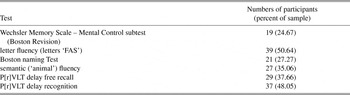
Table 1 displays the numbers of participants who were categorized as single versus multi-domain MCI based on Peterson criteria (Table 1a) and lists the number of MCI participants who scored ≤1.5 standard deviation units on each test (Table 1b). Incidence of single-domain and multi-domain mild cognitive impairment using Peterson criteria (n = 77) Numbers of participants who score ≤1.5 standard deviation units below normative values for each test (n = 77) MCI = mild cognitive impairment; P[r]VLT = Philadelphia (repeatable) Verbal Learning Test.

With respect to single-domain MCI, 34 participants were classified as impaired in one domain of cognitive functioning. Approximately equal numbers of participants showed isolated memory impairment (n = 15) and an isolated dysexecutive MCI profile (MCI; n = 13). Very few patients (n = 6) were assigned into the single-domain naming/lexical retrieval MCI group. An almost equal number of participants were classified with multi-domain MCI on the basis of low scores in at least two domains of cognitive functioning (n = 29). Fourteen participants scored ≤1.5 standard deviation units below normative values on at least one test from all three cognitive domains. Taken together, 43 or 55.84% of the MCI group presented with multi-domain MCI.
Confirmatory Factor Analysis
The confirmatory factor analysis used to validate out predetermined groupings of our six neuropsychological variables into three cognitive domains revealed a goodness of fit test suggesting that the hypothesized three domain structure described above was appropriate (χ2 = 11.67; df = 6; p = .069, ns).
Cluster Analysis
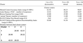
A k-mean cluster analysis (SPSS, version 16) was conducted on MCI participants to identify relatively homogeneous participant groups as described above. Three a priori cluster groups were specified. The results of this analysis are displayed in Table 2.
K-Mean cluster analysis for patients meeting criteria for mild cognitive impairment (n = 77)

P(r)VLT = Philadelphia (repeatable) Verbal Learning Test.
Group 1 (dysexecutive MCI; n = 21) presented with low scores on both executive tests. The largest group was group 2, the so-called mixed group (n = 42). These patients obtained low scores on tests of letter fluency, “animal” fluency, and scored approximately one standard deviation below the mean on both memory test conditions compared with the NC group. Group 3 (aMCI; n = 14) presented with significant memory impairment, suggestive of a primary anterograde amnesia. The average Euclidean distance between each case and its classification center also is shown in Table 2. The greatest distance was obtained between the two single domain MCI groups, that is, the dysexecutive and amnesic groups. Approximately equal distance between cluster centers was obtained between the mixed MCI group versus the dysexecutive and amnesic MCI groups, respectively.
It should be noted that attempts to cluster individuals into two-, four-, and five-cluster solutions either produced groups containing one to two individuals per cluster (e.g., in the five-cluster solution) or variables that loaded across all factors (e.g., in the four-cluster solution). These analyses combined with our confirmatory factor analysis of our predetermined cognitive domains provided evidence for the use of our three-cluster solution.
Neuropsychological Performance
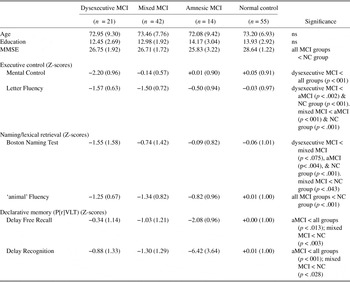
Demographic characteristics for the three MCI groups derived from the cluster analysis and the NC group are displayed in Table 3. Age and education did not differ between groups (Table 3). On the MMSE (F[1,151] = 16.51; p < .001), the NC group obtained a higher score compared with all MCI groups (p < .001). There were no differences between the three MCI groups. Three separate multivariate analyses of variance were conducted to assess for group differences on executive, naming/lexical retrieval, and memory tests.
Group demographics and neuropsychological performance [mean (SD)] for mild cognitive impairment groups derived from cluster analysis (n = 77)

MCI = mild cognitive impairment; MMSE = Mini Mental State Examination; P[r]VLT = Philadelphia (repeatable) Verbal Learning Test.
For executive tests the multivariate effect of group was significant (p < .001). Univariate analyses for both executive tests also were significant (p < .001). Follow-up analyses (Tukey tests) showed that the WMS-Mental Control AcI for the dysexecutive MCI group was lower compared with all other groups (p < .001). No other follow-up comparisons were significant. A different pattern emerged on the letter fluency test. The dysexecutive MCI group generated fewer words compared with the amnesic (p < .002) and NC groups (p < .001); however, there was no difference on this test between the dysexecutive and mixed MCI groups. The mixed MCI group also produced fewer responses compared with the aMCI and NC groups (p < .001).
For naming/lexical retrieval tests the multivariate effect for group was significant (p < .001). Univariate analyses for the Boston Naming Test and the “animal” fluency tests also yielded highly significant effects for the group (p < .001). For the BNT follow-up analyses found that the dysexecutive MCI group obtained a lower score compared with the aMCI (p < .004) and NC groups (p < .001). The mixed MCI group scored lower on this test compared with the NC group. For the “animal” fluency test, all MCI groups generated lower output compared with the NC group (p < .001); however, there was no difference on this measure between the three MCI groups.
For the two P[r]VLT test conditions the multivariate effect for group was significant (p < .001) as were both univariate analyses for both P[r]VLT test conditions (p < .001). Follow-up analyses (Tukey Tests) for the P[r]VLT delayed recall test condition showed that the aMCI group recalled fewer words compared with all other groups (p < .001). The mixed MCI group also recalled fewer words compared with the NC group (p < .003). For the P[r]VLT recognition discriminability index a similar profile emerged. The aMCI group produced a lower score compared with all other groups (p < .001) and the mixed MCI group scored lower on this test condition compared with the NC group (p < .028).
DISCUSSION
In the United States, it is estimated that 4 million people suffer from AD (Hebert et al., Reference Hebert, Scherr, Bienias, Bennett and Evans2003). This number is expected to grow to approximately 13 million by mid century (Hebert, Scherr, Bienias, Bennett, & Evans, Reference Hebert, Scherr, Bienias, Bennett and Evans2003). As disease modifying agents become available, it is critical that dementia is identified as early as possible. Moreover, the figures reported above do not necessarily take into account the influence of risk factors for stroke and concomitant vascular disease. Given that newer research suggests that risk factors for stroke are also risk factors for AD (Dufouil, Alperovitch, Ducros, & Tzourio, Reference Dufouil, Alperovitch, Ducros and Tzourio2003; Fernando & Ince, Reference Fernando and Ince2004; Riekse et al., Reference Riekse, Leverenz, McCormick, Bowen, Teri and Nochlin2004; Szolnoki et al., Reference Szolnoki, Somogyvari, Kondacs, Szabo, Fodor and Bene2004), the figures reported above may be underestimating the prevalence of dementia.
In the current research, we chose a somewhat conservative approach and used a norm-referenced score of ≤ 1.5 standard deviation units below the mean to identify persons with MCI (Table 1). On the other hand, we followed, perhaps, a more liberal path by recruiting into our MCI sample persons who obtained a score of ≤ 1.5 standard deviation units on any of six tests. The reasons for this strategy are two-fold. First, it is not at all clear what cutoff is best, that is, some researchers have used a cutoff of −1.2 standard deviation units to identify persons with MCI (Busse et al., Reference Busse, Hensel, Gühne, Angermeyer and Riedel-Heller2006; Delano-Wood et al., Reference Delano-Wood, Bondi, Sacco, Abeles, Jak and Libon2009). Second, there is no consensus regarding which neuropsychological tests are most effective in identifying MCI. By using a cut off of ≤1.5 standard deviation units we attempted to guard against a type 1 error; however, by permitting a low score on any of six tests, we acknowledge the possibility of making a type 2 error (Brooks, Iverson, & White, Reference Brooks, Iverson and White2007; Brooks, Grant, Holdnack, & Feldman, Reference Brooks, Grant, Holdnack and Feldman2008). Indeed, our rates of discrimination are slightly lower than base rates of low memory performance in older community dwelling adults (Brooks et al., Reference Brooks, Iverson and White2007), but slightly higher than those reported in large-scale epidemiological studies of pathological aging in patients followed through a memory disorders clinic (Edwards, Lindquist, & Yaffe, Reference Edwards, Lindquist and Yaffe2004).
Alternatively, three composite scores representing performance on the six tests described above could have been calculated. We chose not to follow this procedure for two strategic reasons. First, elements of the six neuropsychological tests/paradigms used in the current research are likely to be part of any neuropsychological protocol designed to investigate MCI. Therefore, we justify not using composite scores because we specifically wished to assess how each individual test may be related to each other around the issue(s) of identifying persons with a potential prodromal dementia. Second, one of the goals of the current research was our desire to use the six tests described above to test the prospective hypothesis that it may be possible to identify three distinct MCI subgroups. We acknowledge that our methods may place some limitations on the generalizability of our results. Also, it should be kept in mind that we studied an overwhelming Caucasian group seen at a memory clinic, that is, persons who were self-referred because of their self-perception of a decline in cognitive functioning. This kind of sample may also place some limits on the generalizability of our results when considering the prevalence of MCI subtypes in the general population and/or patients from minority communities.
Despite these caveats, several interesting observations were derived from our three cluster solution. First, the CFA provided support for one of the goals of the current research which was our desire to use the six tests described above to test the prospective hypothesis that it may be possible to identify three distinct MCI subgroups. Second, our results support Petersen’s nosology of single amnesic, single nonamnesic, and multi-domain MCI groups. For example, an aMCI group was clearly identified, although only a small number of participants presented with this profile, suggesting that aMCI may not represent the majority of persons presenting to the memory clinic with subjective cognitive complaints. These aMCI individuals obtained very low scores on tests of delayed free recall and delayed recognition discriminability suggesting the presence of an anterograde amnesia. Performance on most nonmemory tests was largely intact except for comparatively low output on the “animal” fluency test. Low output on semantically guided fluency tests in a group of individuals with anterograde amnesia might suggest the presence of concomitant semantic memory deficits (Adlam, Bozeat, Arnold, Watson, & Hodges, Reference Adlam, Bozeat, Arnold, Watson and Hodges2006; Carew et al., Reference Carew, Cloud, Lamar, Grossman and Libon1997; Dudas, Clague, Thompson, Graham, & Hodges, Reference Dudas, Clague, Thompson, Graham and Hodges2005; Murphy, Rich, & Troyer, Reference Murphy, Rich and Troyer2006). Howieson at al. (Reference Howieson, Carlson, Moore, Wasserman, Abendroth and Payne-Murphy2008) found that patients who were ultimately diagnosed with aMCI initially presented with reduced output on semantically guided fluency tests. When these prior studies are combined with the results of the present study, finer distinctions might be obtainable in individuals presenting with aMCI. For example, it may be possible that different neuropathology underlies aMCI with and without concomitant semantic memory impairment. The course, illness duration, and possible treatment of such subgroups could also be different.
The cluster analysis also produced a group of patients classified into a dysexecutive MCI group unique for their differential impairment on both executive tests; particularly so on the WMS-Mental Control subtest (Boston Revision). In patients with dementia, subcortical vascular disease is often associated with poor performance on this test (Libon et al., Reference Libon, Price, Giovannetti, Swenson, Bettcher and Heilman2008; Price et al., Reference Price, Jefferson, Merino, Heilman and Libon2005). Whether vascular disease is responsible for the differential impairment on executive tests in patients classified as dysexecutive MCI is unknown at the present time. Gainotti, Ferraccioli, Vita, and Marra (Reference Gainotti, Ferraccioli, Vita and Marra2008) found that MCI patients with evidence of subcortical vascular disease performed worse on executive, rather than memory tests. Using diffusion tensor imaging (DTI) Delano-Wood et al. (Reference Delano-Wood, Bondi, Sacco, Abeles, Jak and Libon2009) found MCI patients with higher risks for stroke also presented with greater white matter disease and poorer fractional anisotropy (FA) involving the splenium of the corpus callosum. Our dysexecutive MCI group also displayed mild impairment on both tests related to language functioning, that is, the Boston Naming Test and the “animal” fluency test. Impairment in mental search and retrieval skills associated with mild or subtle vascular disease (Libon et al., 2004) might be the mechanism that explains why the dysexecutive MCI group presented with difficulty on language-related tests. Clearly, this should be the subject of future research.
A large number of MCI patients presented with a rather mixed or heterogeneous profile suggesting impairment across multiple domains of cognitive functioning. The mixed MCI group derived from cluster analysis displayed difficulty on both fluency tests, some impairment on the Boston Naming Test, and reduced performance on the P[r]VLT delayed recognition discriminability index. While deficits associated with mental search and retrieval might underlie these difficulties, the combination of low scores on executive and memory tests begs the question of whether these mixed MCI patients might go on to develop a mixed dementia syndrome. More research is necessary to address this question.
Our data are both convergent and divergent when compared with the cluster solution reported by Delano-Wood et al. (Reference Delano-Wood, Bondi, Sacco, Abeles, Jak and Libon2009). Both studies examined patients from an outpatient memory clinic, and both cluster solutions provide support for Petersen’s nosology of the existence of single domain and multi-domain MCI subtypes. Our data diverge from those of Delano-Wood in that we demonstrate a different composition of clusters. The differences in cluster solution are likely due to the different neuropsychological protocols implemented. A significant limitation of the current research is the lack of corroborating information, that is, data from imaging studies which may have shed some light on the underlying neuropathology associated with each MCI group.
By not using composite Z-scores of neuropsychological test variables, we may have gained some insight into the general sensitivity and specificity of individual neuropsychological tests as related to MCI. For example, performance on both delayed free recall and recognition memory tests may turn out to have considerable specificity given that poor performance on these tests have been linked to gray matter degenerative disease with eventual conversion to AD. Similarly, it may also turn out that either risk factors for stroke or imaging evidence for vascular disease could be directly linked to impaired performance on specific tests of executive control as suggested by Delano-Wood et al. (Reference Delano-Wood, Bondi, Sacco, Abeles, Jak and Libon2009). By contrast, other tests, such as output on the “animal” fluency test were uniformly low across all three MCI groups and may not be useful in the early detection of specific MCI syndromes. Different cognitive constructs associated with specific underlying neuropathology could be responsible for these findings. Hence, performance on fluency tests may be sensitive to overall cognitive impairment in MCI, but may not suggest the presence of a specific type of neuropathology. Issues regarding how neuropsychological test performance is related to possible underlying neuropathology in MCI could have significant treatment implications.
Finally, it has been suggested that neuropsychological assessment is both expensive and time consuming and, therefore, may not be the most parsimonious method for investigating MCI. We disagree. The neuropsychological evaluation used in the current research requires no more than 1 hr to administer and score. The costs for such an evaluation are far less than an MRI study of the brain. Moreover, the quantitative information that has been the subject of the current research, as well as an analysis of errors and processes, which will be the subject of a future report, yield a plethora of information. Future strategies attempting to disambiguate the brain–behavior relationships associated with MCI should combine neuropsychological, imaging, and biomarker data (DeCarli et al., Reference DeCarli, Frisoni, Clark, Harvey, Grundman and Petersen2007; Twamley et al., Reference Twamley, Ropacki and Bondi2006).